Introduction
Since the birth in 1978 of the world’s first baby conceived by in vitro fertilization, assisted reproduction technology (ART) has brought children to thousands of families. Nevertheless, despite 40 years of advancements in ART, clinicians still face challenges such as low rates of embryo implantation, clinical pregnancy, and live birth. Factors influencing the success of ART included female age (Moon et al., Reference Moon, Kim, Jung, Jee, Suh and Lee2000; Ma et al., Reference Ma, Chen, Hu, Dai, Bu and Sun2018), uterine environment (Galliano et al., Reference Galliano, Bellver, Diaz-Garcia, Simon and Pellicer2015), and embryo quality (Siristatidis et al., Reference Siristatidis, Sertedaki and Vaidakis2017). The prediction of embryonic development potential is a critical step in determining the clinical outcomes of ART.
Generally, the factors that influence embryonic developmental potential include the health of the oocytes and sperm and the embryo culture environment. Previous studies have investigated the key factors affecting embryo quality, mainly focusing on oocytes and the culture environment (Swiatecka et al., Reference Swiatecka, Bielawski, Anchim, Lesniewska, Milewski and Wolczynski2014; Kamath et al., Reference Kamath, Mascarenhas, Kirubakaran, Nair and Kulkarni2018). However, the sperm-related factors that affect embryo quality are rarely studied. Currently, male fertility is evaluated through standard semen analysis, including total sperm count, sperm concentration, sperm progressive motility, and sperm morphology. However, semen quality assessment results are within the normal range for c. 15% of infertile men, indicating that semen quality assessment has certain limitations and cannot accurately evaluate male fertility (Chia et al., Reference Chia, Tay and Lim1998; Agarwal and Allamaneni, Reference Agarwal and Allamaneni2005). Sperm DNA fragmentation (SDF) can be added as a potential diagnostic tool in semen quality analysis to produce a more comprehensive evaluation of male fertility (Evenson et al., Reference Evenson, Larson and Jost2002).
Sperm with high SDF not only can survive but also have normal morphology and the ability to fertilize oocytes (Dar et al., Reference Dar, Grover, Moskovtsev, Swanson, Baratz and Librach2013; Boushaba and Belaaloui, Reference Boushaba and Belaaloui2015). Several studies have investigated the effect of SDF on the clinical outcome of ART. However, the conclusions remain controversial. Some studies have concluded that SDF can predict the outcome of assisted reproduction and that high SDF significantly reduces embryo quality and increases abortion rates (Zini et al., Reference Zini, Meriano, Kader, Jarvi, Laskin and Cadesky2005; Enciso et al., Reference Enciso, Pieczenik, Cohen and Wells2012). In contrast, other studies have reported that SDF does not affect embryo quality or clinical outcome and cannot predict the outcome of assisted reproduction (Gat et al., Reference Gat, Tang, Quach, Kuznyetsov, Antes, Filice, Zohni and Librach2017; Sun et al., Reference Sun, Zhang, Li, Liu, Yi, Tian and Liu2018; Antonouli et al., Reference Antonouli, Papatheodorou, Panagiotidis, Petousis, Prapas, Nottola, Palmerini, Macchiarelli and Prapas2019). In these studies, embryo assessment was based on a static morphological score. This score involves the assessment of embryo morphology at specific timepoints. However, the details of embryonic development in the time period between observation points were not clear; therefore, the scoring system has certain limitations and is not able to accurately predict the embryonic development potential (Montag et al., Reference Montag, Liebenthron and Koster2011).
In recent years, with the development and application of time-lapse technology, the above challenges in embryo morphology assessment have been solved. Time-lapse technology involves momentary exposure capture imaging technologies that can accurately record the full embryonic developmental process. Through an intelligent data analysis system, morphokinetic parameters and cleavage patterns were analyzed, and a model of embryo quality assessment was established to predict embryonic development potential (Liu et al., Reference Liu, Chapple, Feenan, Roberts and Matson2016, Reference Liu, Feenan, Chapple and Matson2019). Using this model to select the best embryos for transfer can not only improve the embryo implantation rate but also reduce the number of embryo transfers and prevent multiple pregnancies. Furthermore, the time-lapse imaging approach does not require the embryo to be removed from the incubator at a specific time, therefore reducing the number of times the incubators must be opened or closed and maintaining constant culture conditions to avoid unnecessary stress and fluctuations in environmental factors.
Many factors, such as maternal age (VerMilyea et al., Reference VerMilyea, Ivani, Gvakharia, Boostanfar, Baker and Conaghan2014), the ovarian stimulation protocol selected (Gryshchenko et al., Reference Gryshchenko, Pravdyuk and Parashchyuk2014), culture conditions (Kazdar et al., Reference Kazdar, Brugnon, Bouche, Jouve, Veau, Drapier, Rousseau, Pimentel, Viard, Belaud-Rotureau and Ravel2017), sperm source (Scarselli et al., Reference Scarselli, Casciani, Cursio, Muzzi, Colasante, Gatti, Greco, Greco, Minasi and Greco2018) and SDF, affect the morphokinetic parameters and cleavage patterns of the embryo. However, research on the effects of SDF on morphokinetic parameters and cleavage patterns is very limited and controversial. This study aimed to use sperm chromatin dispersion (SCD) to detect the processed sperm DNA fragmentation rate and to explore whether SDF can affect embryo morphokinetic parameters, cleavage patterns, and embryo quality in intracytoplasmic sperm injection (ICSI).
Materials and methods
Selection of patients
This retrospective analysis included 151 ICSI cycles (1152 embryos) using time-lapse technology from November 2016 to June 2019 at the Reproductive Medicine and Genetics Centre of People’s Hospital of Guangxi Zhuang Autonomous Region. To strictly control the effect of female factors, we adopted the following inclusion and exclusion criteria. Inclusion criteria: (a) maternal age ≤ 35 years old; (b) basic follicle-stimulating hormone (FSH) <10 IU/l; (c) number of basic antral follicles (AFC) ≥ 5; (d) number of metaphase II (MII) oocytes ≥ 5; (e) fresh semen obtained by masturbation on the day of oocyte collection showing <1 million total sperm after processing; (f) male abstinence for 2–7 days prior to sample collection; and (g) peripheral blood karyotyping confirmed normal chromosome complements for both male and female parents. Exclusion criteria: ICSI cycle with surgical sperm extraction. The included patients were divided into two groups on the basis of the SDF rate: SDF < 15% (n = 114) and SDF ≥ 15% (n = 37) (Benchaib et al., Reference Benchaib, Lornage, Mazoyer, Lejeune, Salle and Francois2007; Fei et al., Reference Fei, Huang, Jin and Huang2014). This study was approved by the Ethics Committee of People’s Hospital of Guangxi Zhuang Autonomous Region.
Semen collection and preparation
Fresh semen samples were collected by masturbation on the day of oocyte collection in a dry and sterile cup and placed in a 37°C incubator for liquefaction. Semen quality assessment was performed according to the fifth edition of the WHO guidelines (World Health Organization, 2010). When the semen was completely liquefied, it was added to the upper layer of the gradient solution (40% and 80%, 1:1, SpermGrad, Vitrolife, Goteborg, Sweden). Following centrifugation at 450 g for 15 min, the seminal plasma supernatant was carefully removed, and the pellet was washed in 2 ml of G-IVF PLUS medium (Vitrolife, Goteborg, Sweden). After centrifugation at 450 g for 5 min, the supernatant was removed. The pellet was added to the bottom of the medium containing 0.5 ml of G-IVF PLUS, and the sperm were allowed to swim up for 30 min before ICSI was performed.
SCD test for SDF
SDF was assessed by the SCD technique using the Halosperm kit (Halotech DNA SL). Eppendorf tubes containing 100 μl of agarose were placed in a 90–100°C water bath for 5 min to melt the agarose and then in a 37°C water bath for 5 min. Then, 25 μl of sperm samples were added to the tube and mixed well. Approximately 14 μl of this mixture were dropped on a treated slide provided with the kit and covered with an 18 × 18 mm coverslip. The slides were maintained in a refrigerator at 4°C for 5 min to allow the agarose to set into a microgel with the sperm cells embedded within it. The coverslips were removed gently. The slides were placed horizontally in an acid solution previously prepared by mixing 80 μl of HCl (AD) with 10 ml of distilled water for 7 min. The slides were then immersed in lysis solution for 25 min. After washing with distilled water for 5 min, the slides were dehydrated in increasing concentrations of ethanol (70, 90 and 100%) for 2 min each and then air dried. The slides were then stained by immersion in Wright–Giemsa staining solutions A and B (1:1) for 15 min. SDF was divided into five grades on the basis of halo size: (1) large halo: halo width similar to or higher than the minor diameter of the core; (2) medium halo: halo width smaller than the minor diameter of the core but greater than one-third the minor diameter; (3) small halo: halo width similar to or smaller than one-third of the minor diameter of the core; (4) without halo; (5) no halo and degraded: no halo present and an irregularly or weakly stained core. Spermatozoa with small halos, without halos, and with no halo and degraded were defined as SDF. In total, 500 spermatozoa were assessed for each semen sample, and the percentage of SDF was calculated.
Controlled ovarian stimulation and oocyte collection
All ICSI cycles included ovarian stimulation with a standard long protocol using a gonadotrophin-releasing hormone antagonist and FSH (Gonal-F, Merck Serono, Modugno, Italy). Follicle growth was monitored by B-ultrasound. When one of the dominant follicles was 18 mm or three were 17 mm in diameter, human chorionic gonadotrophin (hCG, Merck Serono, Modugno, Italy) was injected. After 36 h, the follicles were punctured under the guidance of B-ultrasound to retrieve the cumulus–oocyte complexes (COCs). The COCs was washed with G-MOPS PLUS (Vitrolife, Goteborg, Sweden) and then placed into G-IVF PLUS (Vitrolife, Goteborg, Sweden) medium covered with mineral oil (Vitrolife, Goteborg, Sweden), which was pre-equilibrated at 37°C and 6% CO2 overnight. The COCs were then incubated at 37°C, in 6% CO2 and 5% O2 in air for ICSI.
Embryo culture and embryo transfer
After culturing for c. 3 h, the COCs were transferred into 80 IU/ml hyaluronidase (Irvine Scientific, California, USA) preheated to 37°C. The surrounding cumulus cells were removed mechanically by a 140-μm diameter denuding pipette (Cook, Philadelphia, USA) and then transferred into the G-IVF PLUS medium covered with mineral oil, which was pre-equilibrated at 37°C and 6% CO2 in air for c. 1 h before ICSI. The injected MII oocytes were transferred into G-1 PLUS medium (Vitrolife, Goteborg, Sweden), covered with mineral oil in a Primo Vision dish (9-well or 16-well, Vitrolife, Viby, Denmark) and cultured in a Primo Vision (Vitrolife, Budapest, Hungary) incubator at 37°C, 6% CO2, and 5% O2 in air for 3 days. On day 3 (D3) after ICSI, we selected the two best embryos for transfer or freezing, and then the surplus embryo was transferred to G-2 PLUS medium (Vitrolife, Goteborg, Sweden) covered with mineral oil and cultured in a pre-equilibrated Primo Vision dish for 3 days. Images of each embryo were recorded every 10 min across seven equidistant focal planes using Primo Vision capture software.
On D3, two embryos were transferred into the patient’s uterine cavity. The serum hCG concentration test was performed 14 days after embryo transfer. Serum hCG > 10 mIU/ml was defined as chemical pregnancy. Clinical pregnancy was defined as the presence of a gestational sac and heartbeat was confirmed by ultrasound examination at the 4th week after embryo transfer. The implantation rate was calculated as the number of gestational sacs divided by the number of embryos transferred. Miscarriage was defined as pregnancy loss before 12 weeks.
Embryo assessment
The embryo quality and the cleavage stage on D3 were evaluated by referring to the Istanbul consensus (Alpha Scientists in Reproductive Medicine, 2011), which was divided into four grades on the basis of cell number, homogeneity and fragment proportion. Grades 1 and 2 indicate high-quality embryos (≥6 cells), grades 3 and above indicate available embryos, and grade 4 indicates low-quality embryos; blastocyst quality on D5/6 was evaluated by referring to Gardner scoring (Gardner et al., Reference Gardner, Lane, Stevens, Schlenker and Schoolcraft2000). Blastocysts were classified into six stages on the basis of the degree of blastocyst cavity expansion. Inner cell mass or trophectoderm quality was classified as grade A, B or C on the basis of the number of inner cell masses or trophectoderm cells. Blastocysts with a score of ≥ 3BB were defined as high-quality blastocysts, and those with only an inner cell mass or a trophectoderm score of ≥ B were defined as available blastocysts.
Morphokinetic parameters and cleavage pattern
The video data of 2PN embryos from enrolled patients were analyzed. The morphokinetic parameters and cleavage pattern were assessed according to the proposed guidelines for the annotation of dynamic human embryo monitoring (Ciray et al., Reference Ciray, Campbell, Agerholm, Aguilar, Chamayou, Esbert and Sayed2014): the time when the second polar body was completely detached from the oolemma (tPB2), time of appearance of pronuclei (tPNa), time of pronuclei disappearance (tPNf), two to nine discrete cell stages (t2 to t9), first evidence of compaction (tSC), end of compaction process (tM), initiation of blastulation (tSB), expansion of blastocyst (tB) and initiation of hatching process (tHN), the time from 2-cell to 3-cell stage (CC2 = t3–t2), time from 3-cell to 4-cell stage (S2 = t4–t3), time from tPNf to 5-cell stage (T5–tPNf), and time from the 5-cell to the 8-cell stage (S3 = t8–t5) were also calculated.
On the basis of the cleavage pattern, the embryos were divided into five types: (I) direct cleavage (DC) (Athayde et al., Reference Athayde, Chen, Conaghan, Ivani, Gvakharia, Behr, Suraj, Tan and Shen2014), in which the cell directly divides into ≥ 3 cells; (II) reverse cleavage (RC) (Liu et al., Reference Liu, Chapple, Roberts and Matson2014), in which, before the 8-cell stage, two independent cells recombine into one cell or the cytoplasm shrinks but does not divide; (III) chaotic cleavage (CC) (Athayde et al., Reference Athayde, Chen, Conaghan, Ivani, Gvakharia, Behr, Suraj, Tan and Shen2014), in which the cell is divided into ≥ 4 cells with irregular sizes during the first division and produces a large number of cell fragments; (IV) multinucleation (Mn), where Mn occurs in any cell at the 2- or 4-cell stage; and (V) MIX, where the cell exhibits two or more of the above abnormal cleavage phenomena.
Statistical analysis
Statistical analysis was carried out using SPSS Statistics 19.0 software (IBM SPSS Corp., Chicago, IL, USA). Continuous variables are presented as the mean ± standard deviation (SD), and categorical variables are presented as percentages (%). The normal distribution was used for all numerical data using the Kolmogorov–Smirnov test. If the numerical data were consistent with a normal distribution, they were examined using Student’s t-test; otherwise, the Mann–Whitney U-test was used. A chi-squared test was used to compare categorical variables. A P-value < 0.05 was considered to indicate statistical significance.
Results
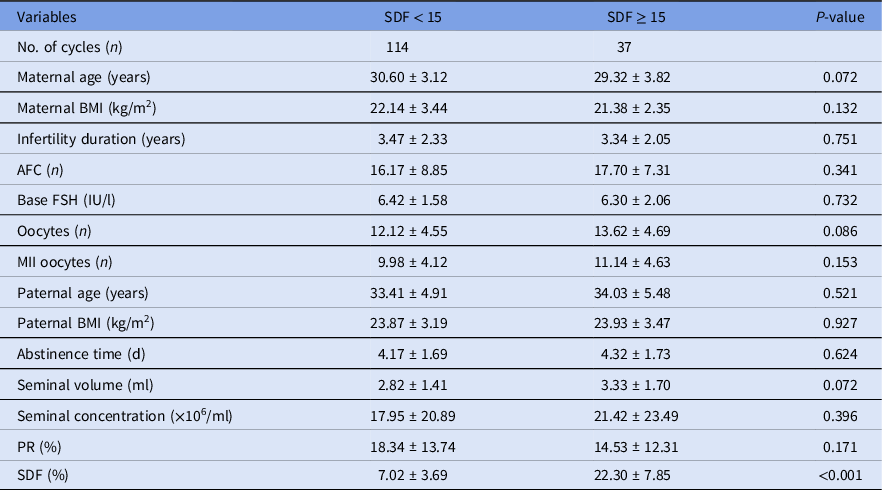
In this study, in total, 151 ICSI cycles were divided into two groups: SDF < 15% (n = 114) and SDF ≥ 15% (n = 37). No significant differences were observed between the two groups in terms of maternal age, maternal body mass index (BMI), infertility years, basic FSH, AFC, paternal age, oocyte number, metaphase II (MII) number, paternal abstinence days, seminal volume, seminal concentration or progressive motility. All variables other than SDF were equally distributed between groups (Table 1).
The patient demographic parameters in different SDF levels

Note: Values are expressed as mean ± SD.
AFC = basic antral follicles; BMI = body mass index; PR = progressive motility; SDF = sperm DNA fragmentation.
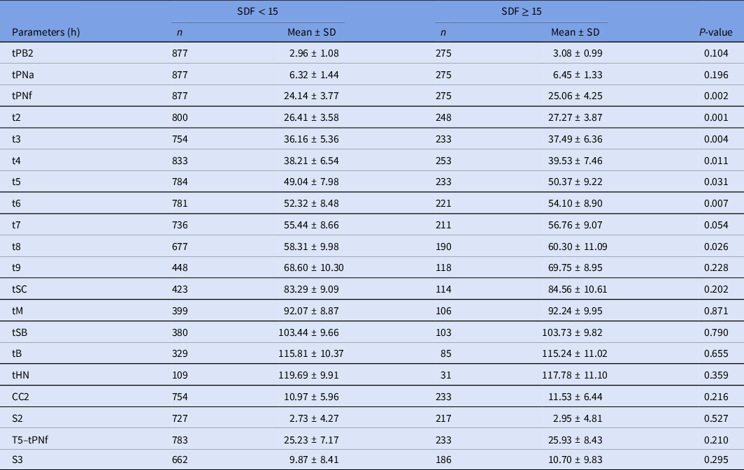
Embryonic development in the SDF < 15% group was significantly faster than that in the SDF ≥ 15% group, with earlier tPNf, t2, t3, t4, t5, t6, and t8 (P < 0.05). No significant difference was found in other morphokinetic parameters between the two groups (Table 2).
Comparison of morphokinetic parameters between two groups

Note: Values are expressed as mean ± SD. SDF = sperm DNA fragmentation; tPB2 = the second polar body is completely detached from the oolemma; tPNa = time of appearance of pronuclei; tPNf = time of pronuclei disappearance; t2 to t9 = two to nine discrete cells; tSC = first evidence of compaction; tM = end of compaction process; tSB = initiation of blastulation; tB = expansion blastocyst; tHN = initiation of hatching process; CC2 = t3–t2; S2 = t4–t3; T5–tPNf = t5–tPNf; S3 = t8–t5.
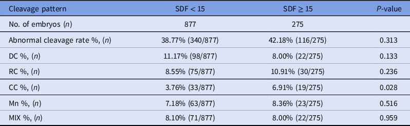
The overall abnormal cleavage rate was similar in both groups. However, the rate of CC was significantly higher in the SDF ≥ 15% group than in the SDF < 15% group (P < 0.05; Table 3).
Comparison of embryo cleavage pattern between two groups

Note: CC = chaotic cleavage; DC = direct cleavage; MIX = cell exhibits two or more of the above abnormal cleavage; Mn = multinucleation; RC = reverse cleavage; SDF = sperm DNA fragmentation.
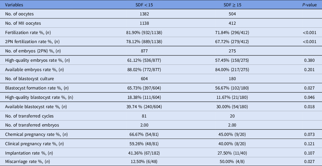
The fertilization and 2PN fertilization rates in the SDF < 15% group were significantly higher than those in the SDF ≥ 15% group (P < 0.05). The proportions of high-quality embryos and available embryos were similar between the two groups. A substantially higher blastocyst formation rate, high-quality blastocyst rate, and available blastocyst rate were observed in the SDF < 15% group than in the SDF ≥ 15% group. Furthermore, the chemical pregnancy, clinical pregnancy and implantation rates decreased with the increase in the SDF level, although no statistical significance was detected. Finally, compared with that of the SDF < 15% group, the miscarriage rate of the SDF ≥ 15% group showed an upward trend (P < 0.05; Table 4).
Comparison of laboratory and clinical outcomes between two groups

Note: PN = pronuclei; SDF = sperm DNA fragmentation.
Discussion
In recent years, many studies on SDF have been performed to predict male fertility and the clinical outcomes of ART. However, conflicting results have been reported. Several studies have indicated that high SDF negatively affected fertilization ability and embryonic development potential (Simon et al., Reference Simon, Lutton, McManus and Lewis2011; Tandara et al., Reference Tandara, Bajic, Tandara, Bilic-Zulle, Sunj, Kozina, Goluza and Jukic2014). In contrast, other studies have stated that oocytes have a stable mechanism for repairing SDF and that SDF was not correlated with fertilization rate, embryo quality, or clinical outcome (Sakkas and Alvarez, Reference Sakkas and Alvarez2010; Bronet et al., Reference Bronet, Martinez, Gaytan, Linan, Cernuda, Ariza, Nogales, Pacheco, San and Garcia-Velasco2012; Anifandis et al., Reference Anifandis, Bounartzi, Messini, Dafopoulos, Markandona, Sotiriou, Tzavella and Messinis2015). By using time-lapse technology, we observed that embryonic development was significantly faster in the SDF < 15% group than in the SDF ≥ 15% group, with shorter times to tPNf, t2, t3, t4, t5, t6, and t8 (P < 0.05). Among these parameters, the timing of t2, t3, t4, and t5 affected blastocyst formation and embryonic development potential (Milewski et al., Reference Milewski, Kuc, Kuczynska, Stankiewicz, Lukaszuk and Kuczynski2015; Storr et al., Reference Storr, Venetis, Cooke, Susetio, Kilani and Ledger2015). Furthermore, the fertilization rate, 2PN fertilization rate, and blastocyst quality were significantly higher in the SDF < 15% group than in the SDF ≥ 15% group. Therefore, high SDF has a negative effect on the morphokinetic parameters and developmental potential of embryos.
In ART, the results of SDF studies have varied due to the application of different fertilization methodologies. In IVF cycles, oocytes can naturally select the sperm, preferentially allowing fertilization by sperm with normal morphology and microstructure. This process weakens the influence of SDF on the outcome of this IVF cycle to some extent, therefore the influence of SDF on clinical outcomes in the IVF cycle is controversial (Esbert et al., Reference Esbert, Pacheco, Vidal, Florensa, Riqueros, Ballesteros, Garrido and Calderon2011; Borges et al., Reference Borges, Zanetti, Setti, Braga, Provenza and Iaconelli2019). In this study, ICSI cycles were chosen as the research object. ICSI bypasses the natural selection of sperm by oocytes, which can lead to a higher frequency of injection of sperm with high SDF. Through this process, the effect of natural selection is reduced as much as possible, therefore ICSI outcomes may better reflect the effects of SDF on embryo morphokinetic parameters.
To date, the single-cell gel electrophoresis assay (comet assay), terminal deoxynucleotidyl transferase-mediated nick end labelling assay (TUNEL), sperm chromatin structure assay (SCSA) and SCD have been used to measure SDF. Studies have shown that these methods produce similar results in terms of detecting SDF (Chohan et al., Reference Chohan, Griffin, Lafromboise, De Jonge and Carrell2006). The SCD assay is a sensitive, specific and economical test because it does not require special and expensive instrumentation. Moreover, some scholars have stated that the SCD assay can be used as the gold standard for the detection of SDF (Fernandez et al., Reference Fernandez, Muriel, Goyanes, Segrelles, Gosalvez, Enciso, LaFromboise and Dejonge2005).
Several studies have reported the relationship between SDF and morphokinetic parameters of embryonic development, but the results were inconsistent. Lundberg et al. (Reference Lundberg, Hambiliki, Sondèn, Akerlund and Bungum2014) divided SDF into four groups: 0–10%, 10.1–20%, 20.1–30%, and >30%. The morphokinetic parameters tPB2, tPNf, t2, and tB were compared among these SDF groups, but no significant difference was observed, and they therefore reported that SDF was not correlated with the morphokinetic parameters of early embryonic development. In contrast, Wdowiak et al. (Reference Wdowiak, Bakalczuk and Bakalczuk2015) showed that SDF has a significant positive correlation with morphokinetic parameters, such as tPNa, tPNf, t2, t3, t4, t5, and tB. A high SDF level in the ICSI cycle was believed to significantly delay the dynamic parameters of embryonic development and affect embryonic development quality and implantation rate. Our study was consistent with this report. Our evidence suggested that the morphokinetic parameters tPNf, t2, t3, t4, t5, t6, and t8 were significantly shorter in the SDF < 15% group than in the SDF ≥ 15% group. Some studies have shown that the delay in embryo morphokinetic parameters caused by a high level of SDF may be due to the time spent by oocytes to repair sperm DNA damage (Scully et al., Reference Scully, Panday, Elango and Willis2019). t2 and S3 were closely related to the formation of high-quality blastocysts and can be used as independent predictors of blastocyst formation (Storr et al., Reference Storr, Venetis, Cooke, Susetio, Kilani and Ledger2015). Concordantly, the high level of SDF negatively affected the embryo morphokinetic parameters and consequently affected the later embryonic development.
The relationship between SDF and abnormal cleavage during embryo development has not been investigated. Our study showed that the abnormal cleavage rates were similar in the SDF ≥ 15% group and the SDF < 15% group. However, among the four types of cleavage patterns, the rate of CC was significantly higher in the SDF ≥ 15% group than in the SDF < 15% group. The CC abnormal cleavage pattern occurs in the first mitosis, with disordered cleavage and the production of numerous cell fragments. The centrosome plays a dominant role in cell division in most species, including humans (but not most rodents) (Sathananthan, Reference Sathananthan1998). After the sperm enters the oocyte, the proximal centriole of the sperm forms the sperm aster, which can lead to the juxtaposition of male and female pronuclei and to the formation of bipolar spindles during mitosis, ensuring the equal separation of sister chromatids into the cytoplasm of the two offspring. Sperm morphological abnormalities, especially deformities in the neck, where the centriole is located, result in abnormal spindles, leading to abnormal embryo mitosis and eventually abnormal cleavage (Stevenson et al., Reference Stevenson, Kramer, Kuhn and Theurkauf2001; Chatzimeletiou et al., Reference Chatzimeletiou, Rutherford, Griffin and Handyside2007; Kalatova et al., Reference Kalatova, Jesenska, Hlinka and Dudas2015). Ugajin et al. (Reference Ugajin, Terada, Hasegawa, Nabeshima, Suzuki and Yaegashi2010) found that ICSI was beneficial to the formation of the sperm aster after ICSI was performed on the sperm with normal midpiece morphology selected by intracytoplasmic morphologically selected sperm injection (IMSI) (Ugajin et al., Reference Ugajin, Terada, Hasegawa, Nabeshima, Suzuki and Yaegashi2010). SDF was directly correlated with sperm deformity rate (Maettner et al., Reference Maettner, Sterzik, Isachenko, Strehler, Rahimi, Alabart, Sanchez, Mallmann and Isachenko2014). Theoretically, a high level of SDF must lead to a high sperm deformity rate and an elevated rate of abnormal cleavage. Based on a previous study, compared with the control group, the embryos from SDF sperm exhibited frequent fragmentation (>25% fragmentation, 61.5% vs. 9.6%, P < 0.001), which significantly reduced blastocyst formation; therefore, CC was closely related to poorer morphology on D3 (Athayde et al., Reference Athayde, Chen, Conaghan, Ivani, Gvakharia, Behr, Suraj, Tan and Shen2014).
After fertilization, the oocyte can repair SDF to a certain extent, which helps to maintain genomic integrity and stability. This mechanism has been shown to be oocyte specific and related to maternal age. However, the repair of SDF by oocytes is not infinite. When SDF is low, the damage can be easily repaired by the oocyte, but when SDF is high, the oocyte repair ability is overwhelmed, and the damage cannot be completely repaired. Extensive DNA fragmentation causes apoptosis, mutations and rearrangements, which subsequently affect embryonic development (Borini et al., Reference Borini, Tarozzi, Bizzaro, Bonu, Fava, Flamigni and Coticchio2006; Martin et al., Reference Martin, John, Bromfield and Brett2018).
The relationship between SDF, fertilization rate, embryo quality and clinical outcome of IVF remains controversial. Some researchers contend that a higher SDF level does not affect the fertilization rate, embryo quality or clinical outcome (Gat et al., Reference Gat, Tang, Quach, Kuznyetsov, Antes, Filice, Zohni and Librach2017; Sun et al., Reference Sun, Zhang, Li, Liu, Yi, Tian and Liu2018; Antonouli et al., Reference Antonouli, Papatheodorou, Panagiotidis, Petousis, Prapas, Nottola, Palmerini, Macchiarelli and Prapas2019). In a retrospective study including 177 IVF–ICSI cycles, embryos were classified into three groups based on the detected values of SDF: 30%, 15–30% and <15%. No significant difference in morphological scores or embryo classification was observed among the three groups. Furthermore, no significant differences were found in pregnancy rates or pregnancy loss (Gat et al., Reference Gat, Tang, Quach, Kuznyetsov, Antes, Filice, Zohni and Librach2017). A prospective study using donor oocytes to study the influence of SDF on embryo development showed no significant correlation between SDF and fertilization rate (r = −0.10, P > 0.05), blastocyst formation rate (r = −0.07, P > 0.05), high-quality blastocyst rate (r = −0.20, P > 0.05) or pregnancy outcome (r = −0.05, P > 0.05) (Antonouli et al., Reference Antonouli, Papatheodorou, Panagiotidis, Petousis, Prapas, Nottola, Palmerini, Macchiarelli and Prapas2019). However, some scholars hypothesized that a high SDF level can significantly reduce the fertilization rate and embryo quality. A previous study with 135 ICSI cycles demonstrated that SDF was negatively correlated with the ICSI cycle fertilization rate (r = −0.433, P < 0.001) (Xue et al., Reference Xue, Wang, He, Mo, Huang, Wang, Mao, Cheng, Huang and Liu2016). A prospective study found that cleavage speed, high-quality embryo rate, blastocyst formation rate, and high-quality blastocyst rate were all significantly lower in the SDF ≥ 30% group than in the SDF < 30% group. The results of the present study showed that the fertilization rate and 2PN fertilization rate in the SDF ≥ 15% group were significantly lower than those in the SDF < 15% group. The elevated SDF significantly affected blastocyst development but not D3 embryos.
The results of this study suggest that, while statistical significance was not achieved with respect to implantation and clinical pregnancy rates, there was a clear tendency for decreases in both when sperm with presumed higher SDF levels were involved in fertilization. More apparent with regard to successful outcomes was the increased rate of miscarriage associated with higher SDF levels. Consequently, we suggested that the inclusion of SDF evaluations as a routine metric of the apparent normality of sperm DNA integrity can have a positive benefit for embryo quality screening and selection for developmentally competent embryos that may reduce the likelihood of miscarriage associated with unrepaired structural DNA defects (e.g. deletions) originating with the male gamete. This finding is consistent with another study involving nearly 5000 IVF/ICSI cycles showing that the early miscarriage rate of patients with SDF > 15% was nearly 5% higher than that of patients with low SDF (Osman et al., Reference Osman, Alsomait, Seshadri, El-Toukhy and Khalaf2015). The potential reasons were as follows: First, SDF ≥ 15% can significantly delay some of the morphokinetic parameters of the embryo, such as t2, t3, t4, and t5, which have a significant correlation with blastocyst development (Milewski et al., Reference Milewski, Kuc, Kuczynska, Stankiewicz, Lukaszuk and Kuczynski2015; Storr et al., Reference Storr, Venetis, Cooke, Susetio, Kilani and Ledger2015). Second, if SDF exceeds the threshold of oocyte repair, the oocyte will be unable to completely repair the sperm defects, which may block embryo development and affect the embryo implantation and miscarriage rates (Borini et al., Reference Borini, Tarozzi, Bizzaro, Bonu, Fava, Flamigni and Coticchio2006). Third, SDF may affect the decondensation process of sperm chromatin, increasing the incidence of sperm aneuploidy and other chromosomal abnormalities and increasing the miscarriage rate (Borges et al., Reference Borges, Zanetti, Setti, Braga, Provenza and Iaconelli2019). Therefore, increasing the detection of male SDF can improve the clinical prediction ability in male infertility patients.
In conclusion, our statistics showed that SDF ≥ 15% can significantly reduce the fertilization rate of ICSI cycles and cause delays in morphokinetic parameters. Compared with the SDF < 15% group, the SDF ≥ 15% group had a significantly higher rate of CC and a substantially reduced blastocyst formation rate and blastocyst quality. A higher SDF negatively affects embryo development. With an increase in the SDF level, the miscarriage rate tended to increase.
Acknowledgements
The authors would like to sincerely thank the embryologist and clinical staff for their interest and cooperation.
Authors contributions
Conceived and designed the experiments: SKW, LTX; analyzed the data: SKW, LTX; performed experiments: SKW, YYH, XBM, ZDL, XHZ, PPW; collected clinical data: WHT, SKW, LTX; wrote the manuscript: SKW, LTX. All authors have reviewed and approved the final version of the manuscript.
Funding
This work was supported by the Natural Science Foundation of Guangxi Zhuang Autonomous Region of China (Grant No. 2019GXNSFAA185056); Research Programme of Guangxi health commission (Grant No. Z20170336); and Key Research and Development Programme in Qingxiu District, Nanning (Grant No. 2018025).
Ethical approval
This study was approved by the Ethics Committee of People’s Hospital of Guangxi Zhuang Autonomous Region.
Disclosure statement
No potential conflict of interest was reported by the authors.