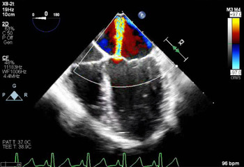
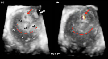
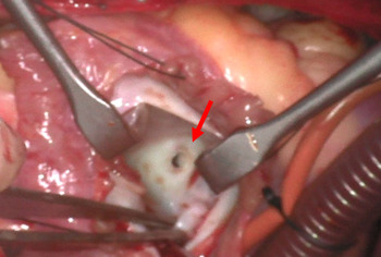
A 13-year-old female had been followed by an outside cardiologist for mitral regurgitation since infancy. It was thought to be caused by a cleft in the anterior leaflet of the mitral valve and was referred to our institution for surgical repair given progressive left atrial dilation. She had no history of endocarditis and no prior surgeries or interventions. Pre-operative transesophageal echocardiography demonstrated eccentric, moderate mitral regurgitation with the jet directed posteriorly (Fig 1, Supplementary Video S1). Forward-facing views of the mitral valve did not clearly define the origin of the regurgitation. Using 3-dimensional echocardiography, we were able to identify a perforation from which the regurgitant jet originated (Fig 2, Supplementary Videos S2 and S3). Surgical inspection confirmed the pre-operative transesophageal echocardiography findings (Fig 3), and the hole was successfully closed via a pledget suture.
Mid oesophagus four-chamber view demonstrating moderate mitral regurgitation originating eccentrically with a posteriorly oriented jet.

( a ) Three-Dimensional reconstruction of the mitral valve as viewed from the left ventricle (LV) in systole. The scallops (A1-3, P1-3), aortic valve (AoV) and zone of apposition (red line) are labelled. The perforation is marked with the red arrow. ( b ) Color Doppler signal is seen as the origination of the regurgitation jet. The perforation is marked with the red arrow.

Intraoperative inspection of the mitral valve revealed congenital perforation of the anterior leaflet of the mitral valve (red arrow).

Previous literature has recommended the use of 3-dimensional echocardiography to better evaluate the mechanism of mitral regurgitation and aid in surgical planning for mitral valve disorders like mitral valve perforation. Reference Bhave, Addetia and Spencer1 Where traditional 2-dimensional echocardiography was unable to accurately make the diagnosis, 3-dimensional echocardiography was able to correctly make the diagnosis and correlated well with surgical findings. 3-dimensional echocardiography should be utilised in unusual cases of congenital mitral regurgitation to aid in an accurate diagnosis.
Supplementary material
To view supplementary material for this article, please visit https://doi.org/10.1017/S1047951121004868
Acknowledgements
The authors would like to thank Dr Mudar Kattash for his aid in diagnosis of this interesting case. The authors would also like to thank Dr N. Adam Stephens for his assistance in acquiring the surgical image.
Financial support
This research received no specific grant from any funding agency, commercial or not-for-profit sectors.
Conflicts of interest
None.