Congenital extrahepatic portosystemic shunts are eponymously called the Abernethy malformation after its first description in 1793. These rare vascular malformations result in portal blood entering systemic veins bypassing the hepatic parenchyma, resulting in varied presentations from asymptomatic hyperammonaemia to hepatic encephalopathy, hepatic nodular hyperplasia, pulmonary hypertension, and hepatopulmonary syndrome.Reference Howard and Davenport 1 The lesion may coexist with visceral heterotaxy, cardiac, renal, bony, and gastrointestinal tract anomalies.
Case report
A 5-year-old boy weighing 16 kg presented with progressively worsening cyanosis and exertional dyspnoea over the preceding few months. There was clubbing and cyanosis with a pulse oximeter recording of 80%. Cardiovascular examination revealed a normal jugular venous pressure, unremarkable precordium, no cardiomegaly, and normal heart sounds with no murmur. Chest radiograph and electrocardiogram were unremarkable. Echocardiogram revealed a structurally normal heart with no evidence of pulmonary hypertension. The presence of pulmonary arteriovenous malformations was suggested by delayed filling of the left atrium beyond three cardiac cycles after right atrial opacification during agitated saline contrast echocardiography. Abdominal ultrasound showed a dilated portal vein with an abnormal fistulous communication measuring 20×12 mm between the portal vein and the intrahepatic portion of the inferior caval vein (Fig 1). CT confirmed the presence of an abnormal portocaval communication, hypoplastic inferior caval vein, and prominent azygos veins (Fig 1). This led to a diagnosis of type II Abernethy malformation with hepatopulmonary syndrome.
Abdominal color Doppler ultrasound ( a ) shows dilated portal vein communicating with a hypoplastic inferior caval vein (IVC). Sagittal CT slice ( b ) shows the portosystemic shunt (dotted arrow) and prominent azygos vein that collateralises the hypoplastic inferior caval vein (IVC) to the superior caval vein (SVC).

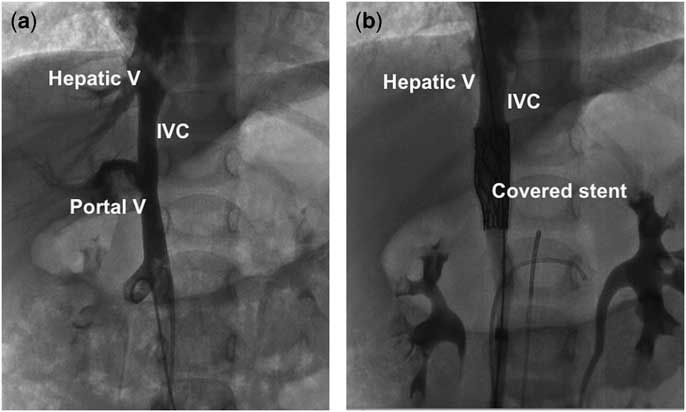
Cardiac catheterisation through two venous and one arterial accesses from the right groin under conscious sedation showed aortic desaturation, normal right heart pressures, and a 5-mmHg gradient between the femoral vein and the right atrium. Femoral venogram showed a hypoplastic inferior caval vein measuring 7 mm, and large decompressing veins coursing upwards to drain into the azygos vein, indicating partially compensated collateralisation (Supplementary video 1). Inferior caval venogram showed a large window-like portocaval communication connecting the dilated portal vein to the hypoplastic inferior caval vein (Fig 2). Selective portal venogram showed normal arborisation of the portal vein and normal hepatic venous drainage. Selective superior mesenteric venogram showed normal tributaries and normal drainage into the portal vein. Aortogram showed normal celiac and superior mesenteric arteries with their branches. Multiple renal arteries were present bilaterally to the upper and lower poles of kidneys.
Inferior caval venogram ( a ) shows the portosystemic communication into the hypoplastic inferior caval vein, managed by a covered stent ( b ). IVC=inferior caval vein.

Superior mesenteric and portal venous pressures were monitored after balloon occlusion of the inferior caval vein, which completely blocked the Abernethy communication. There was no significant elevation of the superior mesenteric venous pressures during the balloon occlusion. Simultaneous angiogram from the portal venous catheter showed adequate intrahepatic portal vein distribution and confirmed complete occlusion of the portocaval shunt (Supplementary video 2). A 34-mm covered CP stent (Numed, Hopkinton, New York, United States of America) mounted over a 12-mm × 4-cm Admiral extreme balloon (Medtronic, Minneapolis, Minnesota, United States of America) was deployed at 12 atmospheres across the portocaval shunt in the inferior caval vein between the hepatic veins and renal veins. Final angiogram confirmed closure of the portocaval shunt (Fig 2). The gradient across the inferior caval vein was fully eliminated. At 1 year of follow-up with oral anti-coagulation, the inferior caval vein was unobstructed with no residual portocaval shunt. There was complete resolution of hepatopulmonary syndrome with normalisation of oximetry readings to 98%.
Discussion
Association of Abernethy malformation with inferior caval vein anomalies
As the development of inferior caval vein and portal vein are known to be complex and closely interlinked to each other, developmental caval vein anomalies may coexist with portal venous anomalies as in our patient.Reference Yoshidome and Edwards 2 Caval anomalies such as interrupted inferior caval vein and double inferior caval veins have been found in patients with portosystemic shunts, congenital absence of portal vein, preduodenal portal vein, and portal continuation of the inferior caval vein.Reference Soni, Ambesh, Sunil and Jena 3 – Reference Bercoff, Colin, Benozio, Janvresse, Berland and Fillastre 5 If the portal vein is absent in patients with inferior caval vein interruption, this is referred as “double interruption”.Reference Le Borgne, Paineau and Hamy 6 When Abernethy malformations occur in patients with significant inferior caval vein hypoplasia, the caval vein gets decompressed by azygos, hemiazygos, and lumbar collaterals, as seen in our patient too.Reference Soni, Ambesh, Sunil and Jena 3
Embryological perspective
The frequent association of Abernethy malformation with inferior caval vein anomalies (with and without left isomerism) gives an insight into its probable embryological basis. This lesion probably represents a spectrum of right-sided venous hypoplasia or agenesis, affecting both the vitelline (portal) and subcardinal (infrahepatic caval vein) venous system. Although the mildest form will be hypoplastic inferior caval vein associated with type 2 Abernethy malformation as in our case, a severe form will be a “double interruption”. Right vitelline vein hypoplasia with poor portal arborisation in the liver leads to shunting of portal blood into systemic veins. Right subcardinal hypoplasia or aplasia will result in inferior caval vein narrowing or interruption, leading to dominance of the supracardinal system that forms the azygos veins. This right venous hypoplasia may occur as an isolated lesion or may be part of a more generalised left isomerism with coexisting cardiac malformations.Reference Bartram, Fischer and Kramer 7 , Reference Doraiswamy and Sivakumar 8 Every patient with interrupted IVC or left isomerism should be investigated for the association of Abernethy malformation.Reference Doraiswamy and Sivakumar 8
Therapeutic options
Therapeutic options depend on site and morphology of the shunt, coexisting congenital anomalies, and hepatic insult, if any.Reference Howard and Davenport 1 Although asymptomatic lesions may be medically followed in the absence of hepatic encephalopathy or hyperammonaemia, the presence of pulmonary vasculopathy is an absolute indication for emergent shunt closure before heart failure and brain abscess complicate future treatment options. The choice between an interventional or surgical shunt closure depends on the anatomy, clinical condition, and available local expertise.Reference Bernard, Franchi-Abella and Branchereau 9 Percutaneous options are preferable whenever the shunt can be closed reliably using an occlusive device without compromising either the portal vein and its branches or the caval vein and its tributaries.Reference Doraiswamy and Sivakumar 8 – Reference Kuo, Miller and Lavine 10 Using a covered stent in an efferent vessel in a patient like ours, who also has a hypoplastic caval vein, is a new rarely used strategy that achieves a twin purpose of relieving the haemodynamic gradient and closing the shunt.Reference Kuo, Miller and Lavine 10 This strategy using covered stents that can be dilated further may also find applications in wide, short window-like shunts where occlusive devices may compromise the lumen. If the portal pressure increases significantly on temporary balloon occlusion, staged interventions may need to be planned.Reference Kuo, Miller and Lavine 10 In contrast to earlier concerns, long-term anti-coagulation after interventions using covered stents or occluders may be needed only in patients with prothrombotic factors.Reference Bernard, Franchi-Abella and Branchereau 9 Complete regression of pulmonary vasculopathy is known following closure of the portosystemic shunt, as shown in our patient.Reference Bernard, Franchi-Abella and Branchereau 9
Conclusions
Developmental anomalies of the inferior caval vein and portal vein may coexist with or without left isomerism. A detailed imaging of the caval vein and portal vein is warranted to identify associated anomalies, as it has significant therapeutic implications. When inferior caval vein hypoplasia occurs in patients with type 2 Abernethy malformation, balloon expandable covered stents achieve the twin purpose of elimination of portosystemic shunt and relieve caval vein gradients.
Supplementary material
To view supplementary material for this article, please visit https://doi.org/10.1017/S1047951118000884
Acknowledgements
None.
Financial Support
This research received no specific grant from any funding agency or from commercial or not-for-profit sectors.
Conflicts of Interest
None.
Ethical Standards
The authors assert that all procedures contributing to this work comply with the ethical standards of the Indian Council of Medical Research and with the Helsinki Declaration of 1975, as revised in 2008, and has been approved by the institutional committees of Madras Medical Mission, Chennai, India.