Introduction
The use of lasers in otology, and in particular cholesteatoma surgery, is common and well accepted. The fibre-guided laser has been shown to allow the cholesteatoma surgeon to preserve the ossicular chain in a systematic manner that is both safe and of benefit to the patient.Reference Hamilton1 Commonly used lasers in otology include the argon, diode and potassium titanyl phosphate lasers, which are transmitted down fibre-optic cables. For laser-assisted cholesteatoma surgery, these lasers have been shown to be suitable devices.Reference Caffier, Marzahn, Franke, Sudhoff, Jovanovic and Haisch2
Laser surgery facilitates ablation of both macroscopic and microscopic cholesteatoma matrix from both the ossicles and other difficult-to-reach areas, whilst limiting mechanical trauma. Although the application of laser in the treatment of cholesteatoma has been shown to decrease residual disease, its use is not risk-free. For example, temporary, delayed, mild facial nerve paresis has been identified, despite the use of a facial nerve monitor.Reference Eskander, Holler and Papsin3
The most commonly used laser fibres in otology in the UK are straight and non-adjustable. The straight laser fibre has limitations, in particular when residual cholesteatoma is in a deep and tightly angled recess, adherent to the deep surface of the ossicular chain and/or near to critical structures such as the facial nerve. In these instances, use of the laser is avoided. ‘Bouncing’ the laser beam around the corner using a mirror is the most commonly adopted approach, but access is relatively limited to about 1 mm of the medial surface of the ossicular chain.
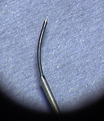
Ophthalmologists have been regularly using curved, adjustable fibre-optic laser carriers for retina surgery. This article describes our experience of using an OcuLight® TX laser with a wavelength of 532 nm. This is used in conjunction with a handheld, curved and adjustable laser fibre (Iridex Adjustable and Intuitive Endo Ocular Probe, model 14573 T; Iridex, Mountain View, California, USA) (Figure 1), to assist in microscopic cholesteatoma surgery using a diode laser.
Curved fibre-optic laser fibre (curved angle, adjustable 0–70 degrees).

Procedure
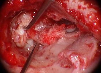
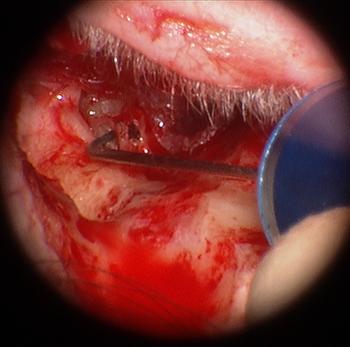
In our unit, a traditional microsurgical approach to cholesteatoma is undertaken, through an endaural incision. Initially, an atticotomy is performed, and then, depending on the extent of the disease, there is progression to an atticoantrostomy or tympanomastoidectomy. Cholesteatoma, once fully exposed, is removed using fine instruments, with further drilling if required (Figure 2). The ossicular chain is left intact if possible. The head of the malleus and body of the incus are removed if invested in cholesteatoma matrix, to gain access to deeper parts of the epitympanum.
Microscopic surgical exposure of cholesteatoma and clearance of macroscopic disease.

Following removal of the majority of the cholesteatoma, a more detailed assessment of the difficult-to-reach or delicate areas is made.
When disease is close or adherent to the ossicular chain (if intact) or facial nerve, or in a hard-to-reach recess, the curved laser fibre is now used to deliver a diode laser beam at a setting of between 500 mW and 1 W, in a pulsed mode, 100 ms on 100 ms off. It is delivered through a visible aiming beam, with a wavelength of 532 nm, and with a penetration depth of 1–3 mm.
The laser comes with a microscope fitted filter for eye protection, allowing the surgeon to work without eye protection. The filter does not alter the colour or contrast of the tissues, and can therefore remain on the microscope at all times.
Like other lasers, it can be used in direct contact, but it is usually preferable to hold it a fraction of a millimetre off. This avoids build-up of char, which can cause the tip of the fibre to adhere to the tissue to which the laser is applied.
Using a mirror to look into difficult areas, whilst simultaneously using the curved laser, allows the surgeon to assess the effectiveness of macroscopic clearance of disease. Further assessment can be made of these areas, with the mirror (after using the laser), to ensure successful eradication or to assist in identifying further disease that needs to be ablated. The curved fibre technique improves the accuracy of energy delivered around a corner compared with ‘bouncing’ the laser beam off a mirror, and decreases the risk of inadvertent damage to nearby structures.
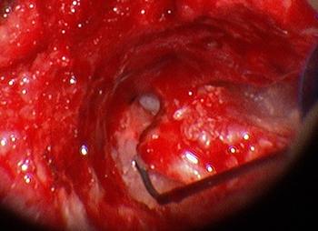
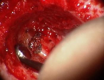
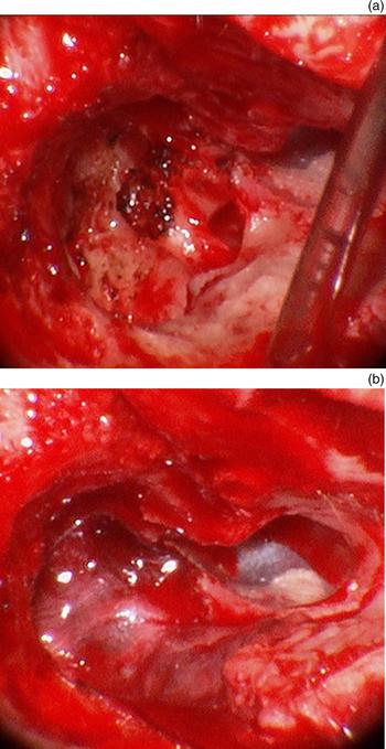
The laser, at the wattage stated above, can be used on potentially carious areas of the ossicular chain that have been in contact with cholesteatoma, ensuring microscopic clearance, whilst avoiding damage to the bone itself (Figures 3 and 4). For the same reason, it can also be safely used for dissecting the sac off a stapes head or disease extending through the stapes crura. In this specific situation, the curved fibre is particularly helpful in enabling dissection with minimal movement of the stapes.
The curved laser safely and effectively ablates disease on the deep surface of the ossicular chain, in this case the incus.

Deep surface of the ossicular chain following laser therapy, showing effective and accurate macroscopic clearance and ‘charring effect’ of disease in mirror.

For dissection of cholesteatoma off the facial nerve canal, suction can be used to provide traction of the sac, whilst adjusting the angle of the fibre to remove disease. The laser can be used in close proximity to the facial nerve, even when dehiscent, but aiming the beam directly at the dehiscent facial nerve is obviously to be avoided. This technique has been employed within our practice with no temporary or permanent weakness of the nerve thus far.
Figure 5 demonstrates the use of the curved laser in close proximity to the facial nerve. Because of the adjustable curvature of the fibre, cholesteatoma can be safely ablated with potentially less risk of temporary or permanent facial nerve palsy.
Safe usage of the laser, adjacent to, but curving away from, the facial nerve (between the lines).

Figure 6 shows appearances pre- and post-grafting with temporalis fascia.
Appearances pre- and post-grafting with temporalis fascia.

The laser has also been used to assist in the dissection of cholesteatoma from the sinus tympani, which is inaccessible to a straight laser fibre.
The fibre tip does not need to be carbonised prior to use, which was the case with the previous diode laser used in the department. When used in intermittent mode, it produces hardly any smoke. The curved fibre can be retracted, which makes it particularly easy to remove any char build-up on the end of the fibre. A further advantage of the retractable curved laser fibre is that, despite the comparative sophistication of the hand piece, it is actually less expensive than the conventional straight laser delivery system routinely supplied for ENT use.
The patient used to illustrate the technique in this paper was followed up one-year post-operatively. There were no post-operative complications, and the mean threshold on pure tone audiogram for between 0.25 kHz and 8 kHz was 11 dB.
For the previous 15 years, the senior author had used a diode laser with fibre delivery using a non-adjustable straight hand piece, with good hearing outcomes in cases suitable for ossicular preservation. The current laser with curved fibres has only been in use in the department for little over a year, and has been employed in around 50 cases. It would therefore be premature to report longer-term hearing outcomes. However, use of the curved fibre has allowed preservation of the incus in a higher proportion of cases with more extensive cholesteatoma extending medial to the ossicular chain, which would previously have been inaccessible using a straight laser handpiece.
Conclusion
Using the curved, adjustable laser fibre for clearance of cholesteatoma can, in certain circumstances, assist in accurate and atraumatic disease removal when perhaps usage of a straight laser fibre would be ineffective or less accurate. This in turn reduces potential damage to delicate structures, such as the facial nerve.
The curved nature of the fibre facilitates more disease eradication, without the need for extra drilling and bone removal. Furthermore, it allows the surgeon to operate in difficult-to-reach areas more safely, accurately and in a contactless fashion.
Acknowledgement
Mr Liam Flood, for advice and review.