Introduction
Successful somatic cell nuclear transfer (SCNT) has been reported in several livestock species. In pigs, however, the viability of SCNT embryos is poor, with an extremely low rate of cloned piglet production (Miyoshi et al., Reference Miyoshi, Sato and Yoshida2006). Improvement in the quality of recipient oocytes and their activation methods would enhance the chance of successful cloning in the pig (Dinnyes et al., Reference Dinnyes, Hirao and Nagai1999). Activation of metaphase II (MII) oocytes is one of the critical steps in improving porcine SCNT (Kim et al., Reference Kim, Lee, Ock, Balasubramanian, Choe and Rho2005). Activation is induced by an increase in intracellular free calcium levels induced by a wide variety of chemical and/or physical stimuli. The artificial activation methods mainly used are based on either the combination of calcium ionophore A23187 or ionomycin with protein phosphorylation or protein synthesis inhibitors (Funahashi et al., Reference Funahashi, Cantley, Stumpf, Terlouw and Day1994; Betthauser et al., Reference Betthauser, Forsberg, Augenstein, Childs, Eilertsen, Enos, Forsythe, Golueke, Jurgella, Koppang, Lesmeister, Mallon, Mell, Misica, Pace, Pfister-Genskow, Strelchenko, Voelker, Watt, Thompson and Bishop2000; Koo et al., Reference Koo, Kang, Choi, Park, Han, Park, Kim, Lee, Son, Chang and Han2000; Roh & Hwang, Reference Roh and Hwang2002; Silvestre et al., Reference Silvestre, Alfonso, Garcia-Mengual, Salvador, Duque and Molina2007), or electric stimulation combined or not with these same inhibitors (Grupen et al., Reference Grupen, Mau, McIlfatrick, Maddocks and Nottle2002; Lee et al., Reference Lee, Tian and Yang2004; Kim et al., Reference Kim, Lee, Ock, Balasubramanian, Choe and Rho2005; Im et al., Reference Im, Seo, Hwang, Kim, Kim, Yang, Yang, Lai and Prather2006). In pig cloning, however, the best procedure for artificial activation is unclear and there are still controversial debates on the use of combined treatments with chemicals for effective development of SCNT embryos (Kim et al., Reference Kim, Lee, Ock, Balasubramanian, Choe and Rho2005).
Another critical factor affecting the development of porcine SCNT embryos is the in vitro oocyte maturation process (Hyun et al., Reference Hyun, Lee, Kim, Kim, Lee, Kim, Lee, Lim, Kang, Lee and Hwang2003; Mastromonaco et al., Reference Mastromonaco, Semple, Robert, Rho, Betts and King2004; Holker et al., Reference Holker, Petersen, Hassel, Kues, Lemme, Lucas-Hahn and Niemann2005). Recipient oocyte contains cytoplasmic factors that have accumulated during maturation critically involved in reprogramming of the transferred donor nucleus during SCNT. In vitro maturation (IVM) of pig oocytes has been successfully achieved, especially at nuclear level, although standard procedures are rather less efficient at cytoplasmic level (Abeydeera, Reference Abeydeera2002). In order to achieve in vitro matured porcine oocytes, media supplemented with serum or pFF have generally been used (Kishida et al., Reference Kishida, Lee and Fukui2004). Serum or pFF, however, contain many unknown factors and there is often considerable variability among sources or even among batches from the same source. Furthermore, there is a possibility that these fluids may contain hidden viruses (Stringfellow & Givens, Reference Stringfellow and Givens2000). In pigs, several reports have demonstrated that it is possible to replace serum or pFF with other defined compounds for maturation medium without reducing the efficiency on IVF or ICSI embryo development (Illera et al., Reference Illera, Lorenzo, Illera and Petters1998; Abeydeera et al., Reference Abeydeera, Wang, Cantley, Rieke, Murphy, Prather and Day2000; Kishida et al., Reference Kishida, Lee and Fukui2004). Recently, in bovine, a lack of serum during oocyte IVM resulted in significantly decreased SCNT embryo development compared with embryos produced by IVF (Mastromonaco et al., Reference Mastromonaco, Semple, Robert, Rho, Betts and King2004). Serum was required, therefore, for IVM when bovine oocytes were to be used for SCNT, but not for IVF (Mastromonaco et al., Reference Mastromonaco, Semple, Robert, Rho, Betts and King2004). Few studies on the effect of IVM medium on pig SCNT embryo development were found in the reviewed literature (Hyun et al., Reference Hyun, Lee, Kim, Kim, Lee, Kim, Lee, Lim, Kang, Lee and Hwang2003; Song & Lee, Reference Song and Lee2007).
The aim of the present work was to study the effect of addition of serum to IVM medium on nuclear maturation and further parthenogenetic and SCNT embryo development. Previously, the effect of three activation protocols based on ionomycin treatment or electric stimuli combined or not with 6-DMAP on parthenogenetic embryo development was also evaluated.
Materials and Methods
Culture media
Unless otherwise stated, all chemicals were purchased from Sigma–Aldrich Química. Media with bicarbonate were covered with mineral oil and equilibrated overnight in a humidified atmosphere containing 5% CO2 and 5% O2 in air at 38.5 °C. Handling medium (HM199) consisted of medium 199 supplemented with 25 mM HEPES, 8% (v/v) heat-inactivated fetal calf serum (FCS; Invitrogen) and antibiotics. Unless otherwise stated, maturation medium (MM199) consists of Medium 199 supplemented with 0.1% (w/v) polyvinyl alcohol (PVA), 0.57 mM cysteine and 10 ng/ml epidermal growth factor (EGF) and antibiotics (Abeydeera et al., Reference Abeydeera, Wang, Cantley, Rieke, Murphy, Prather and Day2000). Embryos were cultured in porcine zygote medium-3 (PZM-3; Yoshioka et al., Reference Yoshioka, Suzuki, Tanaka, Anas and Iwamura2002) supplemented with 3 mg/ml BSA (culture medium; CM).
In vitro maturation and embryo culture
Ovaries were collected from prepuberal gilts at a local abattoir and transported within 45 min to the laboratory in 0.9% (w/v) NaCl supplemented with antibiotics at 37 °C. Cumulus–oocyte complexes (COCs) were obtained by antral follicle puncture (3–7 mm diameter) with an 18 gauge needle. Approximately 50–60 COCs were cultured in 500 μl medium in a 4-well multidish (Nunc) for 44 h. During the first 22 h of culture, COCs were incubated in MM199 supplemented with hormones: 0.1 IU/ml recombinant human-FSH, and -LH (Gonal-F and Luveris respectively, Serono, Spain; Silvestre et al., Reference Silvestre, Alfonso, Garcia-Mengual, Salvador, Duque and Molina2007). Then, COCs were washed twice and cultured in hormone-free MM199 for an additional 22 h. After different treatments (see below), presumptive zygotes were washed twice in CM medium and transferred to a Nunc 4-well multidish containing 500 μl of CM per well. Two per cent of FCS was added on culture medium at day 5 of culture. Both in vitro maturation and culture were performed at 38.5 °C in an atmosphere of 5% O2, 5% CO2 and 100% relative humidity.
Artificial activation
In order to obtain parthenogenetic embryos, three different activation protocols were tested:
1. Chemical activation: MII oocytes were treated with 5 μM ionomycin for 5 min. After ionomycin treatment, oocytes were washed twice and incubated in 2 mM 6-DMAP for 3.5 h (Loi et al., Reference Loi, Ledda, Fulka, Cappai and Moor1998; Silvestre et al., Reference Silvestre, Alfonso, Garcia-Mengual, Salvador, Duque and Molina2007).
2. Electrical activation: oocytes were equilibrated in electropulsing medium (pH ~7.0): 0.3 M mannitol containing 1.0 mM CaCl2.H2O, 0.1 mM MgSO4,6H2O and 5.0 mM HEPES for 1 min before being placed in the electroporator chamber and aligned manually. Pulses were delivered with an electro-cell porator (Multiporator®, Eppendorf). Oocytes received one electrical set consisting of two square wave electrical DC pulses of 1.2 kV/cm for 30 μs each at 1 s apart in a chamber consisting of platinum wire electrodes 0.5 mm apart.
3. Electrical-chemical activation: After electrical activation, oocytes were incubated in 2 mM 6-DMAP for 3.5 h.
Somatic cell nuclear transfer
Donor cells
Pig ears were separated from adult approximately 6 month old animals at a local abattoir. Skin biopsies were obtained from the ears and very thin (0.25–1.0 mm2) tissue pieces were sliced using a scalpel in HM199. Tissue samples were seeded in a 2 h pre-equilibrated 75 cm2 flask containing 10 ml Medium 199 supplemented with 15% (v/v) FCS and incubated in 5% CO2 in air at 95% relative humidity and at 38.5 °C. The culture medium was changed every 4 d. The donor cells used were between passages 1 to 6 of culture and were synchronized in presumptive G0 by serum deprivation for 48 h before nuclear transfer. A single cell suspension of donor cells was prepared by standard trypsination (trypsin–EDTA solution) 1 h before nuclear transfer (Lai & Prather, Reference Lai and Prather2003). The cells were pelleted and resuspended in hormone-free MM199 until their use.
Micromanipulation
Following the Lai & Prather (Reference Lai and Prather2003) method with minor modifications, recipient MII oocytes were stained with 5 μg/ml bisbenzimide in North Carolina State University-23 medium supplemented with 4 mg/ml of BSA (NCSU23-BSA) for 30 min; for the last 10 min, oocytes were incubated in NCSU23-BSA containing 7.5 μg/ml CCB. Enucleation was performed by aspiration of the first polar body and one-quarter of adjacent cytoplasm with the aid of UV light to visualize the MII plate. Enucleation was assessed checking enucleated oocyte under fluorescence microscopy. The maximum exposure time of oocytes to UV light was no more than 5–7 s. After enucleation, oocytes were cultured in NCSU23-BSA for 40–60 min until the deposition of donor cell into the perivitelline space. Enucleated oocyte and donor cell complexes were aligned manually with the aggregation plane of the donor cell and recipient ooplasm parallel to the electrodes and received one electrical set (described above). Only those non-fused were pulsed again, 50 min after the first electrical set. Fused embryos were cultured in CM in the above culture conditions.
Experimental design
In Experiment 1, the effect of three artificial activation protocols (ionomycin plus 6-DMAP and electric pulse with or without an additional treatment of 6-DMAP) on cleavage and embryo development rate and nuclei number of blastocyst was examined. The three artificial activation protocols were described above.
Once the activation protocol was defined, in Experiment 2 the effect of the supplementation of 10% (v/v) heat-inactivated FCS in the maturation medium on nuclear maturation and parthenogenetic and SCNT embryo development rate and nuclei number was studied.
Assessment of nuclear maturation and embryo development
After in vitro maturation, the cumulus-enclosed oocytes were briefly placed in HM199 supplemented with 1 mg/ml bovine testes hyaluronidase and then gently pipetted through a small-bore pipette. Oocytes were scored under stereoscopic vision for presence of the first polar body and therefore corresponding to nuclear matured oocytes. In all cases, cleavage and blastocyst rate were evaluated respectively at 48 and 168 h after activation under a stereomicroscope. Blastocyst quality was assessed by staining with 25 μg/ml bisbenzimide. Fixed and stained blastocysts were mounted on a glass microscope slide in a drop of glycerol and gently flattened with a coverslip. Cell counting was performed under an epifluorescence microscope.
Statistical analysis
At least four replicates were performed per treatment. Nuclear maturation, cleavage or blastocyst rates were analysed by a chi-squared test analysis. When a single degree of freedom was involved, the Yates' correction for continuity was carried out. Result of nuclei cells was analysed by the ANOVA test.
Results
Effect of several oocyte activation procedures
Results of three artificial activation treatments are shown in Table 1. Oocytes activated by ionomycin plus 6-DMAP showed lower cleavage (47.2 vs. 78.5–81.5; p < 0.05) and blastocyst rates (11.3 vs. 29.2–32.1; p < 0.05) than those activated by electrical and electrical plus 6-DMAP treatments. No differences in embryo development rates were observed between both electrical treatments with or without 6-DMAP. The total number of nuclei per blastocyst tended to be higher in both electrical groups than in the chemical group, but this difference was not statistically significant.
Effect of different artificial activation treatment on parthenogenetic embryo development.

a Chemical: oocytes were treated with ionomycin for 5 min and 6-DMAP for 3.5 h. Electrical: MII oocytes received one electrical set consisted of two square wave electrical DC pulses of 1.2 kV/cm for 30 μs each at 1 s apart. Electrical combined with chemical activation: after electrical activation, oocytes were incubated in 6-DMAP for 3.5 h.
b , c Values with different superscript on the same column are significantly different (p < 0.05).
Effects of serum in maturation medium on nuclear oocyte maturation and subsequent parthenogenetic and SCNT embryo development
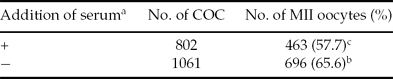
The effects of serum in maturation medium on in vitro oocyte maturation are presented in Table 2. We observed that the serum-free group showed higher nuclear maturation rate than the serum-supplemented group after 44 h of maturation (66 vs. 58%, p < 0.05). The cleavage and embryo development rates of parthenogenetic and NT embryos are shown in Table 3. Regarding parthenogenetic embryo development, serum-free group cleavage rates were significantly higher than those obtained in the serum-supplemented group (81.8 vs. 69.6% respectively; p < 0.05), although these differences were not detected in blastocyst rates or blastocyst nuclei number. Regarding SCNT embryos, no significant differences were observed in cleavage or blastocyst rates among the different experimental groups. However, the SCNT blastocysts obtained after serum-supplementation of IVM medium showed significantly higher nuclei numbers (37.4 vs 19.0 respectively; p < 0.05).
Effect of serum addition on in vitro oocyte nuclear maturation.
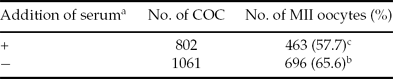
a A 10% heat inactivated fetal calf serum was (+ group) or not supplemented (− group).
b, c Values with different superscript on the same column are significantly different (p < 0.05).
Effect of serum on in vitro parthenogenetic and nuclear transfer embryo development.

a A 10% heat inactivated fetal calf serum was (+ group) or not supplemented (− group).
b Number of parthenogenetic blastocyst (+ group = 24, − group = 16).
c, d Values with different superscript on the same column are significantly different (p < 0.05).
Discussion
When artificial activation is based on only one stimulus, and in order to avoid a fast restoration of the MPF activity and hence the MII arrest, an inhibition of protein kinase activity by 6-DMAP may be performed (Liu & Yang, Reference Liu and Yang1999). However, the beneficial effect of 6-DMAP on activated porcine oocytes is still controversial. Though some authors have found that it enhanced the NT or parthenogenetic embryo development (Grupen et al., Reference Grupen, Mau, McIlfatrick, Maddocks and Nottle2002; Kim et al., Reference Kim, Lee, Ock, Balasubramanian, Choe and Rho2005), other authors did not detect any significant effect of 6-DMAP on blastocyst rates after electrical activation (Roh & Hwang, Reference Roh and Hwang2002; Koo et al., Reference Koo, Chae, Kim, Wee, Song, Lee and Han2005; Kwon et al., Reference Kwon, Park, Yang, Kim and Cheong2007; oocytes IVM at 44 h). Moreover, when a 6-DMAP treatment was applied, a significant increase in chromosomal abnormalities in the NT or parthenogenetic embryos was observed in comparison with electric (Kim et al., Reference Kim, Lee, Ock, Balasubramanian, Choe and Rho2005) or ionomycin (Bhak et al., Reference Bhak, Lee, Ock, Mohana, Choe and Rho2006) treatments. Although we did not check 6-DMAP effects on chromosomes, we did not find a significant increase in parthenogenetic embryo development or embryo quality (cells/blastocyst) after 6-DMAP treatment, and so we decided to use only an electrical activation treatment for the following experiment.
In the present experiment, the effect of serum supplementation of IVM medium on nuclear maturation and further parthenogenetic and SCNT embryo development was studied. Currently, little information on the effect of serum in IVM medium on oocyte viability is available and existing results are controversial. Protein supplementation of IVM medium had different effect on bovine IVF or NT blastocyst embryo development (Mastromonaco et al., Reference Mastromonaco, Semple, Robert, Rho, Betts and King2004). In the present study, we observed a lower nuclear maturation rate in the FCS-supplemented group compared with the FCS-free group. In fact, many studies have shown that the source of protein supplementation affects the kinetics of nuclear maturation and further embryo development (Ali & Sirard, Reference Ali and Sirard2002; Mastromonaco et al., Reference Mastromonaco, Semple, Robert, Rho, Betts and King2004; Krylov et al., Reference Krylov, Kren, Okada, Vackova, Tlapakova and Fulka2005). In addition, we observed a different effect of FCS on IVM medium depending on the type of embryo (parthenogenetic or SCNT). On the one hand, in parthenogenetic embryos we observed a detrimental effect of FCS on the cleavage rate after electrical activation, although this effect disappeared when embryos progressed to the blastocyst stage. According to this, Kishida et al. (Reference Kishida, Lee and Fukui2004) suggested that in vitro pig embryo development after ICSI was delayed after IVM in serum-free IVM medium. It has been reported that the presence of serum in IVM medium modified significantly several transcript levels, which could affect further embryo viability (Watson et al., Reference Watson, De Sousa, Caveney, Barcroft, Natale, Urquhart and Westhusin2000; Calder et al., Reference Calder, Caveney, Sirard and Watson2005). However, others did not detect any delay in bovine IVF embryo development, or differences on cleavage or blastocyst rates, the only significant effects being on blastocyst cell number in favour of the experimental group supplemented by serum (Sagirkaya et al., 2006). On the other hand, in SCNT embryos, no effect of FCS was detected on cleavage or blastocyst rates, although the few blastocysts obtained in the FCS group presented a nuclei number significantly higher than free-FCS group. This great variability between different studies may be due partially to the evident differences among serum batches in some components that affected both cumulus expansion and oocyte maturation (Suzuki et al., Reference Suzuki, Misumi, Ozawa, Noguchi, Kaneko, Ohnuma, Fuchimoto, Onishi, Iwamoto, Saito, Nagai and Kikuchi2006).
In conclusion, electrical pulse followed or not by 6-DMAP was found to be an efficient procedure to artificially activate MII porcine oocytes. Moreover, the addition of serum to oocyte maturation media did not seem to improve parthenogenetic or NT porcine embryo development.
Acknowledgements
This work was partially supported by the Conselleria de Empresa, Universidad y Ciencia de la Generalitat Valenciana (GV05/212) and INIA (RTA2007–0110–00–00). E. García-Mengual was supported by a research grant from the Conselleria de Agricultura, Pesca y Alimentación. The authors would like to thank F. García-Ximénez and R. P. Cervera for their critical comments and advice on the manuscript, N. Macowan for revising the English version of this manuscript and the Mercavalencia Abattoir for their assistance in obtaining porcine ovaries.