Introduction
Porphyrins are aromatic planar macrocycles composed of four pyrrole groups. This class of molecules holds interest in materials development due to their diverse functionality in nature. The π-conjugation of porphyrins gives rise to unique electron mobility which may be controlled by altering certain structural elements. Addition of functional groups at the beta- or meso- positions, controlled intermolecular stacking of the macrocycles, and coordination of different central metal ions all modulate the capability for light absorbance and electron transfer.[Reference Cook, Brewer and Wong-Ng1] Furthermore, the activity of porphyrin molecules is strongly influenced by the surrounding matrix. These properties may be harnessed in a variety of materials contexts. For instance, porphyrins have been used in porous materials for photocatalysis,[Reference Zhao, Ou and Wu2] light harvesting,[Reference So, Wiederrecht, Mondloch, Hupp and Farha3] and sensors.[Reference Ling, Lei, Zhang and Ju4] Layering porphyrin molecules on conducting surfaces like titanium dioxide enables charge separation in solar cells.[Reference Yella, Lee, Tsao, Yi, Chandiran, Nazeeruddin, Diau, Yeh, Zakeeruddin and Gratzel5, Reference Mahmood, Hu, Xiao, Tang, Wang and Zhou6] Oligomerization of porphyrins and addition of linker networks can form molecular electronic components.[Reference Jurow, Schuckman, Batteas and Drain7] Since there are many possible variations of porphyrin structure with different capabilities, it is important to develop improved generalized synthesis methods for porphyrins that yield quantities sufficient to evaluate their behavior when incorporated into solid matrices.
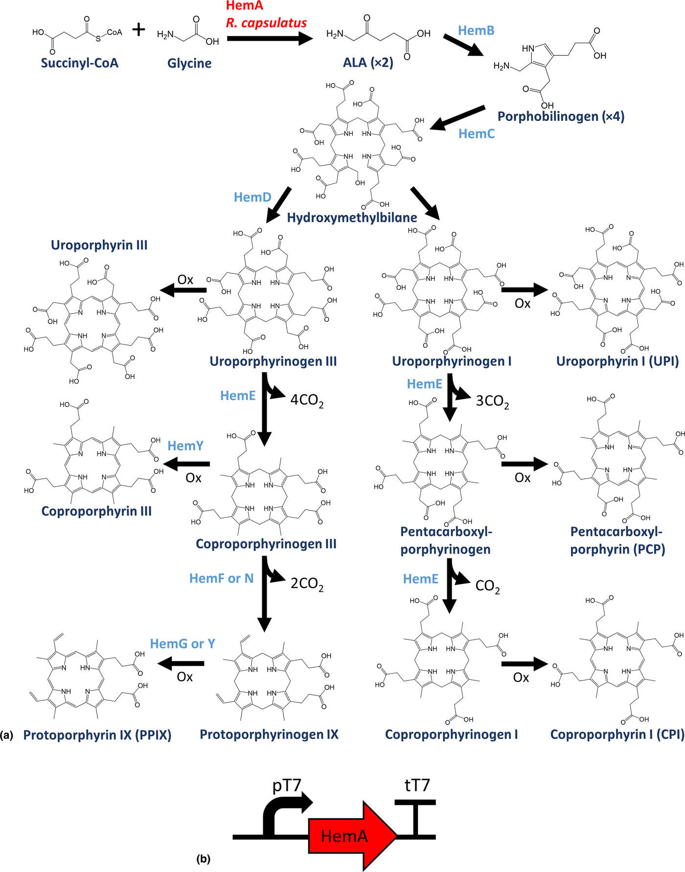
Porphyrins such as heme and chlorophyll serve many functions in living systems and are naturally produced in most organisms. The Shemin pathway (Fig. 1) initiates the biosynthesis of protoporphyrin IX (PPIX).[Reference Shemin and Russell8, Reference Kwon, de Boer, Petri and Schmidt-Dannert9] PPIX and related molecules have been used as a photosensitizer in biomedicine, and are capable of oxidation in solution when exposed to light.[Reference Furuse, Fukuoka, Kato, Horai, Kubota, Kodama, Kusunoki, Takifuji, Okunaka and Konaka10–Reference Ethirajan, Chen, Joshi and Pandey12] Other porphyrins produced via this pathway may also have interesting photocatalytic capabilities, including uroporphyrin, coproporphyrin, and pentacarboxyl porphyrin, as well as downstream heme and chlorophyll derivatives. It is currently very difficult to obtain quantities of these molecules sufficient to evaluate their performance in a materials context. Biosynthesis is a promising approach for the production of these molecules as most organisms possess the necessary enzymes; however, the porphyrin products are natively only produced at very low levels. Several studies have used metabolic engineering to over-produce PPIX in Escherichia coli and decouple the pathway from native gene regulation.[Reference Kwon, de Boer, Petri and Schmidt-Dannert9, Reference Nielsen, Madsen, Seppälä, Christensen, Riisberg, Harrison, Møller and Nørholm13] Simply over-expressing the HemA enzyme from Rhodobacter capsulatus, which catalyzes the synthesis of aminolevulinic acid (ALA), results in greatly increased porphyrin production over the wildtype. The expression of downstream enzymes HemB-F in various combinations led to further increases in porphyrin production and altered the relative concentrations of the different porphyrin intermediates that accumulate in culture.[Reference Kwon, de Boer, Petri and Schmidt-Dannert9, Reference Nielsen, Madsen, Seppälä, Christensen, Riisberg, Harrison, Møller and Nørholm13]
(a) Map of Shemin pathway for porphyrin biosynthesis, (b) diagram of plasmid for overproduction of porphyrins in E. coli.

Metal-organic frameworks (MOFs) are a promising class of porous materials consisting of metal or metal-oxide nodes joined together by multitopic ligands to form self-assembled crystalline frameworks.[Reference Kim, Chen, Reineke, Li, Eddaoudi, Moler, O'Keeffe and Yaghi14] Of emerging interest is the use of MOFs for the remediation of chemical threats including toxic gas adsorption[Reference DeCoste and Peterson15, Reference Barea, Montoro and Navarro16] and chemical warfare agent (CWA) catalytic hydrolysis.[Reference Mondloch, Katz, Isley, Ghosh, Liao, Bury, Wagner, Hall, DeCoste, Peterson, Snurr, Cramer, Hupp and Farha17, Reference Katz, Mondloch, Totten, Park, Nguyen, Farha and Hupp18] Recent advances have utilized the photosensitizing organic linkers of the MOFs NU-1000 (pyrene) and PCN-222 (porphyrin) to photocatalytically degrade sulfur mustard (HD) and its simulant 2-chloroethyl ethyl sulfide (CEES).[Reference Liu, Buru, Howarth, Mahle, Buchanan, DeCoste, Hupp and Farha19, Reference Liu, Howarth, Hupp and Farha20] An excited photosensitizer in its triplet state can be utilized to convert molecular oxygen into singlet oxygen (1O2), which is then utilized to perform soft oxidation processes, such as the conversion of CEES to its sulfoxide product, 2-chloroethyl ethyl sulfoxide (CEESO).[Reference DeRosa21]
NU-1000 [molecular formula Zr6(μ 3-OH)4(μ 3O)4(OH)4(OH2)4(TBAPy)2; TBAPy = 1,3,6,8-tetrakis(p-benzoic-acid)pyrene] has coordinatively unsaturated eight-connected secondary building units (SBUs), leaving four open sites that can be modified. Utilizing molecules containing carboxylic acids to chelate the zirconium sites, the SBUs can be modified via solvent-assisted ligand incorporation (SALI).[Reference Karagiaridi, Bury, Mondloch, Hupp and Farha22–Reference Mondloch, Bury, Fairen-Jimenez, Kwon, DeMarco, Weston, Sarjeant, Nguyen, Stair, Snurr, Farha and Hupp24] These sites have been modified to incorporate a variety of functionalities into the MOF structure, including bulky fullerene photosensitizers for CEES degradation.[Reference Howarth, Buru, Liu, Ploskonka, Hartlieb, McEntee, Mahle, Buchanan, Durke, Al-Juaid, Stoddart, DeCoste, Hupp and Farha25] As the NU-1000 structure has been shown to have the ability to hydrolyze G- and V-agents, enhancing its activity toward sulfur mustard via photocatalysis is of inherent interest.[Reference Mondloch, Katz, Isley, Ghosh, Liao, Bury, Wagner, Hall, DeCoste, Peterson, Snurr, Cramer, Hupp and Farha17]
In this work we harnessed the Shemin pathway in an E. coli host with the goal of building a biosynthesis platform to produce porphyrins of sufficient quantity to examine their photocatalytic function in a heterogeneous MOF material. In addition, we functionalized the MOF NU-1000 with bioderived porphyrin PPIX (PPIX@NU-1000) via SALI. This functionalized porous material with the high surface area was then examined for photocatalytic activity and was found to catalyze the oxidation of CEES similar to PPIX in solution over the entire visible spectrum of light. This is the first instance that bioderived porphyrins have been studied in the context of a MOF for catalysis. This biosynthesis platform represents a scalable route to gain access to many natural porphyrin molecules for materials development.
Materials and methods
Materials
All chemicals were obtained from Sigma Aldrich (St. Louis, MO, USA) at the highest available purity and used without further purification, unless otherwise stated. Polymerase and NEBuilder HiFi DNA assembly kit were purchased from New England Biolabs (Ipswich, MA, USA). Primers and gene fragments were purchased from Integrated DNA Technologies (Coralville, IA, USA).
Plasmids and strains
The plasmid used in this study was mslpl004 which comprises T7 driven expression of hemA encoding the ALA synthase from R. capsulatus (GenBank accession number X53309) and a kanamycin resistance cassette. The hemA gene fragment was purchased and integrated into the polymerase chain reaction-linearized plasmid backbone using NEBuilder HiFi DNA assembly. The complete plasmid sequence is available on GenBank (MK138544). The bacterial strain used for porphyrin synthesis was E. coli BL21 (DE3).
Cell culture
To test overproduction of porphyrins, E. coli was grown overnight in 5 mL of Lysogeny Broth-Lennox (LB-L) medium. For the strain harboring the mslpl004 plasmid, media were supplemented with kanamycin (50 µg/mL). Also, T7 RNA polymerase production was induced by addition of 1 mM isopropyl β-d-1-thiogalactopyranoside (IPTG) at subculture. For small-scale growth curves, cells were sub-cultured with a 1:1000 dilution into 5 mL LB-L in polypropylene 24-well deep well blocks (Axygen Scientific, P-DW-10ML-24C). Cultures were shaken at 37 °C, 100 rpm for 72 h. For each time interval, 150 µL samples were transferred to a 96-well microplate and absorbance (410 nm) and fluorescence (410/633 nm) readings were measured by a Biotek H1 Synergy plate reader. For larger scale production, 1 mL of overnight culture was sub-cultured into 1 L of LB-L in a 2 L flask and cultures were harvested after 72 h.
Porphyrin extraction
Extraction procedures are based on the techniques used by Kwon et al.[Reference Kwon, de Boer, Petri and Schmidt-Dannert9] Cell cultures were centrifuged at 5000 × g for 10 min. The media were decanted and the cell pellet was set aside. A weight of 4 g of dry diethylaminoethyl (DEAE) Sephadex A-25 anion exchange resin (GE Life Sciences 17017001) was added per 1 L of culture media to bind negatively charged porphyrins. The resin was isolated by centrifugation at 4700 × g for 15 min. Media were carefully decanted and discarded. Porphyrins were extracted from the resin by repeated 50 mL washes with methanol adjusted to pH 2–3 with 6 N HCl. The initial resin washes contained deep-red porphyrin pigment, and 50 mL washes were continued until little visible pigment was eluted. Total pooled methanol extract volume was typically 250–300 mL per 1 L culture. The extract was neutralized with 4 M NaOH and filtered prior to further purification steps. Before fractionation, the extract was concentrated by rotary evaporation.
To monitor porphyrin content of the cell pellet, the pellet was extracted with 3:1 mixture of methanol:water for 1 h. This was then centrifuged 4700 × g for 15 min, filtered, and analyzed by liquid chromatography-mass spectrometry (LC-MS).
LC analysis of porphyrins
LC-MS was used to detect porphyrins in either the culture medium or the methanol extract. The instrument used in this study was an Agilent 1290 Infinity II equipped with a diode array detector (DAD) and a 6150 quadrupole mass spectrometer (MS) with electrospray ionization (EI). The column was a BDS Hypersil C18 150 × 2.1 mm, 2.4 µm particle size (Thermo Scientific, Waltham, MA, USA, 28102–152130). The mobile phases were solvent A: 0.1% formic acid in ultrapure water, and solvent B: 0.1% formic acid in methanol. The flow rate was 0.4 mL/min. Porphyrins were separated by elution with a linear gradient transitioning from 100% solvent A to 100% solvent B over 20 min, followed by 100% solvent B for 5 min, and re-equilibration with 100% solvent A for 10 min. Injection volume was 20 µL. Porphyrins were detected by measuring absorbance at 410 nm by the DAD, and tracking the m/z values listed in Table S1 by MSD. Standards for measuring protoporphyrin IX, uroporphyrin I, coproporphyrin I, and pentacarboxyl porphyrin were purchased from Frontier Scientific Inc. (Logan, UT, USA).
Fraction collection
A similar chromatography protocol was developed to inject large volumes and collect eluent fractions. The instrument used was an Agilent 1260 Infinity. The column was a BDS Hypersil C18 150 × 4.6 mm, 2.4 µm particle size (Thermo Scientific, 28120–117300). To isolate pure porphyrin molecules, the elution follows the same protocol used for LC-MS, except the flow rate was 0.3 mL/min. Alternatively, an elution using a step increase in solvent B allowed a shorter protocol to remove salts without separating the organic components of the porphyrin mixture. Injection volumes were 900 µL. Fractions were collected at 30 s intervals. To fractionate large volumes of extract, multiple 900 µL injections were separated, then the equivalent fractions were pooled. LC-MS was used to determine the porphyrin content of each fraction. Porphyrin solutions were dried by rotary evaporation prior to MOF synthesis.
NU-1000 synthesis
NU-1000 was synthesized in a similar manner to a previously reported procedure.[Reference Islamoglu, Otake, Li, Buru, Peters, Akpinar, Garibay and Farha26] To a 8-dram glass vial was added ZrOCl2 · 8H2O (98 mg, 0.3 mmol), benzoic acid (2.0 g, 16.38 mmol), and 8 mL of dimethylformamide (DMF). The vial was closed and the mixture sonicated for 5 min until fully dissolved. The vial was then placed in a preheated 100 °C oven for 1 h. After removing the vial from the oven, the solution was allowed to cool to room temperature. H4TBAPy (40 mg, 0.06 mmol) and trifluoroacetic acid (50 µL, mmol) were then added to the vial, sonicated for 5 min, and placed in a preheated 100 °C oven for 18 h. After removal from the oven and cooling to room temperature, the solution containing the yellow microcrystalline MOF was transferred to a 15 mL centrifuge tube. After centrifugation (6000 rpm, 5 min), the solution was decanted and fresh DMF (7 mL) was added to the tube. Centrifugation was repeated twice with DMF and thrice with acetone. NU-1000 was transferred to a 8-dram vial to which was added 12 mL DMF and 0.5 mL of an 8 M HCl solution. The capped vial was then heated at 100 °C for 24 h. After cooling to room temperature, the powder suspension was centrifuged and washed with DMF and acetone. Residual solvent was removed by heating the powder at 120 °C under vacuum for 18 h.
Solvent-assisted ligand incorporation
NU-1000 was functionalized with PPIX via SALI to form PPIX@NU-1000. PPIX (109 mg, 0.19 mmol) was dissolved in 15 mL of DMF. The solution was added to a 8-dram vial containing NU-1000 (44 mg, 0.021 mmol). The vial was capped and placed in a preheated 65 °C oven for 18 h. After cooling to room temperature, the solvent was carefully decanted and the solid transferred to a 15 mL centrifuge tube. Fresh DMF (7 mL) was added to the tube and centrifuged. This solvent washing process was repeated thrice with DMF and methanol. Residual solvent was removed by heating the powder at 120 °C under vacuum for 18 h.
MOF characterization
Nitrogen isotherm measurements were carried out on a Micromeritics ASAP 2420 at 77 K. Samples were activated at 120 °C for 16 h under vacuum and an outgas rate below 0.05 mmHg/min was achieved. Powder X-ray diffraction (PXRD) data were measured on a Rigaku Miniflex 600 diffractometer at 30 kV, 15 mA (Cu Kα1 radiation, λ = 1.54056 Å) with a scan speed of 5°/min and a step size of 0.05 in 2θ at room temperature. 1H-NMR spectra were recorded on a Varian FT-NMR spectrometer (300 MHz) and data analyzed with MestreNova software. Samples (~5–10 mg) were digested using ~60 µL of D2SO4 and 600 µL of DMSO-d 6 to determine the relative quantity of the organic building block components of each MOF structure. Solid state UV–vis spectra were recorded using a JASCO V-650 Spectrophotometer with an ILV-724 integrating sphere.
Photooxidation of 2-chloroethyl ethyl sulfide
CEES photooxidation experiments were carried out in a similar manner to a previously reported procedure.[Reference Howarth, Buru, Liu, Ploskonka, Hartlieb, McEntee, Mahle, Buchanan, Durke, Al-Juaid, Stoddart, DeCoste, Hupp and Farha25] To a 17 × 83 mm2 glass microwave vial was added 2.2 mg of catalyst, 1 mL of solvent (methanol or acetonitrile), and a stir bar. The vial was capped with a crimp top with septum and the solution was purged with oxygen for 20 min. The internal standard (1-bromo-3,5-difluorobenzene, 10 µL, 0.08 mmol) and CEES (23 µL, 0.2 mmol) were added and an initial 25 µL aliquot was removed, filtered, and diluted with the solvent. The vial was then exposed to the light source while stirring. At various time points, aliquots were removed, filtered, and diluted in a similar manner as the initial aliquot. Reaction kinetics was monitored via gas chromatograph-mass spectrometry (GC-MS).
Results and discussion
Expression of HemA in E. coli results in high levels of porphyrin production
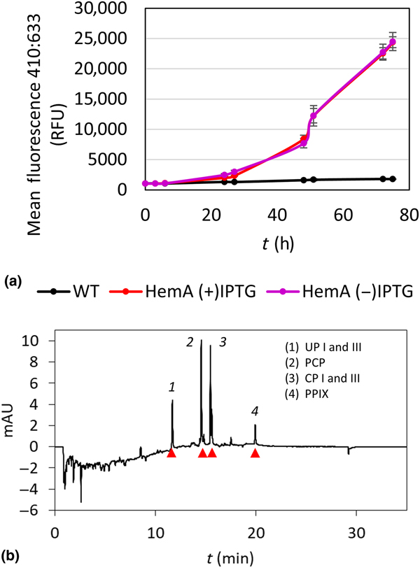
To show that porphyrins may be biosynthesized at sufficient levels to screen material properties, we expressed the 5-aminolevulinate synthase from R. capsulatus (HemA) under the regulation of a T7 promoter with a strong ribosome binding site. Production of HemA protein was confirmed by sodium dodecyl sulfate polyacrylamide gel electrophoresis of whole cell lysate (Fig. S1). Porphyrin molecules are pigments easily detected by fluorescence, enabling rapid screening of porphyrin production in cell culture by microplate reader. Upon HemA expression, greatly increased fluorescence indicated the high-level production of porphyrin molecules in the engineered strain [Fig. 2(a)]. LC-DAD-MS was used to identify and quantify specific porphyrin products. After culturing the bacteria for 72 h, the cells were removed by centrifugation and the media were examined by chromatography. The LC-DAD 410 nm signal showed four main peaks, and two of these were split peaks representing two isomers [Fig. 2(b)]. Using the MSD signals, these products were identified as Uroporphyrin I and III (UP), Coproporphyrin I and III (CP), Pentacarboxyl porphyrin (PCP), and PPIX (Table S1). Standards for UPI, CPI, PCP, and PPIX were obtained to determine the concentration of each molecule (Fig. S2).
(a) Graph fluorescence of cell cultures over time ex: 410/em: 610 nm. “WT” refers to wild-type E. coli that does not harbor a plasmid. “HemA (±) IPTG” refers to E. coli harboring plasmid mslpl004 with or without the isopropyl β-d-1-thiogalactopyranoside (IPTG) inducing molecule, (b) high performance liquid chromatography (HPLC) diode array detector (DAD) chromatograph of media with Shemin pathway products labeled.
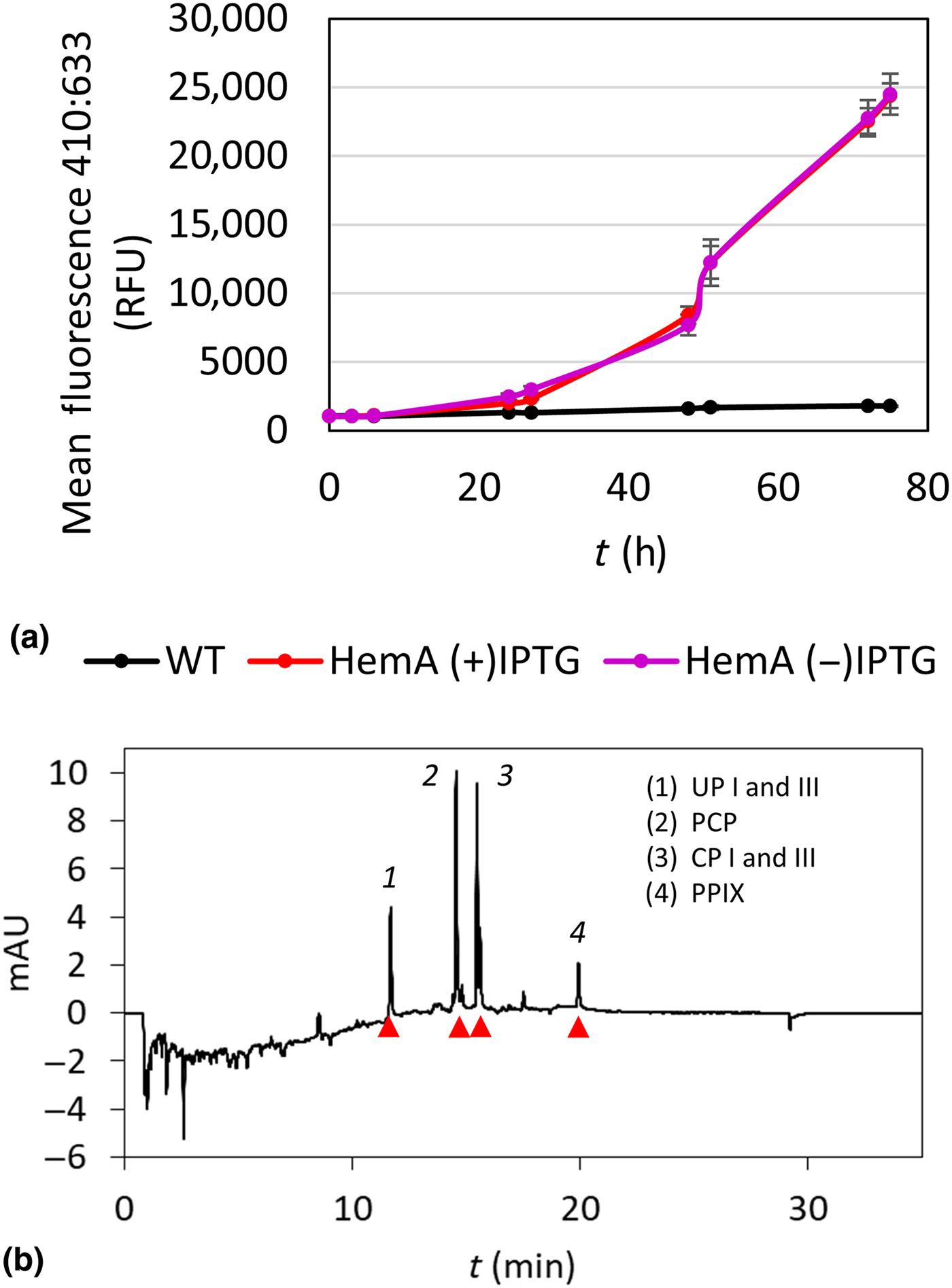
The cell pellets from the same cell cultures were extracted with methanol and analyzed by LC-MS to detect any porphyrins not secreted into the media. This analysis confirmed that the majority of total porphyrin produced is found in the media (Fig. S3), though UP is retained in the cells to a greater extent than the other molecules. Discarding the cell pellet and focusing on product recovery from the media simplifies downstream purification steps.
These findings indicate that simple overexpression of the HemA enzyme resulted in high levels of porphyrin production, and that the majority of porphyrin material may be harvested from media supernatant. The detected porphyrins are a mixture of intermediates and biproducts in the PPIX pathway [Fig. 1(a)]. While many of the molecules produced hold interest as ligands to confer potentially unique properties to materials, they have not been studied in that context until now because of low natural abundance. Engineered biosynthesis enables the production of an array of molecules at sufficient levels to study their contribution to materials properties such as photocatalytic activity. Controlling the expression of the downstream enzymes in the PPIX production pathway can alter the relative abundance of the porphyrin products.[Reference Kwon, de Boer, Petri and Schmidt-Dannert9, Reference Nielsen, Madsen, Seppälä, Christensen, Riisberg, Harrison, Møller and Nørholm13]
Purification of porphyrins
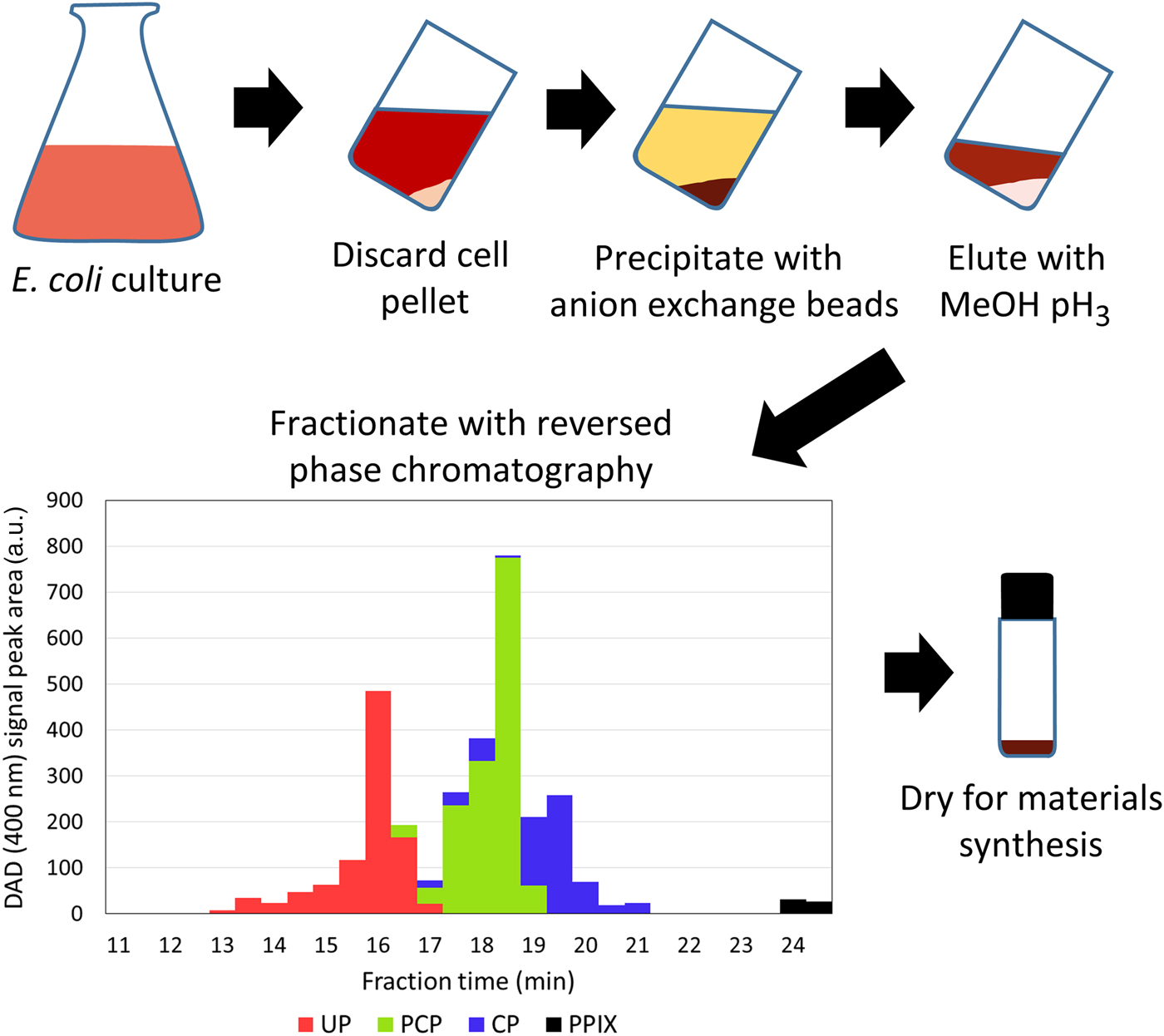
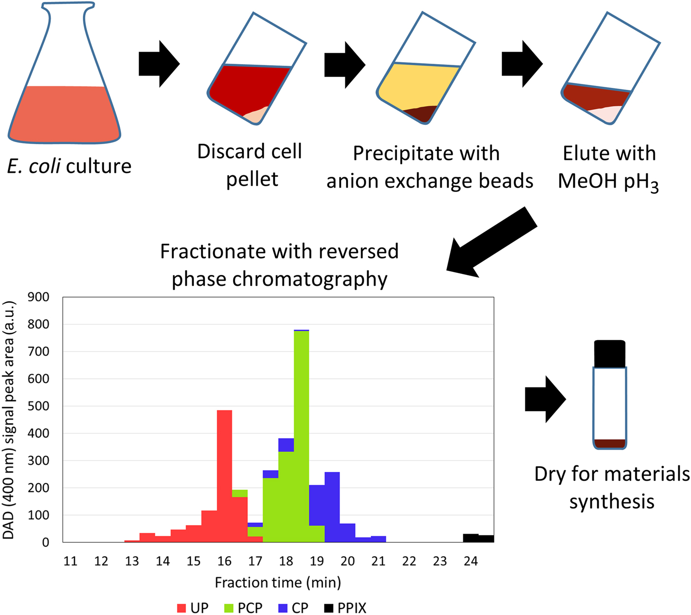
Porphyrins were isolated from the culture medium by binding to DEAE Sephadex anion exchange resin followed by sedimentation of the resin and extraction with acidified methanol. The DEAE extract was analyzed by LC-DAD-MS to assess its purity. The DEAE extract from 1 L of culture medium was found to contain 4 mg UP, 4 mg PCP, 13 mg CP, and 0.04 mg PPIX, as well as some uncharacterized organic biproducts and salts from the acid used to adjust pH. A chromatography fractionation step was added to improve purity. The DEAE methanol extract was neutralized, concentrated by rotary evaporation, and then fractionated with reversed-phase chromatography. The complete purification strategy is depicted in Fig. 3. Depending on the level of purity desired, the chromatography step is either used to quickly remove salts with a step elution or to separate each porphyrin molecule over a longer linear gradient elution protocol. A high degree of purity can be achieved with the linear gradient elution, particularly for PPIX (TABLE I). While it may not be necessary to isolate a single pure porphyrin molecule for all material synthesis strategies; here, we specifically explored the effects of PPIX on NU-1000 for photocatalysis. Future work will explore other porphyrins as well as mixtures of porphyrins to be incorporated in NU-1000 via SALI. Both the strain genotype and the purification process may be tuned to change the distribution of porphyrin products to observe the effects of increasing the abundance of individual molecules.
Schematic of porphyrin purification procedure: Porphyrins are produced in E. coli culture, after centrifugation the cell pellet is discarded and the bulk of the porphyrin product is collected in the medium. Anion exchange resin binds porphyrins in the medium, then is extracted with acidified methanol. Preparative reversed-phase high performance liquid chromatography (HPLC) is used to select fractions containing porphyrin molecules with improved purity. The graph of 400 nm absorbance for the molecules in each fraction illustrates the degree of separation between the porphyrin biproducts uroporphyrin (UP) I and III, pentacarboxyl porphyrin (PCP), coproporphyrin (CP) I and III, and protoporphyrin IX (PPIX). Finally, porphyrin products are dried prior to materials synthesis.
Theoretical quantity and purity of porphyrin within each pooled fraction derived from 1 L of cell culture starting material using the purification strategy outlined in Fig. 3.

These values are calculated based on several 900 µL injection tests of the chromatography procedure.
a Mass percent relative to total mass of porphyrin measured in each fraction.
MOF synthesis and characterization
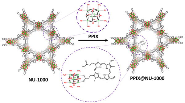
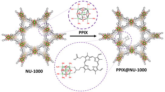
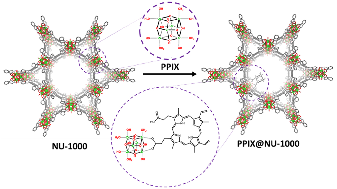
NU-1000 is a MOF composed of H4TBAPy linkers which are connected to SBUs of Zr6(μ 3-OH)4(μ 3O)4(OH)4(OH2)4 nodes forming a porous network of triangular and hexagonal channels with csq topology (Fig. 4). The yellow microcrystalline NU-1000 was modified with PPIX through SALI, generating a brown material termed PPIX@NU-1000 that shows a similar PXRD pattern and FTIR spectrum as the parent MOF (Figs. S4 and S5), indicating that the NU-1000 structure remains intact. Incorporation of PPIX causes a minor loss in BET surface area (2560 versus 2490 m2/g of NU-1000 and PPIX@NU-1000, respectively) and pore size (Fig. S6). Quantification of PPIX within PPIX@NU-1000 was evaluated by 1H NMR. After MOF digestion (i.e. decomposition into building units) 1H NMR suggested that ~1 PPIX molecule was incorporated into the SBU of NU-1000 (Fig. S7). Theoretically, four molecules per SBU can be incorporated into NU-1000 via SALI. However, it has been shown that bulky ligands do not incorporate as freely for steric reasons, reducing the total number of sites that can be modified per SBU. Furthermore, PPIX has two free carboxylic acid groups on the same side of the porphyrin ring, meaning that they could potentially bind to multiple zirconium sites, further reducing the loading per SBU.
The structure of NU-1000 (left) is a mixture of hexagonal and triangular channels for which molecular guest can diffuse through. The organic linker and the SBU are labeled (upper left and upper right circles, respectively). The secondary building unit (SBU) can be modified via solvent-assisted ligand incorporation (SALI) (right) to incorporate compounds with free carboxylic acids, such as protoporphyrin IX (PPIX), on the SBU (bottom circle).
Photocatalytic activity of PPIX@NU-1000
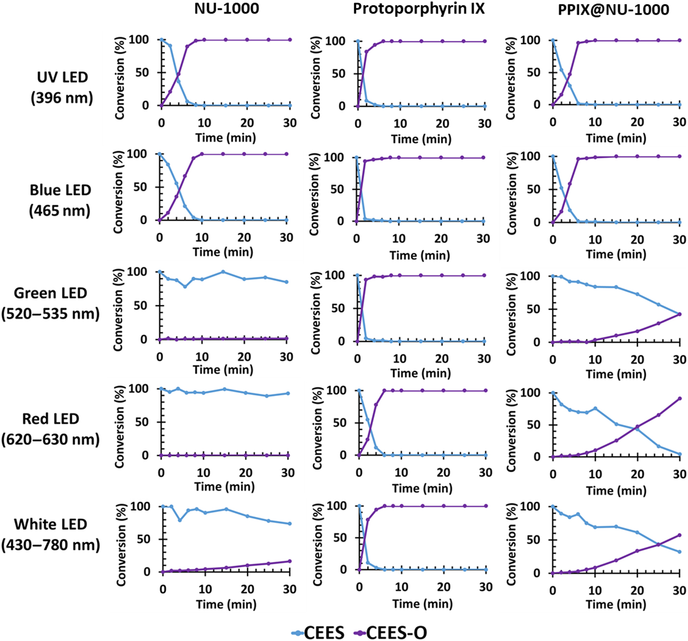
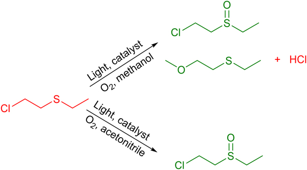
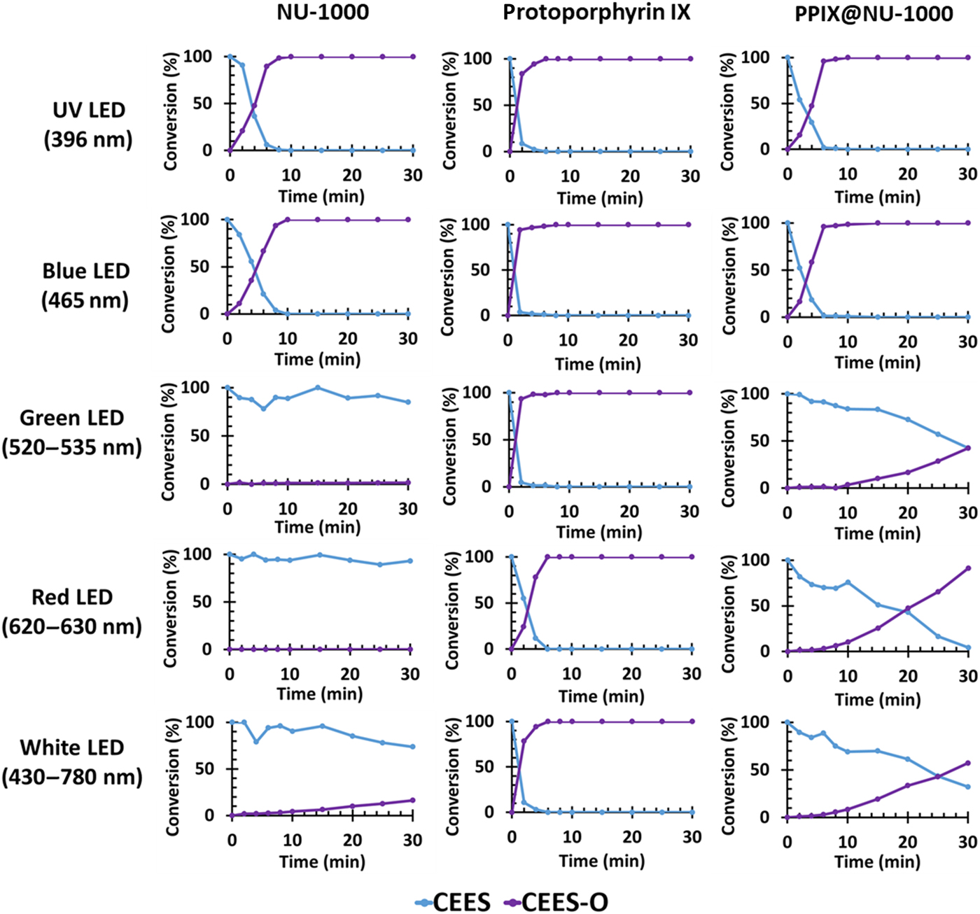
NU-1000 exhibits rapid photooxidation rates of CEES or HD in methanol solutions when exposed to UV light.[Reference Liu, Buru, Howarth, Mahle, Buchanan, DeCoste, Hupp and Farha19, Reference Howarth, Buru, Liu, Ploskonka, Hartlieb, McEntee, Mahle, Buchanan, Durke, Al-Juaid, Stoddart, DeCoste, Hupp and Farha25] PPIX is also an efficient generator of singlet oxygen.[Reference Neamţu, Nădejde, Hodoroaba, Schneider and Panne27] Fig. 5 shows that NU-1000 also performs photooxidation of CEES upon irradiation with blue light. Therefore, PPIX@NU-1000 was examined against CEES in UV and blue light to determine if the presence of PPIX in the MOF would enhance the photocatalytic activity as compared to unmodified NU-1000. The reaction was initially tested in methanol; however, a significant background methanolysis reaction was observed with the formation of up to 30% CEES-OMe after 30 min of reaction upon irradiation with UV light and in the dark with and without the MOF present (Scheme 1, Fig. S8). Interestingly, this side reaction is observed when CEES is placed in methanol in the dark up to 30% conversion almost immediately. In light of these observations it was determined that methanol should not be used for these experiments as the results could prove misleading.
Kinetics plots of the photocatalytic conversion of 2-chloroethyl ethyl sulfide (CEES) to CEES-O by NU-1000, protoporphyrin IX (PPIX), and PPIX@NU-1000 upon irradiation with various wavelengths of light.
In the presence of light, catalyst, and O2, 2-chloroethyl ethyl sulfide (CEES) degrades to a mixture of CEESO and CEES-OMe, producing a side product of HCl in methanol; however, only the 2-chloroethyl ethyl sulfoxide (CEESO) product is observed in acetonitrile. Red compounds are relatively toxic/dangerous, while green compounds are considered decontaminated products.

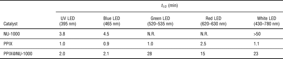
Acetonitrile was instead used as the solvent of choice for subsequent experiments as it does not react with CEES (Scheme 1) and singlet oxygen has a lifetime of ~30 μs in acetonitrile, compared to ~7 µs in methanol.[Reference Merkel and Kearns28] The reaction rates observed in acetonitrile were slightly faster than those previously reported in methanol, likely due to the longer singlet oxygen lifetime. NU-1000 exhibited CEES half-lives of 3.8 and 4.5 min in UV (395 nm) and blue (465 nm) light, respectively.[Reference Liu, Buru, Howarth, Mahle, Buchanan, DeCoste, Hupp and Farha19] PPIX@NU-1000 exhibited enhanced activity compared to the native NU-1000 under both blue and UV LED with half-lives of 2.0 and 2.1 min, respectively (Fig. 5, TABLE II).
Half-lives for photooxidation of CEES in acetonitrile by catalysts upon excitation with various wavelengths of light.

NU-1000 has an absorbance cutoff at approximately 500 nm (Fig. S9), meaning that using light at higher wavelengths will not lead to the generation of singlet oxygen, as indicated by the lack of photocatalytic activity for CEES using these wavelengths of light (Fig. 5, TABLE II). As PPIX absorbs light through the UV and visible regions (Fig. S9), it rapidly degrades CEES using light throughout this range (Fig. 5, TABLE II). It was hypothesized that PPIX@NU-1000 would be active for the photocatalytic degradation of CEES throughout the visible range of light as well, and was therefore assessed under green (530 nm), red (625 nm), and white (430–780 nm) LED conditions as well (Fig. 5). PPIX@NU-1000 reactivity is greatly enhanced as compared to unmodified NU-1000 with half-lives of 28 and 15 min in green and red light, respectively.
While NU-1000 exhibited limited photocatalytic activity beyond its absorption cutoff of 500 nm, the addition of PPIX via SALI allowed for absorption of an expanded region of the visible spectrum. To this end NU-1000 and PPIX@NU-1000 were examined using a white LED (430–780 nm). As a portion of this light can be utilized by NU-1000 ~20% of CEES degraded to CEESO in 30 min. Conversely, PPIX@NU-1000 exhibited a significantly enhanced reaction rate with a half-life of 23 min, presumably due to the utilization of a larger portion of the white light for the production of singlet oxygen (Fig. 5, TABLE II).
There are virtually limitless combinations of metals and organic ligands that greatly alter the properties of MOFs and their interaction with porphyrin functional groups. The ability to examine porphyrin molecules in a variety of MOF scaffolds will greatly accelerate the discovery of materials with useful properties. Immobilizing PPIX in a particle advantageously creates a heterogeneous catalyst from a previously homogeneous counterpart, which can be captured and reused, and also provides the opportunity to develop multifunctional materials (e.g. catalysis, sensing, and adsorption). While unmodified NU-1000 has significant photocatalytic activity, functionalization with PPIX enhances this activity and broadens the spectrum of light absorbed to increase catalysis over a range of wavelengths in the near-UV and visible spectrum. The potential for enhancement of photocatalysis in MOFs is one demonstration of the significant incentive for obtaining high levels of natural porphyins biosynthetically.
Conclusions
Utilization of biosynthetic pathways, such as the Shemin pathway in E. coli, allows for the synthesis of compounds that are not readily achievable via conventional synthetic routes. PPIX, a precursor to chlorophyll and heme B, is an example of compounds that can be synthesized by these means and exploited within materials science. Incorporation of PPIX into NU-1000 via SALI allows for the enhanced photocatalytic generation of singlet oxygen and subsequent degradation of CEES under UV and blue light, as well as expanding the effective spectral range of light that the MOF can utilize through the visible spectrum. Continuing work will explore purification of other porphyrin molecules with free carboxylic acid groups, including others in the Shemin pathway, for their inclusion in NU-1000 or other.
Supplementary material
The supplementary material for this article can be found at https://doi.org/10.1557/mrc.2019.22.
Acknowledgments
Funding was provided by the Director, Edgewood Chemical Biological Center under the authorities and provisions of Section 2363 of the FY 2018 NDAA to develop new technologies, engineer innovations, and introduce game-changing capabilities. This research was conducted while MSL and SJG held an NRC Research Associateship award at ECBC. The authors additionally thank Trevor Glaros and Gabrielle Rizzo for access to and assistance with the fraction collecting chromatography system.