Introduction
Ageing and age-related diseases such as Alzheimer's disease (AD) are a growing medical, social and economic problem. Inflammation and altered immune response are recognized to play a significant role and have become targets for therapeutic intervention. This review will discuss the brain's innate immune system in elderly people and in AD and the response to immunotherapy in animal models and initial clinical trials. Relevant studies published in the last 15 years were identified by searching PubMed, and from the author's own experience having participated in the neuropathological analysis from the Elan vaccination study.
The CNS (intrinsic) innate immune system: what is it and what does it do?
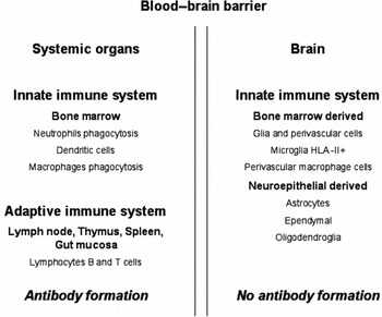
Because the brain is located behind the blood–brain barrier (BBB), there is little evidence for immune surveillance of the brain by cells of the adaptive immune system.Reference Lowenstein1 For this reason the brain developed its own (intrinsic) innate immune system. This CNS innate immune system is based mainly upon resident microglia and to a lesser extent a contribution from astrocytes and ependymal cells that maintain the integrity of the BBB and ventricle lining, respectively. However, macrophages in the perivascular space (surrounding brain blood vessels) also share an embryological origin with monocytes, macrophages and dendritic cells. The perivascular macrophages are also an important part of the systemic innate response to infection.Reference Hauwel, Furon, Canova, Griffiths, Neal and Gasque2, Reference Nguyen, Julien and Rivest3 They provide the first line of defence against systemic pathogens and express all the markers associated with innate immunorecognition of pathogens.Reference Hauwel, Furon, Canova, Griffiths, Neal and Gasque2Figure 1 illustrates the distribution of the adaptive and innate immune systems between the brain and systemic organs. The presence of the BBB resulted in the absence of the adaptive immune system in the CNS. Cells such as the resident glia, especially the microglia, became responsible for delivering the CNS innate immune response.
The diagram shows how the blood–brain barrier provides a physical barrier to prevent the cells of the systemic adaptive immune system entering the brain. The systemic adaptive immune system comprises the bone marrow, lymph nodes, spleen, gut mucosa and thymus. The cellular constituents of the adaptive immune system are B-lymphocytes (producing antibodies) and T-cell lymphocytes. The systemic innate immune system comprises neutrophils, macrophages (phagocytosis) and dendritic cells (antigen-presenting cells or APC) derived from the bone marrow. The CNS innate immune system is composed of glia, microglia (derived from bone marrow), astrocytes, ependyma and oligodendroglia (derived from neuroepithelial precursor cells). Although strictly not within the CNS, there are perivascular and meningeal cells (both derived from the bone marrow) that contribute to the intracranial innate immune response. There are no resident antibody-producing cells in the brain.

Glia provide a range of pattern recognition receptors (PRR) and scavenger receptors (SR) that are capable of distinguishing ‘self’ (host) from ‘non-self’ pathogens and also from aggregated neurotoxic proteins, such as amyloid and prion proteins.Reference Nguyen, Julien and Rivest3, Reference Elward and Gasque4 Scavenger receptors (SR) are able to phagocytose apoptotic cells, modified low density lipoprotein and ‘waste’ material; they include the receptors CD14 and CD36. Astrocytes, ependymal cells and most importantly microglia express a range of highly conserved PRR and SRReference Husemann, Loike, Anankov, Febbraio and Silverstein5–Reference Alarcon, Fuenzalida, Santibanez and von Bernhard10 that provide potential pathways for the detection and clearance of ‘non-self’ (neurotoxic proteins and apoptotic cells) from the CNS.Reference Griffiths, Gasque and Neal11
Microglia are the most important glial population with regard to the expression of SR that can detect pathogens and neurotoxic proteins. In many cases pathogens, apoptotic cells and neurotoxic proteins have been targeted by a group of molecules termed opsonins. The name opsonin is derived from the Greek word meaning sauce or seasoning,Reference Mims, Nash and Stephen12 and includes complement and systemic antibodies that cross the BBB.Reference Nguyen, Julien and Rivest3 The functional importance of an opsonin is that it binds to specific targets, such as amyloid and apoptotic cells to make phagocytosis by microglia more effective.Reference Mims, Nash and Stephen12
One important component of the CNS innate immune system and one potentially relevant to the response of the older brain to infection and clearance of neurotoxic proteins is the complement pathway (CP).Reference Gasque, Fontaine and Morgan13, Reference Gasque, Neal, Singhrao, McGreal, Dean, Van and Morgan14 The activation of the CP provides a number of important molecular signals that assist with the detection and clearance of pathogens, apoptotic cells and neurotoxic proteins from the brain.Reference Griffiths, Gasque and Neal11, Reference Gasque, Neal, Singhrao, McGreal, Dean, Van and Morgan14 However, the CP must be closely regulated to prevent it becoming over activated, resulting in host cell (bystander) damage. This control is provided by the complement regulatory proteins (CRP), which are also expressed by neurons and the glia cells of the innate immune system.Reference Gasque, Fontaine and Morgan13, Reference Griffiths, Neal and Gasque15, Reference Morgan and Meri16
Glia (microglia and astrocytes) are responsible for delivering the CNS innate immune response
Astrocytes are partly within the BBB, ependyma line the ventricle wall and microglia play a vital role in the detection and phagocytosis of pathogens and neurotoxic proteins.Reference Nguyen, Julien and Rivest3 Only microglia and to a lesser extent astrocytes are able to function as antigen-presenting cells in the CNS innate immune system, by virtue of their expression of the HLA-DR molecule (MHC-II).Reference Husemann, Loike, Anankov, Febbraio and Silverstein5 The changing expression of MHC-II, PRR and SR by microglia within the older brain and the effects this has upon the capacity of microglia to remove amyloid protein is discussed below.
Astrocytes are strategically placed in the BBB and share several receptors capable of phagocytosing amyloid with microglia.Reference Husemann, Loike, Anankov, Febbraio and Silverstein5, Reference Wyss-Coray, Loike, Brionne, Lu, Anankov, Yan, Silverstein and Husemann17 Large numbers of reactive astrocytes are present in the amyloid plaque and in vitro have been shown to be actively recruited to these plaques by a cytokine, Monocyte Chemo attractant Protein-1 (MCP-1) expressed by microglia.Reference Ishizuka, Kimura, Igata-yi, Katsuragi, Takamatsu and Miyakawa18 However, astrocytes surrounding the amyloid plaques are unable to remove Aβ, raising the possibility that dysregulation of Aβ removal by astrocytes may be responsible for amyloid accumulation in AD.Reference Wyss-Coray, Loike, Brionne, Lu, Anankov, Yan, Silverstein and Husemann17 Increasing astrocyte-related clearance of neurotoxic proteins including amyloid may be a strategy for preventing neurodegeneration.
Ependyma line the ventricle wall and are therefore located at a strategic position at the boundary between the CSF and brain.Reference Bruni19 Ependymal cells express a range of phagocytic receptors and are capable of regulating the activation of complement in the CSF.Reference Canova, Neal and Gasque20
Microglia play a pivotal role in the efficient function of the CNS innate immune system. They are usually present in a resting state, but following activation they express HLA-DR (MHC-II) and a wide range of phagocytic SR and PRR that are particularity important with regard to clearance of neurotoxic proteins such as amyloid.Reference Husemann, Loike, Anankov, Febbraio and Silverstein5 The range of SR and PRR expressed by microglia and the other glia include:
(1) a receptor for the first component of the CP, C1q. This is an opsonin and can bind to a variety of proteins including amyloid. It is found in areas of tissue damage and promotes phagocytosis of amyloid by microglia.Reference Webster, Yang, Margol, Garzon-Rodriguez, Glabe and Tenner21
(2) two receptors, CR3 and CR4, which recognize the opsonins C3, iC3b and C4, and bind to ‘non-self’ targets such as Aβ.Reference Ross22, Reference Reichert and Rotshenker23
(3) the scavenger receptors, CD14 and CD36, which are all capable of recognizing apoptotic cells and capable in vitro of clearing neurotoxic proteins such as Aβ.Reference Gregory6, Reference El Khoury, Moore, Means, Leung, Terada, Toft, Freeman and Luster8, Reference Fassbender, Walter, Kühl, Landmann, Ishii, Bertsch, Stalder, Muehlhauser, Liu, Ulmer, Rivest, Lentschat, Gulbins, Jucker, Staufenbiel, Brechtel, Walter, Multhaup, Penke, Adachi, Hartmann and Beyreuther9, Reference Bamberger, Harris, McDonald, Husemann and Landreth24, Reference Liu, Walter, Stagi, Cherny, Letiembre, Schulz-Schaeffer, Heine, Penke, Neumann and Fassbender25
(4) the Fc receptor, which detects the Fc part of immunoglobulin protein (antibodies).Reference Song, Tanaka, Cox and Lee26, Reference Komine-Kobayashi, Chou, Mochizuki, Nakao, Mizuno and Urabe27
Microglia can express either an inflammatory or protective response. They are myeloid in origin and migrate into the CNS during development, sharing this characteristic with macrophages in the perivascular spaces around CNS blood vessels, systemic macrophages, dendritic cells and monocytes.Reference Santambrogio, Belyanskaya, Fischer, Cipriani, Brosnan, Ricciardi-Castagnoli, Stern, Strominger and Riese28 The reaction of microglia towards neurotoxic proteins is of great importance, as this will determine whether the CNS innate immune system mounts a protective or destructive response. Stimulation of in vitro microglia by cytokine macrophage colony-stimulating factor (M-CSF) or granulocyte-stimulating factor (GM-CSF) produces microglia expressing CD11c+ (macrophage/inflammation) or microglia that are CD11c+ (dendritic antigen-presenting/anti-inflammation) cells, respectively.Reference Liu, Walter, Stagi, Cherny, Letiembre, Schulz-Schaeffer, Heine, Penke, Neumann and Fassbender28, Reference Monsonego and Weiner29 CD11b+ express complement, IL-1β and TNFγ to promote an inflammatory immune response, whereas CD11c+ are capable of presenting antigens to T-cells and expressing anti-inflammatory cytokines.Reference Monsonego and Weiner29, Reference Weiner and Frenkel30 CD11c+ and CD11b+ microglia are both present in AD and are able to capture Aβ. CD11c+ microglia migrate to the local cervical lymph nodes to induce T-cell activation and generate Aβ-reactive T-cells.Reference Monsonego and Weiner29 This aspect of microglial function is of relevance to the generation of antibodies by the peripheral adaptive immune system against CNS Aβ following vaccination.Reference Weiner and Frenkel29
Complement pathways are proteins expressed by all glia and promote the CNS innate immune response
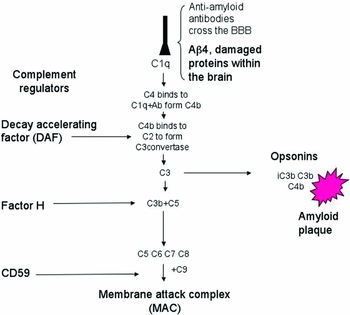
The complement system is composed of the classical, alternative and lectin pathways.Reference Gasque, Fontaine and Morgan13, Reference Gasque, Neal, Singhrao, McGreal, Dean, Van and Morgan14 The components of the first two pathways are expressed by most brain cell types and this includes neurons and glia. Activation of the CP results in the formation of opsonins (molecules that help identify targets for phagocytosis), chemo-attractant molecules and the cytolytic membrane attack complex (MAC) that lyses pathogens and host cells such as neurons.Reference Gasque, Neal, Singhrao, McGreal, Dean, Van and Morgan14, Reference Griffiths, Neal and Gasque15 Activation of the classical CP relies upon the first component, C1q, acting as a receptor for a wide range of ligands found in areas of brain damage. They include amyloid, apoptotic cells, hypoxic neurons, myelin and antibody–antigen complexes.Reference Elward and Gasque4, Reference Korb and Ahearn31, Reference Nicholson-Weller and Klickstein32 The result of C1q binding to these ligands is the activation of the CP generating the complement proteins C3b and iC3b. These molecules are important opsonins and capable of targeting pathogens and amyloid, increasing their phagocytosis by the resident glia.Reference Gasque, Neal, Singhrao, McGreal, Dean, Van and Morgan14, Reference Reichert and Rotshenker23 The alternative CP (AP) is activated by microbes and together with the classical pathway also generates C3b and iC3b. These two opsonins are recognized by the receptors CR3 and CR4, expressed by microglia, and the Kolmer cells found in the choroid plexus.Reference Gasque, Neal, Singhrao, McGreal, Dean, Van and Morgan14, Reference Singhrao, Neal, Rushmere, Morgan and Gasque33 The final common pathway shared by CP and AP is the generation of membrane attack complex (MAC), a cytolytic complex of proteins capable of lysing most cell types.Reference Elward and Gasque4, Reference Gasque, Fontaine and Morgan13, Reference Gasque, Neal, Singhrao, McGreal, Dean, Van and Morgan14Figure 2 provides a simplified classical pathway showing activation of C1q, and generation of the opsonins C3i, C3b and C4 with the final steps in the pathway generating MAC.
Simplified version of the classical complement pathway. The first component C1q can be activated by anti-Aβ4 antibodies, Aβ4 amyloid-damaged proteins. This leads to a series of activated complement components with the most important step, C3, producing the opsonins C3b, iC3b and C4 and the final steps in the pathway generating membrane attack complex (MAC). At crucial steps along the pathway the complement regulatory proteins DAF, Factor H and CD59 prevent activation and the generation of C3b, iC3b and MAC is reduced.

Uncontrolled activation of the CP would result in widespread bystander host damage, so the individual stages in the CP are regulated by complement regulatory proteins.Reference Gasque, Fontaine and Morgan13, Reference Gasque, Neal, Singhrao, McGreal, Dean, Van and Morgan14, Reference Morgan and Meri16 These proteins are expressed by glia and neurons and can be divided into cell membrane-bound and soluble regulators. Their potential therapeutic importance controlling any CNS inflammation is discussed later.
The adaptive immune system, cytokines and T-cells
An important feature of the T-cell response to systemic infection and vaccination is the differentiation of helper CD4+ T-cells into two subsets of effector cells, either the Th1 or Th2 population. The distinction depends upon the range of cytokines expressed by these two populations and their contribution to the immune response following systemic infection. The inflammatory cytokines (IL-2 and TNFα), cytotoxic T-cells and macrophage activation are all associated with the Th1 population, whereas the anti-inflammatory cytokines IL-4, IL-10 and activation of B-cells (with antibody production) are related to the Th2 population.Reference Abbas, Lichtman and Pillai34 With ageing, the balance between the Th1 and Th2 response begins to change, with more emphasis on the Th1 or inflammatory biased population of cells.Reference Giunta, Fernandez, Nikolic, Obregon, Rrapo, Town and Tan35 The effects of ageing produce a change in the Th1/Th2 balance and this must be taken into consideration when predicting the consequences of vaccination in an older population.
The adaptive and innate immune response in older people
As a generalization, antigen challenge of the older adaptive immune system changes from an anti-inflammatory (Th2) to a pro-inflammatory response (Th1). This change coincides with an age-related expansion of the pro-inflammatory, CD28-CD8+ T-cell population,Reference Almanzar, Schwaiger, Jenewein, Keller, Herndler-Brandstetter, Würzner, Schönitzer and Grubeck-Loebenstein36 reduced serum immunoglobulin concentrations with lower affinity, decreased numbers of B cellsReference Yang, Stedra and Cerny37, Reference Franceschi, Bonafe and Valensin38 and deficient MHC-II class antigen presentation by ageing dendritic and microglia.Reference Perlmutter, Scott, Barron and Chui39 The age-related increase in the number of antigen-experienced cells and a decrease of the naive T-cell population, results in a overpopulation of ineffective memory T-cells preventing an effective response to systemic antigens and vaccination.Reference Saurwein-Teissl, Lung, Marx, Gschösser, Asch, Blasko, Parson, Böck, Schönitzer, Trannoy and Grubeck-Loebenstein40
CD8+ T-cells have a decreased capacity to secrete the cytokine interferon IFNγ, whereas helper CD4+ cells demonstrate a reduced responsiveness to both antigen stimulation and capacity to assist B-cell antibody production.Reference Ouyang, Wanger and Voehringer41 These effects reflect the failure to maintain intrinsic T-cell function and not merely exhaustion due to an ageing process.Reference Swain, Cllise-Dwyer and Haynes42 Overall, these changes in the composition of the T-cell population in the older person, together with increased numbers of CD28-CD8+, tip the immune system response into the Th1 phenotype. This is associated with increased pro-inflammatory cytokine levels and reduced number of plasma cells and antibody production following systemic infection, compared with younger individuals.Reference Giunta, Fernandez, Nikolic, Obregon, Rrapo, Town and Tan35, Reference Frasca, Riley and Bloomberg43–Reference Herrero, Marques, Lloberas and Celada45 This change in the balance from Th2 to Th1 in the older population will have implications for successful antibody production following systemic vaccination.Reference Saurwein-Teissl, Lung, Marx, Gschösser, Asch, Blasko, Parson, Böck, Schönitzer, Trannoy and Grubeck-Loebenstein40
Dendritic cells and microglia are members of the innate immune system and are responsible for presenting antigens to T-cells. Their antigen-presenting capacity declines with age, adding to the reduced effectiveness of T-cell response to antigen stimulation in older people.Reference Swain, Cllise-Dwyer and Haynes42 Stimulation of the ageing CNS innate immune system also increases the expression of pro-inflammatory cytokines (TNFβ, IL-1α, IL-6 and IL-8) and decreases the level of anti-inflammatory IL-10 by activated microglia. A similar response to the bacterial wall constituent lipopolysaccharide protein (LPS) administration in older animals was present in macrophages and blood monocytes.Reference Giunta, Fernandez, Nikolic, Obregon, Rrapo, Town and Tan35, Reference Fagiolo, Cossarizza, Scala, Fanales-Belasio, Ortolani, Cozzi, Monti, Franceschi and Paganelli46 This increase of pro-inflammatory properties in the older adaptive and innate immune systems has been termed ‘inflammaging’ by Franceshi and colleagues.Reference Franceschi, Bonafe and Valensin38, Reference Aspinal47, Reference Allman and Miller48 It represents a low-grade chronic upregulation of the CNS innate immune system and may in fact be a ‘prodrome to developing AD’ in the older population.Reference Franceschi, Bonafe and Valensin38
Brain ageing, microglial priming and inflammation
In a mouse model of prion disease, a subsequent systemic inoculation with LPS resulted in an inflammatory response greater than when the same inoculation was given to mice without the prion-related disease.Reference Combrinck, Perry and Cunningham49, Reference Dilger and Johnson50 This elevated inflammatory response was a consequence of substantial inflammatory cytokine release by microglia, resulting in considerable behavioural change and acceleration of the severity of the neurodegeneration.Reference Dilger and Johnson50 These experiments in rodents prompted the hypothesis that concurrent neurodegeneration provides a sensitizing or ‘priming mechanism’ of the elderly microglia.Reference Perry, Cunningham and Holmes51 Any subsequent systemic stimuli, such as systemic infection, provides a triggering stimuli to further increase the microglial release of inflammatory cytokines, promoting further neurodegeneration.Reference Dilger and Johnson50 Microglial priming results in an up-regulation of MHC-I and -II,Reference Godbout, Chen, Abraham, Richwine, Berg, Kelley and Johnson52 complement receptors (CR3) and scavenger receptors such as CD14. All these parameters have been shown to increase in older humans, non-human primates and aged dogs, and they represent components of the ‘inflammaging’ response described by Franceshi.Reference Franceschi, Bonafe and Valensin38
In an analogous way to neurodegeneration, brain ageing is proposed to act as a ‘priming’ stimulus for microglia, probably through the increased levels of pro-inflammatory cytokines, as found in the elderly CNS.Reference Dilger and Johnson50, Reference Perry, Cunningham and Holmes51, Reference Godbout, Chen, Abraham, Richwine, Berg, Kelley and Johnson52 Levels of systemic LPS significantly increased the expression of microglial cytokines in the ageing brain compared with young animals, contributing to an age-related reduction in memory and cognitive performance.Reference Godbout, Chen, Abraham, Richwine, Berg, Kelley and Johnson52 Of interest are the observations showing increased frequency of depressive symptoms in older people with systemic illness. These effects have been attributed to the increased inflammatory cytokine response following transient stimulation of the ageing CNS innate immune system following systemic infection.Reference Dantzer, O'Connor, Freund, Johnson and Kelley53
Microglia in the older brain are, therefore, in a heightened state of readiness, ‘primed’ to respond to systemic infection and presence of antibodies, with an exaggerated inflammatory response. This is of therapeutic importance, as both impaired cognitive performance in older people and increased cognitive decline in AD, are associated with concurrent episodes of systemic infection.Reference Holmes, El-Okl, Williams, Cunningham, Wilcockson and Perry54 With each episode of systemic infection, bacterial wall components cross the BBB and activate primed microglia increasing the likelihood of neuronal injury. Similarly, vaccination with antibody formation will potentially have an increased pro-inflammatory effect in the ageing CNS as the result of stimulating already primed microglia, compared with the response of microglia in the younger brain.Reference Perry, Cunningham and Holmes51, Reference Godbout, Chen, Abraham, Richwine, Berg, Kelley and Johnson52, Reference Holmes, El-Okl, Williams, Cunningham, Wilcockson and Perry54
Neurotoxic protein clearance is compromised in the older brain
Microglia are of mixed origin.Reference Santambrogio, Belyanskaya, Fischer, Cipriani, Brosnan, Ricciardi-Castagnoli, Stern, Strominger and Riese28, Reference Simard, Soulet, Gowing, Julien and Rivest55 The resident population are mesodermal in origin and migrated into the brain during early development, whereas a second population of microglia are derived from the bone marrow. This population enters the CNS after an appropriate stimulus, usually following CNS infection, trauma or neurodegeneration.Reference Simard, Soulet, Gowing, Julien and Rivest55, Reference Simard and Rivest56 In young transgenic mice with AD, microglia recruited from the bone marrow are specifically attracted to Aβ in amyloid plaques and are more effective at phagocytosis of this protein than resident microglia.Reference Simard, Soulet, Gowing, Julien and Rivest55, Reference Simard and Rivest56 However, with ageing this capacity declines, despite large numbers of microglia surrounding amyloid plaques.Reference Perlmutter, Scott, Barron and Chui39, Reference Simard, Soulet, Gowing, Julien and Rivest55 In older individuals with AD a similar problem is noted. Despite the large numbers of microglia clustering around amyloid plaques, little amyloid is present within these cells and, in cases where it is, the amyloid remains in a non-degraded form.Reference Frackowiak, Wisniewski, Wegiel, Merz, Iqbal and Wang57
One explanation for the apparent failure of microglia to effectively remove amyloid in older mice or from AD brains is the reduced expression of SR and PR receptors by ageing microglia, compared with younger animals.Reference Holmes, El-Okl, Williams, Cunningham, Wilcockson and Perry54, Reference Hickman, Allison and El Khoury58 This effect was explained as the consequence of elevated levels of pro-inflammatory cytokines, TNF-α and IL-1β, in the surrounding ageing brain, inhibiting the expression of SR by microglia. Gene expression studies of brain regions with high Aβ levels demonstrated an over-expression of several genes associated with inflammatory cytokines and complement proteins.Reference Hickman, Allison and El Khoury58 This inflammatory response is associated with little change in the levels of mRNA of several phagocytic receptors (CD36, CR3),Reference Hickman, Allison and El Khoury58 all capable of phagocytosing Aβ. The reduced clearance of Aβ by ageing microglia results in extracellular amyloid accumulation, with further free radical formation and associated neuronal injury.Reference Gregory6, Reference Bamberger, Harris, McDonald, Husemann and Landreth24 The high levels of pro-inflammatory cytokines present in the elderly brain inhibits the expression of microglial PRR and SR (CD36 and CD14).Reference Hickman, Allison and El Khoury58, Reference Fiala, Cribbs, Rosenthal and Bernard59
Finally, the presence of the inflammatory cytokine IFNγ in vitro and in the elderly CNS has been found to stimulate the formation of Aβ from brain APP. The production of IFNγ by primed microglia, primed CNS, or through transport of this inflammatory cytokine across the BBB following systemic infection, has been shown to stimulate the breakdown of APP, further increasing Aβ formation in the ageing CNS.Reference Blasko, Marx, Steiner, Hartmann and Grubeck-Loebenstein60
Alzheimer's disease, ageing and inflammation
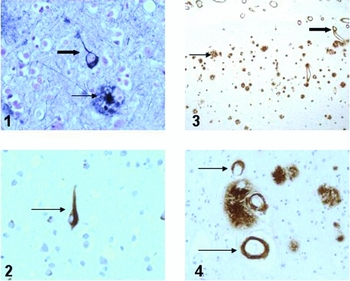
AD is the commonest cause of dementia, resulting from an accumulation of extracellular amyloid (Aβ) and intraneuronal tau protein. Aβ is derived from the breakdown of the transmembrane protein APP, as the result of the two enzymes β and γ secretase.Reference Duyckaerts, Dickson and Dickson61 The Aβ peptide forms β pleated fibrillary amyloid sheets to produce neuritic amyloid plaques.Reference Duyckaerts, Dickson and Dickson61 Over-production of Aβ results in its accumulation as an extracellular amyloid in the form of Aβ peptides composed of non-soluble, fibrillary Aβ1–42 (a peptide of between 1 and 42 amino acids) and truncated N-terminal forms of Aβ4 peptide. The other main amyloid peptide (Aβ1–40) is forty amino acids in length. It is more soluble than Aβ1–42 and is found in cortical blood vessel walls.Reference Weller, Subash, Preston, Mazanti and Carare62 Tau accumulates as hyperphosphorylated tau in paired helical filaments in both axons, dendrites (dystrophic neurites) and as neurofibrillary tangles within neuronsReference Duyckaerts, Dickson and Dickson61 (see Figure 3 illustrating the neuropathological findings in typical Alzheimer's disease). Amyloid neuritic plaques also contain pro-inflammatory microglia (CD11b+) and astrocytes both express inflammatory cytokines, tumour necrosis factor (TNFα and INFγ), MCP, nitric oxide and numerous complement components.Reference Weller, Subash, Preston, Mazanti and Carare62–Reference Veerhuis, Janseen, Hoozemans, DeGroot, Hack and Eikelenboom65 Microglia express a range of potentially important opsonin and phagocytic receptors capable of clearing amyloid from the brain,Reference Gregory6, Reference Webster, Yang, Margol, Garzon-Rodriguez, Glabe and Tenner21, Reference Bamberger, Harris, McDonald, Husemann and Landreth24, Reference Fiala, Cribbs, Rosenthal and Bernard59, Reference Chen, Iribarren, Hu, Chen, Gong, Cho, Lockett, Dunlop and Wang66, Reference Paresce, Ghosh and Maxfield67 but they are prevented from phagocytosing amyloid by the surrounding inflammatory environment, as demonstrated in animal models of AD and older brain (see above).Reference Hickman, Allison and El Khoury58 A vital part of any therapeutic programme designed to prevent neurodegeneration and cognitive decline in AD requires effective phagocytosis of Aβ4 by microglia.
Neuropathology of Alzheimer's disease. 1, AD brain stained with Bielschowsky stain, demonstrating neuritic amyloid plaque and neurofibrillary tangle (thick arrow); 2, a neurofibrillary tangle stained with an antibody against tau protein; 3, cerebral cortex from AD stained with anti-Aβ4 antibodies, demonstrating amyloid plaques (thin arrow) and blood vessels with congophilic angiopathy (CAA) in their walls (thick arrow); 4, high-power section from AD stained with anti-Aβ antibodies showing amyloid plaques and small diameter blood vessels with CAA.

Animal models of AD and Aβ vaccination
The experimental approach to vaccine development adopted the mechanism used by the systemic immune response to remove pathogens from the CNS. Following exposure to a pathogen, the systemic immune system generates systemic antibodies that cross the BBB and opsonize pathogens such as viruses and assists their removal from the brain by microglia.Reference Mims, Nash and Stephen12 This approach was exploited by using a vaccine, based upon an amyloid peptide to stimulate the production of systemic antibodies and their transport across the BBB, permitting them to bind to amyloid plaques, provoking phagocytosis of amyloid proteins by microglia.Reference Schenk, Barbour, Dunn, Gordon, Grajeda, Guido, Hu, Huang, Johnson-Wood, Khan, Kholodenko, Lee, Liao, Lieberburg, Motter, Mutter, Soriano, Shopp, Vasquez, Vandevert, Walker, Wogulis, Yednock, Games and Seubert68, Reference Schenk, Hagen and Seubert69
Young PDAPP (platelet-derived growth factor promoter expressing amyloid precursor protein) mice that gradually accumulate Aβ amyloid as they age, were vaccinated with an Aβ peptide composed of 1–42 amino acids, in conjunction with an adjuvant. This regimen resulted in the formation of polyclonal anti-amyloid antibodies that crossed the BBB and prevented Aβ plaque formation.Reference Schenk, Barbour, Dunn, Gordon, Grajeda, Guido, Hu, Huang, Johnson-Wood, Khan, Kholodenko, Lee, Liao, Lieberburg, Motter, Mutter, Soriano, Shopp, Vasquez, Vandevert, Walker, Wogulis, Yednock, Games and Seubert68 In older PDAPP mice with pre-existing amyloid plaques, Aβ immunization resulted in amyloid clearance by microglia. This effect was also present in other mouse models of AD. In these, amyloid removal following vaccination and peripherally administrated antibodies prevented cognitive decline.Reference Schenk, Hagen and Seubert69–Reference Bard, Cannon, Barbour, Burke, Games, Grajeda, Guido, Hu, Huang, Johnson-Wood, Khan, Kholodenko, Lee, Lieberburg, Motter, Nguyen, Soriano, Vasquez, Weiss, Welch, Seubert, Schenk and Yednock72Ex vivo studies using either PDAPP mouse or AD tissue cultured with a monoclonal antibody 3d6 (IgG2a) resulted in the activation of microglia and increased clearance of Aβ through Fc receptor-mediated phagocytosis.Reference Bacskai, Kajdasz, McLellan, Games, Seubert, Schenk and Hyman73–Reference Koenigsknecht-Talboo, Meyer-Luehmann, Parsadanian, Garcia-Alloza, Finn, Hyman, Bacskai and Holtzman76
Antibodies can gain access to the older brain and neurons
A vital requirement for an antibody to provide a therapeutic effect is its capacity to cross the BBB and enter the brain.Reference Mims, Nash and Stephen12 However, most neurotoxic proteins are intraneuronal, so gaining access to the neuronal cytoplasm is a further vital requirement. IgG can enter the brain through regions deficient in the BBB and also through adsorptive transcytosis.Reference Zlokovic, Deane, Sallstrom, Chow and Miano77 In AD mice, the permeability of the BBB is raised, increasing the amount of circulating IgG that can enter the brain.Reference Poduslo, Curran, Wengenack, Malester and Duff78 Intraneuronal antibodies raised against tau proteins have been found within neurons, probably the result of pinocytosis (receptor-mediated endocytosis) using neuronal Fc receptors.Reference Asuni, Boutajangout, Quatermain and Sigurdsson79 Once inside the neuron, an antibody will gain access to the lysosome–endosome pathway, where it will bind to the relevant neurotoxic protein and trigger its degradation.Reference Asuni, Boutajangout, Quatermain and Sigurdsson79, Reference Masliah, Rockenstein and Adame80 These observations support the capacity of systemic antibodies not only to enter the brain, but also to become internalized into the same neurons that contain neurotoxic proteins.Reference Fabian81, Reference Mohamed, Mosier, Zou, Siklós, Alexianu, Engelhardt, Beers, Le and Appel82
Opsonins, complement and vaccination
The potential contribution made by the CNS innate immune system, in particular the CP, to amyloid clearance on the older CNS is well documented.Reference Reichwald, Danner, Wiederhold and Staufenbiel83 Experiments using complement-inhibited AD mice demonstrated increased Aβ deposition, whereas mice with increased complement activation had reduced amyloid deposition and increased microglial activation.Reference Wyss-Coray, Yan, Lin and Lambris84, Reference Wyss-Coray and Mucke85 One explanation for the clearance of Aβ by the CP is the generation of the opsonin C3, which localizes on the amyloid plaque.Reference Eikelenboom, Bate, Van Gool and Hoozemans64, Reference Veerhuis, Janseen, Hoozemans, DeGroot, Hack and Eikelenboom65, Reference Reichwald, Danner, Wiederhold and Staufenbiel83 The presence of C3 provides a more ‘appetizing’ target for the CR3 and CR4 receptors expressed by microglia.Reference Gasque, Neal, Singhrao, McGreal, Dean, Van and Morgan14, Reference Reichert and Rotshenker23 In older AD mice that were also C3 deficient, amyloid plaque numbers increased, microglia expressed more inflammatory cytokines and neuronal loss was higher than in wild-type animals.Reference Maier, Peng, Jiang, Seabrook, Carroll and Lemere86, Reference Zhou, Fonseca, Pisalyaput and Tenner87 Several of these features were age related. Complement deficiency in young mice did not have any effects upon cortical amyloid accumulation.Reference Reichwald, Danner, Wiederhold and Staufenbiel83, Reference Maier, Peng, Jiang, Seabrook, Carroll and Lemere86 This could reflect the less inflammatory brain environment in young mice allowing microglia to function more effectively.
The ‘peripheral sink’ effect: an alternative explanation for amyloid clearance
An alternative explanation for amyloid removal, independent of microglia phagocytosis, was the ‘peripheral sink’ model proposed by DeMattos et al.Reference DeMattos, Bales, Cummins, Dodart, Paul and Holtzman88 Following vaccination, the presence of systemic anti-Aβ antibody was associated with an increase in plasma Aβ levels and reduced amyloid load in the CNS. The mechanism responsible for the efflux of brain amyloid into systemic circulation was described as the ‘peripheral sink’ effect, and it did not rely upon microglial phagocytosis of amyloid.Reference DeMattos, Bales, Cummins, Dodart, Paul and Holtzman88 The mechanism responsible for this efflux was related to the capacity of anti-Aβ antibodies to solubilize the fibrillary amyloid within the amyloid plaques.Reference Solomon, Koppel, Frankel and Hanan-Aharon89 Soluble amyloid is subsequently removed from the CNS via perivascular and CSF pathways and finds its way into the plasma.Reference Weller, Subash, Preston, Mazanti and Carare62, Reference Weller90, Reference Preston, Steart, Wilkinson, Nicoll and Weller91 Despite this interesting explanation for amyloid efflux from the brain into plasma following immunization, quantitative studies in transgenic animals have provided less convincing results.Reference Levites, Smithson, Price, Dakin, Yuan, Sierks, Kim, McGowan, Reed, Rosenberry, Das and Golde92 The rapid rise of the Aβ in plasma following anti-Aβ inoculation was explained by the increase of the half-life of Aβ when bound to the anti-Aβ antibody. There was no evidence for Aβ efflux from the brain into plasma during the short time interval when the antibody was not bound to Aβ. Furthermore, only a very small percentage of the total plasma antibody was found to enter the brain and this was not accompanied by a decline in total brain amyloid. In fact, this study found that only 3–4% of the total brain amyloid could be sequestered by plasma monoclonal anti-Aβ antibodies.Reference Levites, Smithson, Price, Dakin, Yuan, Sierks, Kim, McGowan, Reed, Rosenberry, Das and Golde92
Vaccination in older people and the systemic response to Aβ: some surprises
The effectiveness of vaccination using an Aβ peptide designed to remove amyloid from the mouse brain depends upon the production of systemic anti-Aβ antibodies and the enhancement of microglial phagocytosis. T- and B-cell immunity in older humans generally decreases with age, resulting in a greater susceptibility to infection and a reduced systemic antibody response to vaccination.Reference Grubeck-Loebenstein, Della Bella, Iorio, Michel, Pawelec and Solana93 Predictably, stimulation of lymphocytes from AD patients with APP did not result in their proliferation.Reference Treib, Ransmayr, Sgonc, Lassman and Grubeck-Loebenstein94 Similarly, in BS SJL mice (transgenic for human APP), immunization with the peptide Aβ1–40 resulted in both T-cell hyporesponsiveness and the failure to produce anti-Aβ antibodies.Reference Monsonego, Maron, Zota, Selkoe and Weiner95 The inability to produce systemic anti-amyloid antibodies and T-cell hyporesponsiveness could reflect the consequences of chronic exposure to APP in mice and Aβ in humans.Reference Monsonego, Maron, Zota, Selkoe and Weiner95
However, stimulation of T-cells from older humans and AD patients with synthetic amyloid peptide sequences 16–33 (located within the middle of the Aβ1–42 peptide) surprisingly produces a strong Aβ-reactive T-cell response and included both anti- and pro-inflammatory phenotypes (Th1 and Th2). The increased T-cell reactivity was restricted to the Aβ peptide and not merely an overall increase in T-cell response to a wide range of antigens.Reference Monsonego, Zota, Karni, Krieger, Bar-Or, Bitan, Budson, Sperling, Selkoe and Weiner96 One explanation for the persistence of this population of Aβ-reactive T-cells in the older person is the capture of brain Aβ by microglia of the CD11c+ phenotype and their subsequent migration to local cervical lymph nodes.Reference Hauwel, Furon, Canova, Griffiths, Neal and Gasque2, Reference Fassbender, Walter, Kühl, Landmann, Ishii, Bertsch, Stalder, Muehlhauser, Liu, Ulmer, Rivest, Lentschat, Gulbins, Jucker, Staufenbiel, Brechtel, Walter, Multhaup, Penke, Adachi, Hartmann and Beyreuther9 Within the cervical lymph node, microglia adopt an antigen-presenting role and present Aβ to local T-cells.Reference Monsonego and Weiner29 This initiates T-cell activation, resulting in an increase in the number of systemic Aβ-reactive T-cells.Reference Monsonego and Weiner29, Reference Cserr, Harling Berg and Knopf97
Vaccination and brain inflammation
Vaccination with a ‘self’ protein such as Aβ requires the addition of an adjuvant to reduce ‘self’ tolerance and provoke a reliable systemic immune response against the Aβ. As shown by Mosonego in an older population, the predominant T-cell epitope in the Aβ peptide is located in the middle of the 1–42 region, whereas the B-cell epitope is in the amino-terminal of the peptide.Reference Monsonego and Weiner29, Reference Monsonego, Zota, Karni, Krieger, Bar-Or, Bitan, Budson, Sperling, Selkoe and Weiner96 The Elan AN1792 vaccination trial used a dose of 225 micrograms of Aβ (1–42), the same peptide as that in the AD amyloid plaque, in conjunction with adjuvant, a saponin QS-21 (50 micrograms).Reference Bayer, Bullock, Jones, Wilkinson, Paterson, Jenkins, Millais and Donoghue98 Phase I included older people with mild or moderate AD (n = 80) and began in 2000. It resulted in only 23% of individuals producing anti-amyloid antibodies, although this increased to 53% following additional injections.Reference Bayer, Bullock, Jones, Wilkinson, Paterson, Jenkins, Millais and Donoghue98 In the subsequent study (n = 372), 20% developed an antibody response, but the trial was halted when 6% of cases developed a meningoencephalitis. In all cases this was unrelated to the anti-Aβ antibody titre.Reference Orgogozo, Gilman, Dartigues, Laurent, Puel, Kirby, Jouanny, Dubois, Eisner, Flitman, Michel, Boada, Frank and Hock99 Neuropathology from two cases with a clinical diagnosis of meningoencephalitis found the inflammation was composed of T-cells in the meninges and macrophages within the white matter. In both of these two cases amyloid plaque clearance was present, but there was no evidence of T-cell-mediated demyelination, as found for example in multiple sclerosis.Reference Nicoll, Wilkinson, Holmes, Steart, Markham and Weller100, Reference Ferrer, Boada Rovira, Sánchez Guerra, Rey and Costa-Jussá101 However, a subsequent case with no encephalitis also found evidence for amyloid plaque clearance.Reference Masliah, Hansen, Adame, Crews, Bard, Lee, Seubert, Games, Kirby and Schenk102
The development of meningoencephalitis was apparently surprising because, in the experimental animal studies, inflammation was reported in only Aβ-vaccinated double transgenic mice, expressing both APP and the inflammatory cytokine IFNγ.Reference Monsonego, Imitola, Petrovic, Zota, Nemirovsky, Baron, Fisher, Owens and Weiner103 The presence of up-regulated IFNγ expression increased the numbers of meningeal and parenchymal CD4+ T-cells, together with increased numbers of activated microglia within the Aβ plaques. Stimulation of microglia by IFNγ in young mice, in contrast to older humans, results in stimulation of amyloid phagocytosis, but also an increase of T-cells infiltrating the CNS.Reference Monsonego, Imitola, Petrovic, Zota, Nemirovsky, Baron, Fisher, Owens and Weiner103 The production of excessive IFNγ following stimulation of already primed ageing microglia would increase the influx of T-cells into the brain.Reference Monsonego, Imitola, Petrovic, Zota, Nemirovsky, Baron, Fisher, Owens and Weiner103
In older humans, the presence of a meningoencephalitis following active vaccination has several possible explanations:
(1) the presence of increased numbers of systemic Aβ-reactive T-cells in elderly people migrating into the CNS and initiating an inflammatory response with a subsequent T-cell response to myelin.Reference Monsonego and Weiner29, Reference Monsonego, Zota, Karni, Krieger, Bar-Or, Bitan, Budson, Sperling, Selkoe and Weiner96
(2) the use of a full-length Aβ peptide (16–33 amino acids) as a vaccine is particularly effective at stimulating Aβ-reactive T-cell proliferation in the systemic circulation.
(3) vaccination producing anti-Aβ antibodies further activates the already ‘primed’ microglia in the older CNS to produce an accelerated Th1 pro-inflammatory response, associated with IFNγ, IL-1β, IL-6 and the activation of complement.Reference Perry, Cunningham and Holmes51, Reference Dantzer, O'Connor, Freund, Johnson and Kelley53, Reference Monsonego, Imitola, Petrovic, Zota, Nemirovsky, Baron, Fisher, Owens and Weiner103 The increased expression of pro-inflammatory cytokines, for example IFNγ, would increase the risk that some vaccinated older individuals will develop an encephalitis.
Aβ vaccination, antibodies and neuropathology
In animal models of AD, anti-Aβ antibodies generated against the N-terminal part of the peptide were most effective at amyloid plaque removal.Reference Schenk, Hagen and Seubert69, Reference McLaurin, Cecal, Kierstead, Tian, Phinney, Manea, French, Lambermon, Darabie, Brown, Janus, Chishti, Horne, Westaway, Fraser, Mount, Przybylski and St George-Hyslop104 The therapeutic and clinical importance of generating antibodies against Aβ following vaccination was initially shown by Hock.Reference Hock, Konietzko, Papassotiropoulos, Wollmer, Streffer, von Rotz, Davey, Moritz and Nitsch105 In thirty individuals with AD who were all vaccinated with Aβ42, only twenty produced significant serum levels of anti-Aβ. The same antibodies recognized Aβ in the amyloid plaques, as shown by incubating the individuals’ sera containing the anti-Aβ antibodies with AD mouse tissue.Reference Hock, Konietzko, Streffer and Tracy106 More importantly, these individuals were also shown to have a reduced rate of cognitive decline, preserved hippocampus function, and a long-lasting anti-Aβ42 production, together with the presence of anti-Aβ antibodies in the CSF.Reference Hock, Konietzko, Papassotiropoulos, Wollmer, Streffer, von Rotz, Davey, Moritz and Nitsch105
Neuropathological examination of three individuals who died during the initial stages of the Elan trial found that cortical amyloid plaque removal after vaccination was associated with and without T-cell inflammation.Reference Nicoll, Barton and Bouche107 In each case the cause of death was not related directly to the effects of vaccination upon the CNS. Each of the three cases was shown to have generated a range of antibodies including anti-Aβ40, anti-Aβ42 and, most importantly, antibodies against the truncated N-terminus amyloid peptide.Reference Nicoll, Barton and Bouche107 The truncated N-terminal amyloid proteins are able to aggregate more rapidly and are regarded as more neurotoxic than the full-length amyloid. For these reasons, a successful vaccination protocol must provide clearance of truncated N-terminal amyloid from the brain.Reference Lee, Bard, Johnson-Wood, Lee, Hu, Griffith, Black, Schenk and Seubert108
Most antibodies generated by Aβ peptide vaccination in AD individuals bind to the N-terminal region of Aβ, and it is therefore unlikely that they will bind to the amyloid plaques containing mainly the N-terminal truncated form of Aβ.Reference Lee, Bard, Johnson-Wood, Lee, Hu, Griffith, Black, Schenk and Seubert108 Although this could not be directly examined, in the three cases studied by Nicol and colleagues,Reference Nicoll, Barton and Bouche107 large areas of cortex were completely devoid of any amyloid plaques, inferring that the N-terminal truncated variety of fibrillary or non-fibrillary Aβ had in fact been removed following vaccination.
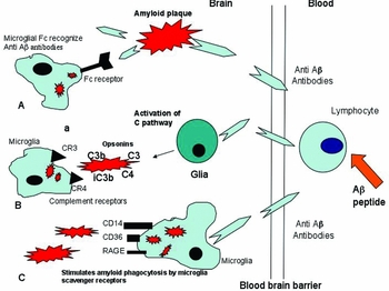
Neuropathological studies therefore demonstrated similarities between the response to vaccination in AD and animal models of AD.Reference Schenk, Hagen and Seubert69 At least two potential pathways exist to remove amyloid plaque, either by phagocytosis of amyloid by microglia following anti-Aβ production,Reference Paresce, Ghosh and Maxfield67, Reference Schenk, Hagen and Seubert69, Reference Bard, Cannon, Barbour, Burke, Games, Grajeda, Guido, Hu, Huang, Johnson-Wood, Khan, Kholodenko, Lee, Lieberburg, Motter, Nguyen, Soriano, Vasquez, Weiss, Welch, Seubert, Schenk and Yednock72–Reference Bacskai, Kajdasz, Christie, Carter, Games, Seubert, Schenk and Hyman74, Reference Wilcock, Rojiani, Rosenthal, Levkowitz, Subbarao, Alamed, Wilson, Wilson, Freeman, Gordon and Morgan109 or anti-Aβ binding to amyloid plaques producing solubilization and efflux from the CNS via a perivascular pathway into the systemic circulation.Reference DeMattos, Bales, Cummins, Dodart, Paul and Holtzman88, Reference Solomon, Koppel, Frankel and Hanan-Aharon89 The three possible pathways regarding the involvement of microglial phagocytosis and clearance of Aβ following vaccination, are illustrated in Figure 4.
This diagram shows the three possible pathways responsible for phagocytosis of Aβ4 by microglia following vaccination with Aβ4 peptide. A, anti-Aβ amyloid antibodies produced following vaccination bind to the amyloid plaque. Microglia expressing Fc receptors recognize the anti-Aβ amyloid antibodies. This results in phagocytosis of amyloid by microglia. B, anti-Aβ antibodies stimulate glia to produce complement; this results in the generation of the opsonins C3 and iC3b, binding to amyloid plaque. Microglia expressing CR3 and CR4 receptors detect the opsonins C3 and iC3b and this results in phagocytosis of plaque amyloid. C, microglia are activated by anti-Aβ antibodies, and this results in the phagocytosis of plaque amyloid by a wide range of potential scavenger receptors (CD14, CD36, RAGE).

Long-term effect of vaccination in AD
Subsequent post-mortem analysis of nine cases within the follow-up of the first Elan trial found definite amyloid plaque removal, but no therapeutic effect preventing cognitive decline.Reference Holmes, Boche, Wilkinson, Yadegarfar, Hopkins, Bayer, Jones, Bullock, Love, Neal, Zotova and Nicoll110 Histological examination of the nine Aβ42 vaccinated cases found variable levels of microglial phagocytosis of amyloid and absence of tau-containing dystrophic neurites, but no effect upon intraneuronal, neurofibrillary tangle numbers. There was no relationship between the AN1792 dose and either amyloid load or degree of plaque removal. Although no relationship between the degree of amyloid removal and antibody titre was found, immunization was able to stimulate a long-term process with amyloid plaque removal still in progress five years after first vaccination.Reference Holmes, Boche, Wilkinson, Yadegarfar, Hopkins, Bayer, Jones, Bullock, Love, Neal, Zotova and Nicoll110 Despite the presence of serum anti-Aβ antibody, this was not able to prevent progression of dementia, raising the possibility that amyloid does not in fact play a major part in the neurodegeneration. However, there are alternative explanations for the findings:
(1) Aβ plaques might only initiate, but not maintain, neurodegeneration. For example, older individuals can have similar amyloid plaque numbers to those found in AD, but remain cognitively intact.Reference Davis, Schmitt, Wekstein and Markesbery111
(2) amyloid plaque load does not change as the clinical signs deteriorate, as demonstrated by in vivo imaging.Reference Klunk, Mathis, Price, Lopresti and DeKosky112
(3) plaque removal can occur after days in animal models of AD. The Elan study found amyloid plaques were removed by 4 months, but extensive amyloid plaque removal is only present 5 years after immunization, suggesting that amyloid removal is a slow process.Reference Holmes, Boche, Wilkinson, Yadegarfar, Hopkins, Bayer, Jones, Bullock, Love, Neal, Zotova and Nicoll110
(4) vaccination might not reduce the neurotoxic oligomeric form of Aβ, allowing further neurodegeneration independently of the fibrillary amyloid.Reference Patton, Kalback, Esh, Kokjohn, Vickle, Luehrs, Kuo, Lopez, Brune, Ferrer, Masliah, Newel, Beach, Castaño and Roher113
(5) vaccination with the full-length Aβ peptide and adjuvant increases the activation of the innate immune system.Reference Weiner and Frenkel30 This resulted in an increased Th1 pro-inflammatory response, reducing microglial capacity for amyloid removal.Reference Pride, Black and Hagen114
In summary, amyloid removal has been demonstrated in vaccinated AD individuals and is a protracted process, associated with variable levels of serum anti-Aβ antibodies. However, amyloid removal does not prevent cognitive decline.
The cerebral circulation and amyloid removal after immunization
Amyloid clearance via perivascular pathways in the elderly brain
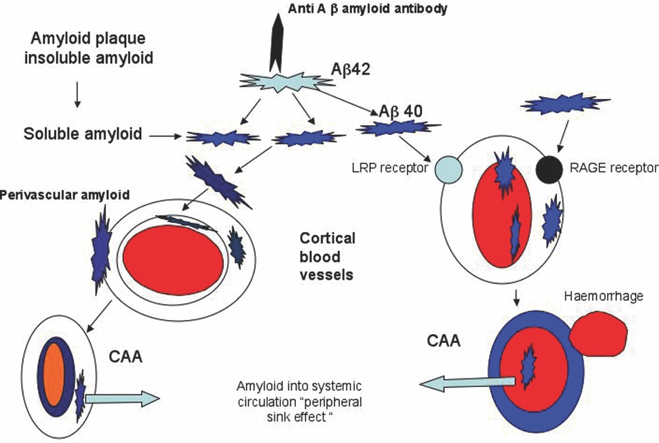
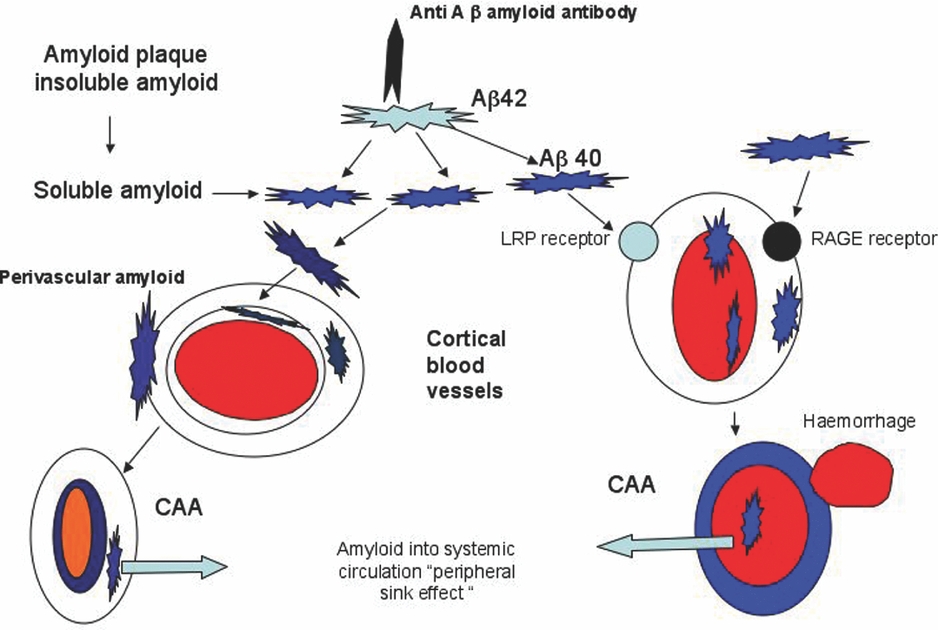
In the older non-AD brain, one potential route for soluble Aβ to exit from the cortex is by accumulating in the interstitial fluid (ISF) in the perivascular space that surrounds blood vessel walls. From the perivascular space, the Aβ can gain access to the venous circulation and CSF.Reference Weller, Subash, Preston, Mazanti and Carare62, Reference Weller90, Reference Preston, Steart, Wilkinson, Nicoll and Weller91, Reference Boche, Zotova, Weller, Love, Neal, Pickering, Wilkinson, Holmes and Nicoll115 Pulsation of the adjacent blood vessel wall will supply the motive force to propel the fluid within the perivascular space into the venous circulation and CSF. However, as the artery wall becomes less compliant with age, the efficiency of pulsatile flow decreases, with an accumulation of Aβ in the ISF. This has two consequences: blockage of the perivascular space and an accumulation of Aβ in the vessel wall as congophilic angiopathy (CAA).Reference Weller, Subash, Preston, Mazanti and Carare62 This angiopathy represents an accumulation of both Aβ40 and Aβ42 peptides in the wall of cortical and meningeal arteries, creating thickening and weakening of the wall, resulting in cortical haemorrhage.Reference Weller90, Reference Boche, Zotova, Weller, Love, Neal, Pickering, Wilkinson, Holmes and Nicoll115Figure 5 shows after vaccination the alternative pathways for soluble amyloid to either become deposited in the cerebral vessel wall or within the perivascular space surrounding cerebral vessels.
This diagram shows the pathways proposed to explain the increased numbers of cerebral vessels demonstrating congophilic angiopathy (CAA) following Aβ4 vaccination. The presence of amyloid in the vessel wall can result in haemorrhage into the surrounding brain. Anti-Aβ antibodies bind to the insoluble amyloid plaque. This produces soluble amyloid, which diffuses via interstial fluid into the perivascular space and eventually into the adjacent cerebral blood vessel wall. The alternative explanation is accumulation of soluble amyloid through receptor-mediated transport (LRP and RAGE) into the wall of the blood vessel with subsequent localization in the wall. In both cases, amyloid is able to enter the systemic circulation by passing along the perivascular space into the local venous circulation and then into the cerebral venous circulation. The alternative explanation is the amyloid within the lumen of the cerebral vessels enters the cerebral venous circulation and contributes to the ‘peripheral sink effect’.
Vaccination and perivascular amyloid removal
One consistent observation in the immunized AD cases was an increase in CAA,Reference Boche, Zotova, Weller, Love, Neal, Pickering, Wilkinson, Holmes and Nicoll115 with a significant decrease in the cortical amyloid plaque load. This relationship is consistent with the proposal that fibrillary Aβ in the plaque is solubilized as the result of an anti-Aβ antibody binding.Reference Solomon, Koppel, Frankel and Hanan-Aharon89 The soluble amyloid is deposited in the perivascular space and is then deposited in adjacent blood vessel walls to produce CAA. This explanation is consistent with the finding that the more soluble variety of Aβ, Aβ40 (as well as Aβ42), was found with greater frequency in the wall of blood vessels in AD cases receiving vaccination.Reference Weller, Subash, Preston, Mazanti and Carare62, Reference Boche, Zotova, Weller, Love, Neal, Pickering, Wilkinson, Holmes and Nicoll115 Despite the presence of CAA in the immunized AD individuals, this did not increase the formation of CAA-related microhaemorrhages.Reference Orgogozo, Gilman, Dartigues, Laurent, Puel, Kirby, Jouanny, Dubois, Eisner, Flitman, Michel, Boada, Frank and Hock99, Reference Holmes, Boche, Wilkinson, Yadegarfar, Hopkins, Bayer, Jones, Bullock, Love, Neal, Zotova and Nicoll110, Reference Boche, Zotova, Weller, Love, Neal, Pickering, Wilkinson, Holmes and Nicoll115 These observations are consistent with the proposal that Aβ can be removed from the CNS via the perivascular pathway and independently of microglial involvement.Reference Weller, Subash, Preston, Mazanti and Carare62 This possibility is illustrated in Figure 5, showing the efflux of soluble amyloid along the perivascular space into the local venous circulation.
Transportation of amyloid across the BBB
Clearance of Aβ from the vessel wall into the systemic circulation is also promoted through enzyme degradation and transportation across the BBB by the enzymes neprilysin and insulin.Reference McDermott and Gibson116 The receptor for advanced glycosylation end-products (RAGE) is a multi-ligand receptor (a member of the immunoglobulin family) that is a signal transducing cell-surface receptor for Aβ and, together with the lipoprotein receptor protein (LRP-1), is located on the blood vessel wall. Figure 5 shows the location of these receptors on the blood vessel wall and the accumulation of amyloid following vaccination Both these receptors increase soluble amyloid transport from the CNS across the BBB into the systemic circulation and CSF.Reference McDermott and Gibson116 LRP-1 preferentially clears Aβ40 and, on this basis, the development of therapeutic agents to enhance the binding of Aβ to LRP-1 is being investigated.Reference Shibata, Yamada, Kumar, Calero, Bading, Frangione, Holtzman, Miller, Strickland, Ghiso and Zlokovic117 Promising results have been obtained using statins and other agents to block Aβ/RAGE interaction, making higher levels of Aβ available to bind to the more efficient LRP-1 transport pathway.Reference Shibata, Yamada, Kumar, Calero, Bading, Frangione, Holtzman, Miller, Strickland, Ghiso and Zlokovic117 Whilst clinical trials of statins for prevention of AD and for slowing cognitive decline in established dementia have shown no clinical benefits, trials of RAGE inhibitors are being initiated.Reference Jones, Kivipelto, Feldman, Sparks, Doody, Waters, Hey-Hadavi, Breazna, Schindler and Ramos118, Reference Neugroschl and Sano119
Passive immunization: a safe alternative to active immunization?
Intracerebroventricular (ICV) injection of anti-Aβ antibody into the ventricle of transgenic AD mice is able to reduce the number of amyloid plaques, restore pre-synaptic injury and reduce microglial activation. Within 24 hours after ICV injection, the antibody is distributed throughout the hippocampus and cortex, but it is entirely removed after a time interval of between 36 hours to 4 days. One major drawback to this approach is that amyloid reappears 4–8 weeks later, but amyloid angiopathy, perivascular haemorrhage and inflammation have not been reported.Reference Chauhan, Siegel and Lichtor120
Intraventricular infusion of anti-Aβ, although effective at clearing brain amyloid, is unlikely to prove to be a practical therapy. A different approach, but again using passive immunization, involves the infusion of a specific range of humanized anti-Aβ antibodies and immunoglobulins. One report from a Phase II trial using this approach with human intravenous infusion of immunoglobulins containing anti-Aβ antibodies showed some improvement in cognitive testing.Reference Dodel, Du, Depboylu, Hampel, Frölich, Haag, Hemmeter, Paulsen, Teipel, Brettschneider, Spottke, Nölker, Möller, Wei, Farlow, Sommer and Oertel121
Despite the advantage of passive administration of anti-Aβ antibodies, it is still possible for the presence of these antibodies in the CSF and systemic circulation to activate the ‘primed’ microglia in the older CNS, resulting in unwanted brain inflammation. Nevertheless, passive immunization can be terminated if serious side-effects are encountered.
Vaccination for the clearance of non-Aβ intracellular protein aggregates in neurodegenerative disease
Intrahippocampal injections of anti-Aβ monoclonal antibody into transgenic mice that develop both amyloid and tau lesions initially removed intra- and extracellular Aβ deposits and several days later cleared early somato-dendritic, non-phosphorylated tau.Reference Oddo, Billing and Kesslak122 However, the amyloid reaccumulated 30 days after the injection of anti-Aβ, soon followed by hyperphosphorylated tau (similar to human NFT) 15 days later. This response is related to the neuron's proteasome (a cellular organelle responsible for degrading misfolded proteins), initial removal of amyloid from the proteasome allowing subsequent removal of non-phosphorylated intra-neuronal tau.Reference Oddo, Billing and Kesslak122 Despite the reported lowering of the amyloid load and some evidence for the loss of tau in dystrophic neurites, the number of intraneuronal NFT was not significantly reduced. These findings indicate that Aβ-directed therapy is unlikely to permanently reverse intraneuronal, hyperphosphorylated tau accumulation once it has been established.Reference Hutton and McGowan123
Vaccination with an active tau phosphorylated protein in P301L, tangle-producing mice reduced tau aggregation and produced anti-tau antibodies that entered the CNS, and this was found to be an age-related effect. In young mice, anti-tau antibodies reduced the level of hippocampal tau and slowed behavioural decline to a greater degree than the same parameters in elderly mice.Reference Asuni, Boutajangout, Quatermain and Sigurdsson79 Interestingly, immunization of alpha-synuclein transgenic mice with human alpha-synuclein peptide produced antibodies against this protein, which decreased intracellular accumulation of alpha-synuclein aggregates by stimulating its degradation through a lysosomal pathway.Reference Masliah, Rockenstein and Adame80 These experiments raise the possibility that amyloid and tau removal in AD require the production of both anti-amyloid and anti-tau antibodies.Reference Hutton and McGowan123 Vaccines based upon the generation of anti-tau antibodies could play a potential role in treating the tauopathies, for example progressive supra-nuclear palsy, corticobasal degeneration and frontotemporal lobe dementia linked to chromosome 17.
In contrast to the experiments designed to produce antibodies against amyloid and tau, the generation of anti-prion antibodies in mice has been generally unsuccessful, with a failure to generate anti-prion antibodies after vaccination with prion proteins.Reference Sigurdsson, Brown, Daniels, Kascsak, Kascsak, Carp, Meeker, Frangione and Wisniewski124 Similarly, passive administration of anti-prion monoclonal antibodies also failed to prevent prion-related disease in mice. Unlike Aβ and tau, prion proteins are widely distributed throughout the body and this contributes to both B- and T-cell tolerance of vaccinated prion proteins, preventing the generation of systemic anti-prion protein antibodies. A full discussion of the experiments designed to reduce self tolerance following vaccination with prion proteins is provided by Heppner.Reference Sigurdsson, Brown, Daniels, Kascsak, Kascsak, Carp, Meeker, Frangione and Wisniewski124, Reference Heppner and Aguzzi125
Complement in neurodegeneration: molecules to promote amyloid clearance and reduce inflammation
Complement is synthesized by glia and neurons and was first demonstrated to be localized on neuritic amyloid plaques in AD by Eikelenboom.Reference Weller, Subash, Preston, Mazanti and Carare62–Reference Eikelenboom, Bate, Van Gool and Hoozemans64 Activation of the complement system in AD and Pick's disease is well documented.Reference Yasuhara, Aimi, McGeer and McGeer126–Reference Singhrao, Neal, Gasque, Morgan and Newman128 The first component, C1q, binds to fibrillary but not soluble Aβ amyloid and activates microglial synthesis of inflammatory cytokines and further glial expression of the opsonins C3, iC3b and C4.Reference Gasque, Neal, Singhrao, McGreal, Dean, Van and Morgan14, Reference Eikelenboom, Bate, Van Gool and Hoozemans64, Reference Veerhuis, Janseen, Hoozemans, DeGroot, Hack and Eikelenboom65 The opsonins C3 and C4 are detected by two well-established receptors CR3 and CR4, both expressed by activated microglia, astrocytes and Kolmer cells.Reference Ross22, Reference Reichert and Rotshenker23, Reference Nicholson-Weller and Klickstein32 Both these receptors recognize target proteins, such as Aβ opsonized with C3b and iC3b.Reference Nicholson-Weller and Klickstein32 There is also evidence that the CP is activated when C1q binds to the acute phase proteins, serum amyloid and C-reactive protein, both of which function as opsonins and bind to fibrillary amyloid.Reference Gasque, Neal, Singhrao, McGreal, Dean, Van and Morgan14
The protective function of the complement system in neurodegenerative disease is confirmed by the observation identifying reduced levels of phagocytosis of Aβ in AD transgenic mice with complement deficiency.Reference Wyss-Coray and Mucke85 The explanation for this effect is the failure to generate C3 and other opsonins that locate amyloid plaques as the result of complement deficiency.Reference Wyss-Coray and Mucke85 One explanation for the clearance of amyloid following vaccination with Aβ peptide is C1q binding to anti-Aβ antibodies that have crossed the BBB and attached themselves to an amyloid plaque. This results in the activation of the CP, with increased expression of the opsonins C3 and iC3b, promoting subsequent amyloid removal by microglia expressing the CR3 and CR4 receptors.
Complement regulatory proteins provide targets to regulate CNS inflammation
Uncontrolled activation of the CP results in MAC attack of bystander neurons and increased severity of the disease process.Reference Gasque, Neal, Singhrao, McGreal, Dean, Van and Morgan14In vitro and in vivo data indicate that astrocytes, microglia and oligodendrocytes are protected from the effects of direct bystander complement lysis, because they express high levels of complement regulator proteins (CRP).Reference Gasque, Neal, Singhrao, McGreal, Dean, Van and Morgan14, Reference Morgan and Meri16, Reference Singhrao, Neal, Gasque, Morgan and Newman128, Reference Griffiths, Neal, Fontaine, Das and Gasque129 To counter the effects of complement activation, CRP are expressed by microglia in at least two neurodegenerative diseases, AD and Pick's disease,Reference McGeer and McGeer63, Reference Veerhuis, Janseen, Hoozemans, DeGroot, Hack and Eikelenboom65, Reference Singhrao, Neal, Gasque, Morgan and Newman128 but are not expressed by astrocytes and neurons.Reference Singhrao, Neal, Rushmere, Morgan and Gasque33, Reference Griffiths, Neal, Fontaine, Das and Gasque129 An important regulator of the CP is factor H (FH), a protein that is strongly expressed by neurons and regulates inflammation by inhibiting complement activation.Reference Gasque, Neal, Singhrao, McGreal, Dean, Van and Morgan14, Reference Griffiths, Neal, Fontaine, Das and Gasque129 Administration of intra-peritoneal FH to animals with experimental autoimmune encephalitis reduced inflammation, axonal injury and leucocyte infiltration.Reference Griffiths, Neal, Fontaine, Das and Gasque129
Factor H deficiency is associated with up-regulation of complement activation in plasma, causing renal injury due to the deposition of complement and inflammatory cells on the basement membrane of kidney.Reference de Cordoba and de Jorge130, Reference Haines, Hauser, Schmidt, Scott, Olson, Gallins, Spencer, Kwan, Noureddine, Gilbert, Schnetz-Boutaud, Agarwal, Postel and Pericak-Vance131 A similar observation was found in AD and age-related macular degeneration, two degenerative diseases associated with a polymorphism (Y402H) in FH.Reference Zetterberg, Landgren, Andersson, Palmér, Gustafson, Skoog, Minthon, Thelle, Wallin, Bogdanovic, Andreasen, Blennow and Zetterberg132, Reference Hye, Lynham, Thambisetty, Causevic, Campbell, Byers, Hooper, Rijsdijk, Tabrizi, Banner, Shaw, Foy, Poppe, Archer, Hamilton, Powell, Brown, Sham, Ward and Lovestone133 The polymorphism is located within the binding sites for heparin and C-reactive protein, altering the effectiveness of FH to inhibit C.Reference Honda, Itoh, Yoshimoto, Ohno, Hinoda and Imai134 In a recent proteomic study involving AD plasma, FH was shown to be the best marker of disease, with highly elevated levels in AD compared with control cases.Reference Strohmeyer, Ramirez, Cole, Mueller and Rogers135, Reference Rinne, Brooks, Rossor, Fox, Bullock, Klunk, Mathis, Blennow, Barakos, Okello, Rodriguez Martinez de Liano, Liu, Koller, Gregg, Schenk, Black and Grundman136 The same protein is found in the amyloid plaque in AD tissue and this raises the possibility that a failure of either FH regulation, or ‘dysregulation’, increases brain inflammation due to Aβ chronically activating the CP with further neuronal injury. Therapeutic control of CRPs provides a potential target to reduce CNS inflammation, although this is unlikely to be brain specific as the CRPs are found throughout the body.
Conclusion
The older brain accumulates neurotoxic proteins and these proteins stimulate local production of inflammatory cytokines, reducing the capacity of the local innate immune system to clear neurotoxic proteins. Furthermore, the microglia in the older innate immune system in the brain are in a ‘primed state’, which increases their expression of inflammatory cytokines following stimulation by neurotoxic proteins (Aβ) and systemic infection. Vaccination of transgenic AD mice with amyloid peptides consistently produced anti-Aβ antibodies and this was associated with a reduction of brain amyloid due to microglial phagocytosis. Following active vaccination with an amyloid peptide in older individuals with AD, systemic anti-amyloid antibody production was inconsistent, but in a small number of individuals anti-Aβ antibody production stimulated the removal of amyloid plaques. This effect was the result of a combination of activating microglial phagocytosis and stimulating efflux of amyloid from the plaque into local blood vessels and eventually into the systemic circulation. In some older people vaccination initiated brain inflammation, possibly due to either increasing the systemic Th1 response, or an over-stimulation of the already primed microglia (innate immune system). Vaccination programmes based upon passive immunization infusing monoclonal anti-Aβ antibodies into the systemic circulation have had some success, as have experiments using active vaccination with tau and alpha-synuclein, promoting the clearance of these proteins from the CNS.
An alternative to removing neurotoxic proteins by generating systemic antibodies is the reduction of brain inflammation to reduce neurodegeneration. Another approach is the therapeutic regulation of the CNS complement system to prevent bystander attack of host cells and the stimulation of opsonins to facilitate the clearance of neurotoxic proteins. However, the regulation of CP is not brain specific and this approach will potentially inhibit the protective functions of complement activation elsewhere in the body. These are exciting times for the treatment and prevention of dementia in the older population including development of in vivo imaging methods to identify amyloid accumulation and clearance following therapeutic trials.Reference Rinne, Brooks, Rossor, Fox, Bullock, Klunk, Mathis, Blennow, Barakos, Okello, Rodriguez Martinez de Liano, Liu, Koller, Gregg, Schenk, Black and Grundman136 There are now some grounds for guarded optimism in the prevention of Alzheimer's disease, based upon the experimental and clinical advances discussed in this review.
Conflict of interest
The author has an interest in brain immunity and infection and has recently collaborated with colleagues in Bristol, Cardiff and Southampton investigating the neuropathological changes in participants involved with the Elan AD vaccination study.