Introduction
Collision tumours are rare entities defined as neoplastic lesions with two or more histologically and morphologically different tumours that maintain distinct borders within the same organ. It can be of two benign tumours, two malignant tumours, or a benign and malignant tumour.Reference Bulte, Hoegler and Khachemoune1 Collision tumours are more frequently found in female patients, with most cases occurring in the fifth to seventh decades of life. Often, only one component of these collision tumours is recognised during pre-surgical evaluation, and the other component is an incidental histopathological finding.Reference Negura, Ianole, Danciu, Preda, Iosep and Dănilă2
Collision tumours are found in various organs, such as the kidneys, colon, stomach, ovaries, colon, and lungs; they are extremely rare in the thyroid and represent only < 1 per cent of thyroid cancers.Reference Negura, Ianole, Danciu, Preda, Iosep and Dănilă2–Reference Abbi, Hameed, Jiang, De Las Casas and Schwann7 Among thyroid collision tumours, co-occurrence of medullary and papillary carcinomas is the most frequent.Reference Bojoga, Stănescu and Badiu8 There are several theories to explain the pathogenesis of collision tumours, including stem cell theory, random effect theory, tumour predisposition, and hypotheses about common genetic behaviour, such as germline mutation of rearranged-during-transfection proto-oncogene in medullary thyroid carcinoma and papillary thyroid carcinoma co-existence.Reference Bojoga, Stănescu and Badiu8
The medullary component of thyroid collision tumours is more aggressive than the papillary component, and medullary thyroid carcinomas in collision tumours are more aggressive than medullary thyroid carcinoma-only tumours.Reference Negura, Ianole, Danciu, Preda, Iosep and Dănilă2 The management of collision tumours is more challenging than that of individual tumours because of different biological aggressiveness, prognoses, and treatment options; therefore, management needs to evolve through critical evaluation of the disease course.Reference Bojoga, Stănescu and Badiu8
There is uncertainty regarding the clinicopathological features of collision tumours. Management guidelines for these tumours are poorly defined. Therefore, in the present study, we aimed to understand the characteristics of these tumours. We retrospectively analysed thyroid collision tumours with components of medullary thyroid carcinoma andpapillary thyroid carcinoma, confirmed by histopathology, and analysed the demographic features, clinical characteristics, management, and clinical outcomes of these patients.
Materials and methods
This was a retrospective study of collision tumours of the thyroid in 24 patients conducted in a tertiary care centre between 2009 and 2022. The inclusion criteria were cases of collision tumours of medullary thyroid carcinoma with papillary thyroid carcinoma and/or follicular thyroid carcinoma proven by histopathological diagnosis and those who had already undergone total thyroidectomy. The exclusion criteria were patients with insufficient histological material, those who received pre-operative radiotherapy, and those aged less than 18 years. Clinical details of the patients were obtained from their electronic medical records.
All patients were evaluated by a multidisciplinary team, and therapy decision making was performed based on available guidelines updated at that time and expert individualised remarks from the team. Pre-operative and post-operative ultrasonographies were performed by a senior radiologist using high-frequency linear probes. Serum thyroglobulin and calcitonin assays were performed in standard institutional laboratories using radioimmunoassay/chemiluminescence immunoassay. The normal range for serum thyroglobulin post-operative patients with thyroid-stimulating hormone (TSH) stimulation was considered to be less than 1 ng/ml, while post-treatment on thyroid suppression therapy was given as per their American Thyroid Association risk category and checked at follow-up visits.
Pre-surgery fine needle aspiration cytology (FNAC) and post-surgery histopathology samples were handled by a team of senior pathologists specialised in head–neck oncology. Post-surgery radioactive iodine scans were performed on planar double-headed gamma camera 24 h after administration of 1 mCi/37 MBq of radioactive iodine. Single-photon emission computed tomography/computed tomography (CT) was performed if needed. For post-radioactive iodine therapy, follow-up scans were acquired in a similar manner using 3–5 mCi/(111–185) MBq radioactive iodine activity 2–3 days prior to administration.
For somatostatin receptor positron emission tomography (PET) 1–2 mCi/(37–74) MBq of 68Ga-DOTATATE/NOC was administered 1 hour prior to acquisition, and all patients were scanned vertex to mid-thigh on PET-CT scanner. Radioactive iodine therapy (from 30 to 250 mCi) was administered in the high-dose therapy ward after decision by the team to abide by all national radiation regulatory parameters. The team consisted of a head–neck oncology specialist surgical oncologist, medical oncologist, and radiation oncologist for each therapy plan and follow up.
Results
Our retrospective study was conducted in patients with medullary thyroid carcinoma over a period of 15 years. Twenty-four of the 470 patients were found to have collision tumours, which implies a rate of occurrence of 5.1 per cent in medullary thyroid carcinoma cases.
Demography and clinical characteristics
Among 24 patients diagnosed with thyroid collision tumours, the mean age of the patient was 50.7 years (range 26–90 years) with a male-to-female ratio of 1:1.4 (10 males and 14 females). All the patients presented with swelling in the front or on either side of the neck.
Investigations
Pre-operative ultrasound data were available for 10 patients. Of these, five collision tumours were detected by ultrasonography. Three papillary thyroid carcinoma (two of which were micro-papillary thyroid carcinomas) and two medullary thyroid carcinoma were missed by ultrasonography.
Pre-operative serum calcitonin was available for nine patients, and the mean value was 6476 pg/ml (range, 1080–20,842 pg/ml). According to histopathology, among 28 lesions of medullary thyroid carcinoma, 16 were in the right lobe (57.14 per cent), 11 in the left lobe (39.28 per cent), and one in the isthmus of the thyroid (3.57 per cent).
Among 24 patients with collision tumours of the thyroid, in most cases (20 of 24; 83.3 per cent) the average size of the medullary thyroid carcinoma component was larger than that of the papillary thyroid carcinoma component. Among the 25 papillary thyroid carcinomas, 14 lesions were in the right lobe (56 per cent), 10 were in the left lobe (40 per cent), and one lesion was in the isthmus (4 per cent).
Eighteen patients (75 per cent) presented with lymph node metastases. Among them, 16 (88.8 per cent) patients had the medullary thyroid carcinoma component. Four patients (22.2 per cent) had the papillary thyroid carcinoma component, and two patients (11.1 per cent) had metastases from medullary thyroid carcinoma and papillary thyroid carcinoma.
Thirteen patients had extrathyroidal extension (54.16 per cent). Among the 24 patients, only two (8.3 per cent) presented with distant metastasis to the lung and skeletal system.
Treatment and follow up
All patients underwent total thyroidectomy, with or without lymph node dissection. Eight patients underwent radioactive iodine scan. Thyroid hormone was withdrawn and a low-iodine diet followed for 3–4 weeks to attain a TSH value of >30 mIU/ml before the radioactive iodine scan according to the institutional protocol. Imaging was acquired 24 hours after oral administration of 1 mCi (37 MBq) of radioactive iodine (I-131). A wide-field-of-view gamma camera with a high-energy parallel-hole collimator was used for imaging, employing a 20 per cent window centred at 364 keV. Images were acquired for 15 min, and the percentage of radioactive iodine uptake was calculated using a thyroid uptake probe.
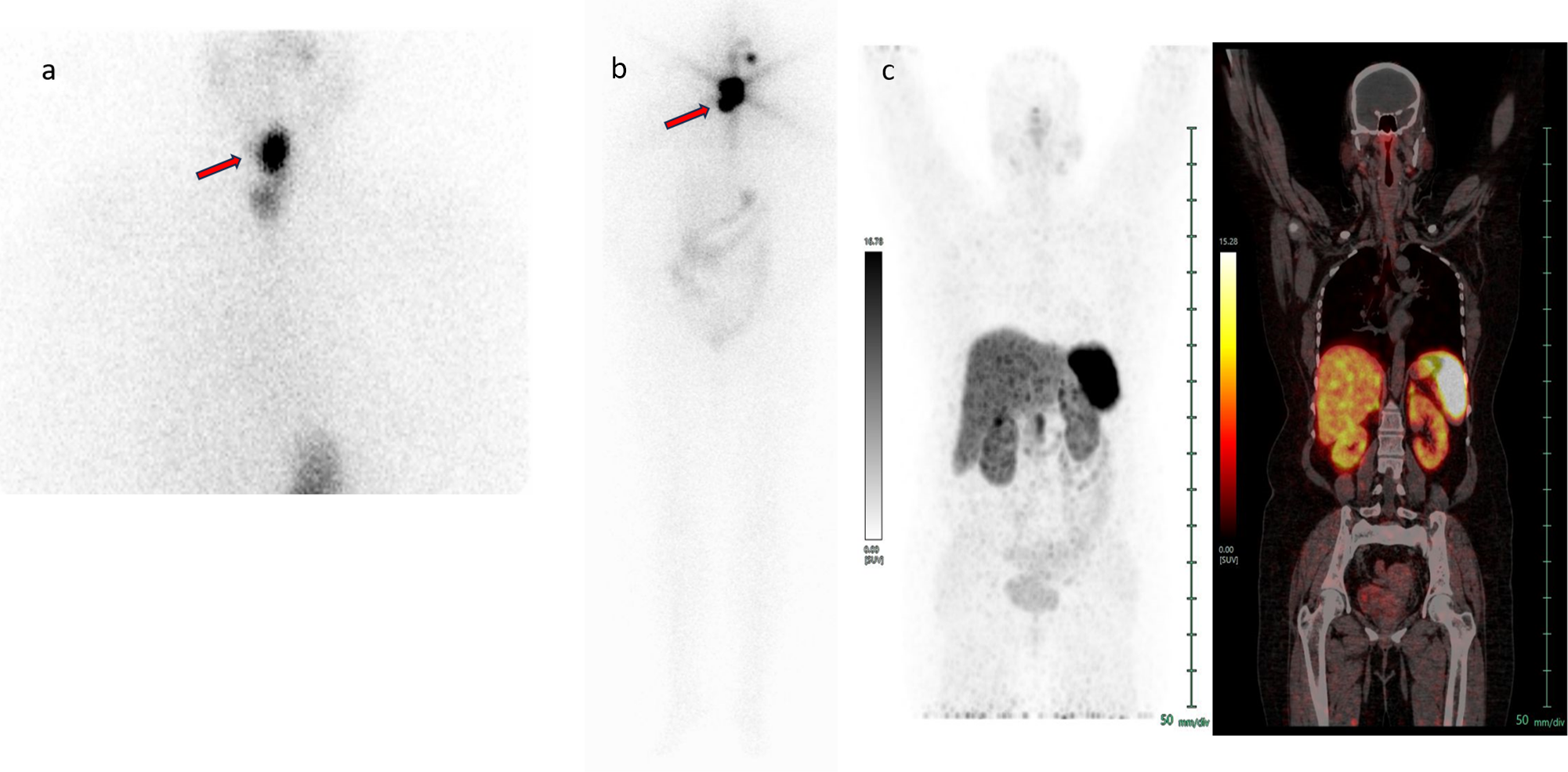
Seven patients showed neck uptake. Four of these seven patients had lymph node metastasis (three from medullary thyroid carcinoma and one from papillary thyroid carcinoma). Patients without lymph nodal metastasis from papillary thyroid carcinoma received around 50–100 mCi (1.85–3.7 GBq) of radioactive iodine. (Figure 2) Patients with lymph node metastasis from papillary thyroid carcinoma received around 150 mCi (5550 MBq) of radioactive iodine, as per institutional protocol.
A 43-year-old female patient with histopathology report of right lobe of thyroid lesion 1 showing invasive encapsulated follicular variant of papillary thyroid carcinoma, right lobe of thyroid lesion 2 showing medullary thyroid carcinoma with capsular invasion, and left lobe lesion showing differentiated papillary thyroid carcinoma, classical type. The patient underwent a total thyroidectomy. Post-operative stimulated thyroglobulin was 8.23 ng/ml and serum calcitonin was 0.53 pg/ml. 1mCi (37 MBq) radioactive iodine scan showed a (depicted by arrow) bifocal abnormally increased tracer uptake noted in the neck (a), whereas the 68Ga-DOTATATE PET-CT scan showed no abnormal somatostatin receptor-expressing lesion noted in the whole-body scan (c). Patient received 146 mCi (5402 MBq) of high-dose radioactive iodine therapy, and a post-therapy scan showed abnormal bifocal tracer uptake (depicted by arrow) in the neck region (b).

A negative iodine scan was found in one patient with papillary microcarcinoma component. A radioactive iodine scan performed after 6 months revealed no obvious radioactive iodine uptake in all patients who received therapy, suggestive of complete ablation to iodine therapy. Three patients with advanced-stage medullary thyroid carcinoma underwent external-beam radiotherapy (RT) of the neck. Irrespective of the treatment received, all patients received a suppression dose of thyroxine and were followed up with tumour markers (serum thyroglobulin and serum calcitonin).
Follow up was for a mean period of 7.7 years (range: five months to 15 years). Of the 24 patients, 15 patients were disease-free (62.5 per cent) at the time of analysis, eight patients were living with the disease (33.33 per cent) and one patient died due to an aetiology other than primary disease. One patient had locoregional recurrence of medullary thyroid carcinoma three years after primary diagnosis and treatment.
Eight patients underwent 68Ga-DOTATATE/CT on follow up, based on the attending oncologists’ referral. Whole-body somatostatin receptor-based PET-CT scan was performed after intravenous injection of 2–3 mCi (74–111 MBq) of 68Ga-DOTATATE, using a whole-body full-ring dedicated PET-CT camera. A whole-body low-dose (50 mA, 120 kVp) CT scan was acquired for attenuation correction and anatomical colocalisation. Images were reconstructed using an iterative algorithm.
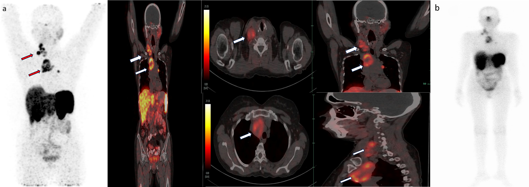
The patient with a calcitonin level of 20,862 pg/ml showed somatostatin receptor-expressing regional nodal disease. (Figure 3) Another two patients showed non-somatostatin receptor expressing regional lymph nodes and non-somatostatin receptor expressing bilateral lung nodules at the levels of calcitonin 370 pg/ml and 1333 pg/ml, respectively. Five patients whose mean calcitonin level was 32.81pg/ml (range: 0.4–84.19 pg/ml) did not show any residual disease.
A 46-year-old female patient presented with a histopathological report of medullary carcinoma of the right lobe and papillary carcinoma of the thyroid of the left lobe. She had received radioactive iodine at 104 mCi (3848 MBq) 10 years previously. A post-therapy scan revealed focal abnormal tracer uptake in the neck region. Subsequently, the patient had defaulted for 10 years. The patient presented with neck swelling for last 1 year. Ultrasonography the of neck outside with FNAC of the right supraclavicular swelling showing medullary carcinoma thyroid. Serum calcitonin level was 20,842 pg/ml. 68Ga DOTATATE PET-CT scan showed somatostatin receptor expressing right cervical level III/IV lymph nodes and mediastinal lymph nodes (depicted by arrows); Krenning’s score: 3 (a). The patient received 188 mCi (6956 MBq) of 177Lu-DOTATATE IV in view of Krenning’s score of three lesions. A post-therapy scan showed tracer uptake in the cervical and mediastinal lymph nodes (b).

Discussion
Collision tumours of the thyroid gland, although rare, increasingly are being reported on histopathological examination. Among medullary thyroid carcinomas, simultaneous medullary thyroid carcinoma/DTC increased from 2.7 per cent (in 1988–1997) to 12.3 per cent (in 2003–2008).Reference Wong, Kazaure, Roman and Sosa9
Several hypotheses have been postulated to explain the development of collision tumours. Lax et al. proposed that they originate from a single pluripotent precursor cell. In the case of medullary and papillary thyroid cancers, the ultimobranchial body represents the embryological nest for such stem cells.Reference Lax, Beham, Kronberger-Schönecker, Langsteger and Denk10 Another proposed origin of collision tumours is ‘chance theory’, which postulates independent origin of two tumours and mere chance occurrence of one tumour next to an unrelated primary tumour.Reference Lamberg, Reissel, Stenman, Koivuniemi, Ekbolm and Mäkinen11, Reference Tanaka, Yoshimi, Kanai, Mori, Nagai and Fujii12 A third hypothesis proposes that the presence of first tumour alters the tumour microenvironment, which facilitates development of the second primary tumour.Reference Thomas, Mittal, Rane, Bal, Patil and Ankathi13
Our study showed that females were slightly more predominantly affected (58.3 per cent). Other studies (Ryan et al.,Reference Wong, Kazaure, Roman and Sosa9 Abdullah et al.Reference Abdullah, Qaradakhy, Ahmed, Salih, Omar and Kakamad14) also showed a female predominance (77.77 per cent and 75 per cent, respectively). The study conducted by Ryan et al.Reference Wong, Kazaure, Roman and Sosa9 showed that the mean age of occurrence was 53.4 years, a finding quite similar to that obtained in our study (50.7 years). Ages were 27–84 years in their study,Reference Wong, Kazaure, Roman and Sosa9 whereas in our study, ages were 26–90 years. All patients in our study presented with anterior neck swelling, which is the usual finding in other reported studies.Reference Wong, Kazaure, Roman and Sosa9
A retrospective analysis conducted by Kim et al.Reference Kim, Gong, Kim, Kim, Hong and Kim15 showed that 19 per cent of the patients with medullary thyroid carcinoma presented with concomitant papillary thyroid carcinoma. They also showed that the incidence of concurrent papillary thyroid carcinoma in patients with medullary carcinoma thyroid, Graves’ disease, and follicular carcinoma thyroid are similar. Kim et al. concluded that the concurrent occurrence of medullary thyroid carcinoma and papillary thyroid carcinoma might be a simple coincidence.Reference Kim, Gong, Kim, Kim, Hong and Kim15
A retrospective study conducted by Thomas et al.Reference Thomas, Mittal, Rane, Bal, Patil and Ankathi13 in a tertiary care centre showed that collision tumours comprised 4.7 per cent of all medullary carcinoma thyroid cases diagnosed over 10 years. Co-occurrence of medullary thyroid carcinoma and papillary thyroid carcinoma was the most common type of collision tumour. They also mentioned that the papillary thyroid carcinoma component of collision tumours, which is most frequently a microcarcinoma and unifocal, is usually undiagnosed pre-operatively. Therefore, they stressed the importance of adequate sampling, especially for grossly normal lobes, for accurate diagnosis. Their findings also supported the chance theory of co-occurrence.Reference Thomas, Mittal, Rane, Bal, Patil and Ankathi13
A study conducted by Biscolla et al.Reference Biscolla, Ugolini, Sculli, Bottici, Castagna and Romei16 investigated 196 cases of medullary thyroid carcinoma, of which 27 (13.8 per cent) were collision tumours of medullary thyroid carcinoma and papillary thyroid carcinoma. Our study showed that 24 of 470 patients (5.1 per cent) were diagnosed with collision tumours of medullary thyroid carcinoma with papillary thyroid carcinoma. Biscolla et al.Reference Biscolla, Ugolini, Sculli, Bottici, Castagna and Romei16 also stated that the presence of papillary thyroid carcinoma with medullary thyroid carcinoma did not change the outcome of medullary thyroid carcinoma, which is similar to our findings. Additionally, rearranged-during-transfection gene assessment did not show any common mutations in these collision tumours.Reference Biscolla, Ugolini, Sculli, Bottici, Castagna and Romei16
Our study showed that 75 per cent of the patients with collision tumours presented with lymph node metastasis. Among these, 80 per cent were from the medullary thyroid carcinoma component, which is comparable to the incidence of lymph node metastasis in sporadic medullary thyroid carcinoma with palpable neck nodules (70 per cent).Reference SA, Asa, Dralle, Elisei, Evans and Gagel17 In our study, the incidence of distant metastasis was 8.3 per cent, which was comparable to the incidence of distant metastasis in sporadic medullary thyroid carcinoma with a palpable neck nodule (10 per cent).Reference SA, Asa, Dralle, Elisei, Evans and Gagel17
Owing to the rarity of collision tumours, treatment guidelines are poorly defined, and these tumours are generally managed as two separate entities.Reference Thomas, Mittal, Rane, Bal, Patil and Ankathi13 If a medullary thyroid carcinoma diagnosis is made on FNAC, baseline measurements of serum calcitonin and carcinoembryonic antigen are indicated. If calcitonin levels are greater than 500 pg/ml or if there is clinical suspicion of metastatic disease, further imaging studies are needed. Total thyroidectomy is the definitive treatment for primary disease. Therapeutic central and lateral compartment neck dissection was performed for lymph node metastasis. Prophylactic neck dissection should be performed based on the T stage of the primary disease and serum calcitonin levels. According to American Thyroid Association risk stratification, adjuvant treatment by radioactive iodine therapy should be performed if the papillary thyroid carcinoma component falls in the intermediate- and high-risk categories. Radioactive iodine plays no role in medullary thyroid carcinoma management.
Adjuvant RT for differentiated thyroid cancer is recommended in patients with gross residual or unresectable locoregional disease. External-beam RT has not been routinely considered as an adjuvant therapy after complete resection of the gross disease. Cervical lymph node involvement alone is not an indication for adjuvant external-beam RT.Reference Kiess, Agrawal, Brierley, Duvvuri, Ferris and Genden18 For the medullary thyroid carcinoma component, adjuvant external-beam RT was carried out for unresectable gross residual disease, gross extra-thyroidal extension, macroscopic multifocal disease, macroscopic tumour invasion, microscopic residual disease and multiple lymph nodal metastasis and extra-nodal spread in nodal metastasis.Reference Brierley and Tsang19
Measurement of serum calcitonin and carcinoembryonic antigen should be performed approximately three months after surgery. If the values are normal, they should be repeated 6–12 months later.Reference Maia, Siqueira, Kulcsar, Tincani, GMFS and Maciel20 Persistently elevated serum calcitonin and carcinoembryonic antigen levels were suggestive of residual/recurrent disease. In such cases, anatomical and functional imaging such as somatostatin receptor-based PET-CT is recommended; the advantage of functional imaging being that it is a whole-body imaging and enables better diagnosis of tumour recurrence/metastasis which could be missed by anatomical imaging. Although Kim et al.Reference Kim, Gong, Kim, Kim, Hong and Kim15 suggested that collision tumours behave more aggressively than singleton tumours, we found that metastatic incidence and survival rates are similar to those of singleton pathology.
We consider our sample size to be limited considering the rarity of the disease, and fewer data are available from the medullary thyroid carcinoma-predominant group that showed metastatic disease. The retrospective nature of this study is another limitation.
• Management guidelines are poorly defined for thyroid collision tumors
• Lymph node metastasis is common in collision tumors; among them, metastasis from a medullary thyroid carcinoma component is more common
• Prognosis of the disease primarily depended upon tumor aggressiveness of medullary thyroid carcinoma
• For residual/metastatic lesions positive on somatostatin receptor-positron emission tomography, definitive treatment options such as re-exploration, tyrosine kinase inhibitors, adjuvant chemotherapy, and peptide receptor radionuclide therapy should be considered
• Follow up with biochemical and imaging markers (serum thyroglobulin and serum calcitonin with neck ultrasound sonography, radioactive iodine scan and somatostatin receptor-based positron emission tomography-computed tomography) for both tumors should be carried out according to risk category).
Based on the currently available literatureReference Negura, Ianole, Danciu, Preda, Iosep and Dănilă2, Reference Thomas, Mittal, Rane, Bal, Patil and Ankathi13, Reference SA, Asa, Dralle, Elisei, Evans and Gagel17, Reference Maia, Siqueira, Kulcsar, Tincani, GMFS and Maciel20 and our experience, we have devised a simplified scheme for managing these tumours (Fig. 1), which suggests that decision making should start at the pre-operative stage in the form of suspicious multisite FNAC and tumour marker values (both serum thyroglobulin and serum calcitonin). However, in most cases, the diagnosis of a collision tumour is established by cautious post-operative histopathological examination.
A proposed simplified schema for managing collision tumours of thyroid. Abbreviations: ATA = American Thyroid Association; EBRT = external beam radiation therapy; MTC = medullary thyroid carcinoma; PRRT = peptide receptor radionuclide therapy; PTC = papillary thyroid carcinoma; RAI = radioactive iodine; SSTR = somatostatin receptor; Tg = thyroglobulin; TKI = tyrosine kinase inhibitor; USG = ultrasonography.

Here, treatment protocols for both tumour types are considered, primarily based on the disease extent and pathological risk category. Imaging evaluation of both tumour components with radioactive iodine scan and somatostatin receptor-based PET should be performed meticulously to improve sensitivity for the detection of any residual/metastatic disease. Further treatment plans based on multiple available treatment options for both tumour types should be decided using a multidisciplinary approach. Considering the aggressive nature of medullary thyroid carcinoma compared to its differentiated/papillary thyroid carcinoma counterparts, when residual/metastatic lesions are positive on somatostatin receptor-PET, aggressive options such as re-exploration, tyrosine kinase inhibitors, adjuvant chemotherapy and/or peptide receptor radiotherapy should be considered based on the disease stage; simultaneously, radioactive iodine scan should be performed to look for another counterpart.
Implementing and sequencing both therapies should not always follow the individual guidelines of one counterpart; in fact, it should be decided on a case-to-case basis by a multidisciplinary team. On commencement of these therapies, follow up (both imaging and tumour markers) of both counterparts should be performed irrespective of their risk category and baseline staging. Here, we recommend follow-up ultrasonography, radioactive iodine scan, and serum thyroglobulin level, along with somatostatin receptor-PET and serum calcitonin level based on the risk category. Standard thyroid hormone suppression therapy will go along hand in hand with this surveillance. No test is a replacement of other components. For example, the ultrasonography neck should not replace the tumour markers or PET examination. The duration and frequency of these follow-up visits should also be decided on a case-to-case basis by the team.
Conclusion
In summary, thyroid collision tumours in a medullary thyroid carcinoma setting are relatively uncommon, in which the most common combination is medullary thyroid carcinoma with papillary thyroid carcinoma. This is more commonly observed in women than in men. This tumour is usually undiagnosed pre-operatively, and histopathology is the main modality for accurate diagnosis.
Lymph node metastasis is common in cases of collision tumours; among these, metastasis from the medullary thyroid carcinoma component is the most common. Although lymph node metastasis from both medullary thyroid carcinoma and papillary thyroid carcinoma in the same patient has been documented, its incidence is rare. The prognosis of the disease depends on the aggressiveness of the medullary thyroid carcinoma. Follow up of both components should be performed biochemically using serum tumour markers (i.e. thyroglobulin and calcitonin) and imaging, especially molecular imaging approaches in addition to neck ultrasonography (i.e. radioactive iodine scan and somatostatin receptor imaging PET-CT scan) according to the risk category, tumour marker profile, and staging of the disease.
Competing interests
None declared.