Introduction
Rock coatings of considerable chemical diversity occur extensively on the Earth's surface as accumulations of materials on rock surfaces brought from external sources (Dorn, Reference Dorn1998). Chemical coatings and the mechanisms of their genesis are of particular interest in astrobiology, as they have been observed on the surface of Mars and have the potential to serve as biomarkers (Strickland, Reference Strickland1979). Using the rock-coating classification described by Dorn (Reference Dorn1998), this study investigates the role of bacteria in the formation of sulphate crusts, aluminium glazes, and iron–manganese films on boulder surfaces (Fig. 1). In addition, it promotes rock coatings as a high-priority astrobiology target for current and future missions on Mars.
Rock-coating types used in this study. (A) Rust-coloured Fe/Mn film with possible green/yellow jarosite crust residue. (B) Sulphate crust, jarosite depicted, often found on the unexposed underside of boulders. (C) White, aluminium glazes commonly found along stream beds and in the form of streaks on the eastern valley wall.

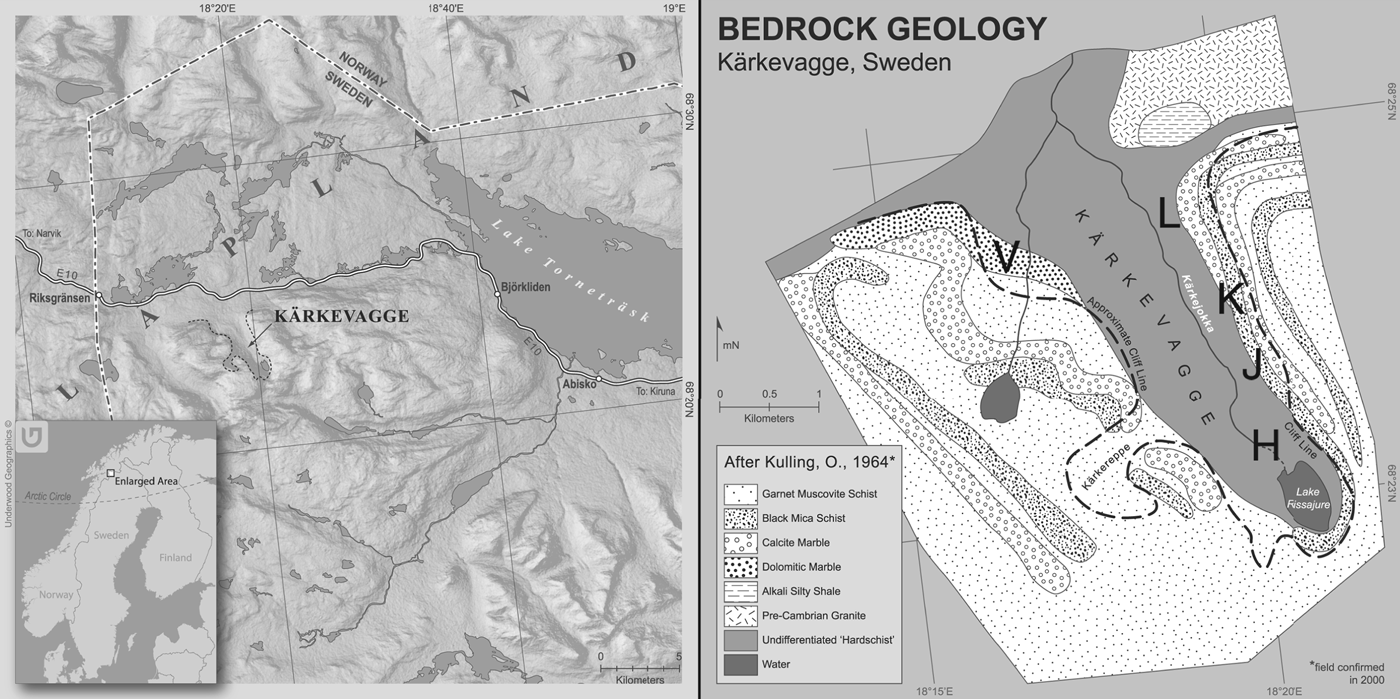
The coatings investigated in this study were collected from Kärkevagge, in Swedish Lapland. Kärkevagge is a glacially eroded U-shaped valley in Swedish Lapland, adjacent to the Norwegian border (68°26′N and 18°18′E) (Fig. 2). The valley is bounded by steep bedrock walls, with upper valley walls dominated by beds of resistant garnet mica schist (Dixon et al. Reference Dixon, Darmody, Schlyter and Thorn1995). Lower valley walls are predominately quartz mica schist dominated, and separating the two schist units is thinly bedded marble. Finely disseminated pyrite is found throughout the valley and thought to be a primary source of sulphur that is incorporated into the rock coatings, along with sulphate ions found in streams (Darmody et al. Reference Darmody, Thorn and Dixon2007). The valley floor is approximately 600 m above sea level (masl) at its mouth and rises to 800 masl at its head (Dixon et al. Reference Dixon, Thorn and Darmody2008). While mean annual air temperature in Kärkevagge is in the vicinity of −2 °C (Thorn et al. Reference Thorn, Schlyter, Darmody and Dixon1999), investigation of shallow soil and bedrock/soil interface temperatures reveal thermal regimes on daily and hourly scales that approximate −15 °C (Thorn et al. Reference Thorn, Darmody, Dixon and Schlyter2001). The majority of the precipitation in the valley is in the form of snow (50–75%) with depths ranging from 0.75 to 1.5 m over much of the year. Total annual precipitation is approximately 800 mm, as measured at the Riksgränsen-Katterjåkk station to the west of Kärkevagge (Eriksson, Reference Eriksson1982).
Left: Location of Kärkevagge in greater Swedish Lapland. Right: Geological map of Kärkevagge with sampling areas shown.
An early study of geomorphological processes operating in the valley was undertaken by Rapp (Reference Rapp1960) who identified what he originally described as ‘lime crusts’ and ‘rust coatings’ on bedrock surfaces. Recent studies have determined these to be alumina glazes (composed of basaluminite and gypsum) and Fe/Mn films, respectively (Dixon et al. Reference Dixon, Darmody, Schlyter and Thorn1995, Reference Dixon, Thorn, Darmody and Campbell2002, Reference Dixon, Thorn and Darmody2008; Darmody et al. Reference Darmody, Thorn and Dixon2007). We focus on three coating types in this study classified using nomenclature from Dorn (Reference Dorn1998): sulphate crusts are dominated by jarosite [KFe3(SO4)2(OH)6] composed of approximately. 70% O, 1% Si, 10% S, 4% K and 15% Fe and minor amounts of gypsum [CaSO4.H2O] composed of approximately 69% O, 2% Al, 1% Si, 12% S, 14% Ca and 2% Fe (Darmody et al. Reference Darmody, Thorn and Dixon2007). The aluminium glazes are predominantly basaluminite, an amorphous aluminium oxyhydroxide sulphate [Al4(SO4)(OH)10.H2O] consisting of approximately 52% Al2O3, 10% SiO2, 11% SO3, 1% K and Ca, and 0.1% Na, Mn and Mg (Darmody et al. Reference Darmody, Thorn and Dixon2007). Iron films are predominantly iron oxyhydroxides with compositions of 44% Fe, 9% Al, 6% Mg, 17% Si, 1.4% S, 0.3% K, 0.08% Ca and 0.05% Mn. Iron films are composed of primarily goethite and hematite. The Fe in the coatings is thought to be sourced from pyrite oxidation, subsequent bedrock weathering and release of Fe into the hydrologic system (Dixon et al. Reference Dixon, Thorn, Darmody and Campbell2002).
Kärkevagge represents a potential martian micro-environmental analogue: the valley is cold, relatively dry and has generally acidic water chemistry. Geochemically and mineralogically, the valley is a strong analogue with abundant sulphates and iron dominated mineralogies. As rock coatings by definition are composed of materials transported to the rock surface, parent rock lithology is insignificant in the development of the coating, and thus is not considered in the argument for Kärkevagge as a geochemical and mineralogical analogue to Mars.
While pure water is unstable on the surface of Mars, theoretical and experimental approaches have demonstrated that sulphate-rich brines can remain stable over extensive longitudinal ranges between 0° and 30° latitude because of lowered freezing points and evaporation rates (Chevrier & Altheide, Reference Chevrier and Altheide2008; Altheide et al. Reference Altheide, Chevrier, Nicholson and Denson2009). Models show that sulphate brines derived from acid–sulphate weathering similar to that operating in Kärkevagge have the potential to deposit hematite, jarosite and gypsum at the martian surface and shallow subsurface (Chevrier & Altheide, Reference Chevrier and Altheide2008; Chevrier & Rivera-Valentin, Reference Chevrier and Rivera-Valentin2012). Similarly, Chevrier & Rivera-Valentin (Reference Chevrier and Rivera-Valentin2012) have demonstrated that some sulphates and chlorides may remain fluid at and near the martian surface under temperature conditions similar to those of Kärkevagge on south-facing crater walls between 30° and 50°. Likewise, sulphate-reducing bacteria (SRB) have been shown to survive in many of these sulphate brines at concentrations as high as the eutectic (Marnocha et al. Reference Marnocha, Chevrier and Ivey2011). Rock coatings have been observed on the surface of Mars since the Viking landers (Strickland, Reference Strickland1979) and by the Mars Exploration Rovers (MER) (Krinsley et al. Reference Krinsley, Dorn and DiGregorio2009). Because of the distinct appearance of rock varnish and its strong association with biomineralizing microbes, rock varnish has frequently been suggested as a potential biosignature on Mars (Dorn, Reference Dorn1998; Barnouin-Jha et al. Reference Barnouin-Jha, Murchie, Johnson, Bell and Morris2000; Bishop et al. Reference Bishop, Murchie, Pieters and Zent2002; Allen et al. Reference Allen, Probst, Flood, Longazo, Schelble and Westall2004; Murchie et al. Reference Murchie, Barnouin-Jha, Barnouin-Jha, Bishop, Johnson, McSween and Morris2004; Krinsley et al. Reference Krinsley, Dorn and DiGregorio2009). However, despite the widespread occurrence of gypsum, jarosite and iron oxides (Christensen et al. Reference Christensen, Bandfield, Clark, Edgett, Hamilton, Hoefen, Kieffer, Kuzmin, Lane and Malin2000; Klingelhöfer et al. Reference Klingelhöfer2004, Reference Klingelhöfer, DeGrave, Morris, Alboom, Resende, Souza, Rodionov, Schröder, Ming and Yen2007; Squyres et al. Reference Squyres2004; Bibring et al. Reference Bibring2005, Reference Bibring2006; Gendrin et al. Reference Gendrin2005; Swayze et al. Reference Swayze, Ehlmann, Milliken, Poulet, Wray, Rye, Clark, Desborough, Crowley and Gondet2008), other rock coatings compatible with known martian mineralogies have been largely ignored as potential locations for life on Mars.
A number of mechanisms for bacterial biomineralization have been suggested (Kleinmann et al. Reference Kleinmann, Crerar and Pacelli1981; Ghiorse, Reference Ghiorse1984; Beveridge & Fyfe, Reference Beveridge and Fyfe1985; Beveridge, Reference Beveridge1989; Ghiorse & Ehrlich, Reference Ghiorse and Ehrlich1992; Konhauser, Reference Konhauser1997; Dorn, Reference Dorn1998; Kappler et al. Reference Kappler, Schink and Newman2006; Benzerara et al. Reference Benzerara, Miot, Morin, Kappler and Obst2008; Konhauser et al. Reference Konhauser, Lalonde and Phoenix2008, Reference Konhauser, Kappler and Roden2011; Benzerara & Miot, Reference Benzerara and Miot2011; Petrash et al. Reference Petrash, Gingras, Lalonde, Pecoits and Konhauser2012). Many of these mechanisms involve the scavenging and concentration of relevant chemical species (Fe, Mn, S, etc.) from the surrounding environment within the extracellular polymeric substances (EPS). These trapped ions can then serve as nucleation points for reactions that lead to the precipitation of minerals. While this occurs, microbial metabolisms (e.g. iron and sulphide oxidation) can simultaneously affect local pH, thus affecting the overall chemistry and sometimes leading to additional precipitation by promoting stable conditions for those minerals (e.g. jarosite) (Konhauser, Reference Konhauser1997; Petrash et al. Reference Petrash, Gingras, Lalonde, Pecoits and Konhauser2012).
Iron oxides and hydroxides can form through bacterially induced processes (e.g. bacterial EPS as nucleation sites), or bacterial controlled processes, in which bacterial oxidation of Fe(II) produces large quantities of precipitated iron (Konhauser, Reference Konhauser1997). Microbial oxidation of Fe(II) can occur and compete with abiotic oxidation in acidic and neutral conditions (Kappler et al. Reference Kappler, Pasquero, Konhauser and Newman2005; Reference Williams, Buck, Soukup and MerklerWilliams et al.; Reference Varnali and EdwardsVarnali & Edwards). Concentration of Fe(II) and co-reacted ions, pH and other environmental factors contribute to determination of which mineral is produced and remains stable (Zachara et al. Reference Zachara, Kukkadapu, Fredrickson, Gorby and Smith2002; Roh et al. Reference Roh, Zhang, Vali, Lauf, Zhou and Phelps2003). Biomineralization products can include goethite, hematite, magnetite and siderite (Banfield et al. Reference Banfield, Welch, Zhang, Ebert and Penn2000; Zachara et al. Reference Zachara, Kukkadapu, Fredrickson, Gorby and Smith2002; Roh et al. Reference Roh, Zhang, Vali, Lauf, Zhou and Phelps2003; Larese-Casanova et al. Reference Larese-Casanova, Haderlein and Kappler2010), where goethite and hematite are the dominant minerals in Fe/Mn films in Kärkevagge.
Precipitation of sulphates occurs through a series of biological and abiotic reactions, with biological influence from Fe2+ oxidation and hydrolysis (Kleinmann et al. Reference Kleinmann, Crerar and Pacelli1981; Clarke et al. Reference Clarke, Konhauser, Thomas and Bottrell1997; Konhauser, Reference Konhauser1997). This process has been described as follows (Kleinmann et al. Reference Kleinmann, Crerar and Pacelli1981):
 $${\rm FeS}_{\rm 2} + {\rm 3}.{\rm 5O}_{\rm 2} + {\rm H}_{\rm 2} {\rm O} \to {\rm Fe}^{{\rm 2} +} + {\rm 2H}^ + + {\rm 2SO}_{\rm 4}^{^{{\rm 2} -}} $$
 $${\rm Fe}^{{\rm 2} +} + 0.{\rm 25O}_{\rm 2} + {\rm H}^ + \to {\rm Fe}^{{\rm 3} +} + 0.{\rm 5H}_{\rm 2} {\rm O}\; $$
 $${\rm FeS}_{\rm 2} + {\rm 14Fe}^{{\rm 3} +} + {\rm 8H}_{\rm 2} {\rm O} \to {\rm 15Fe}^{{\rm 2} +} + {\rm 2SO}_4^{{\rm 2} -} + {\rm 16H}^ + $$
 $${\rm Fe}^{{\rm 2} +} + {\rm 2}.{\rm 5H}_{\rm 2} {\rm O} + 0.{\rm 25O}_{\rm 2} \to {\rm Fe}\left( {{\rm OH}} \right)_{\rm 3} + {\rm 2H}^ + $$
where initial oxidation of sulphides occurs through biotic or abiotic means (reaction 1), Fe2+ is oxidized through microbial oxidation (reaction 2). This is followed by accelerated oxidation of sulphides (reaction 3) and finally the production of a precipitate from oxidation and hydrolysis of Fe2+ (reaction 4) (Kleinmann et al. Reference Kleinmann, Crerar and Pacelli1981). Ferric hydroxysulphate and jarosite may precipitate in acidic environments (Lazaroff et al. Reference Lazaroff, Sigal and Wasserman1982; Brady et al. Reference Brady, Bigham, Jaynes and Logan1986), whereas ferric hydroxides precipitate at higher pH (Carlson & Schwertmann, Reference Carlson and Schwertmann1980; Brady et al. Reference Brady, Bigham, Jaynes and Logan1986).
It is through these mechanisms of biologically controlled and biologically induced biomineralization that we suggest bacteria may play a role in the genesis of rock coatings in Kärkevagge. Studies of rock coatings in Kärkevagge until now have been from an abiotic, geochemical perspective. However, microbes and other organic materials have been reported in previous work (Dixon et al. Reference Dixon, Darmody, Schlyter and Thorn1995). Thus, it has suggested that they may play a role in rock-coating genesis.
This work represents the first study to investigate the bacterial communities inhabiting the rock coatings from Kärkevagge and the communities’ metabolic potential for biomineralization processes and rock-coating formation. Given the geochemical and mineralogical analogue that Kärkevagge represents for Mars, and the observations of rock coatings on Mars, we propose rock coatings as high-priority targets for astrobiology on Mars and present the results of our initial investigations of the bacterial communities of rock coatings in Kärkevagge.
Methods
Sample collection
Rock debris displaying accumulations of the principal rock-coating types was collected from field sites associated with sampling transects established along the length of the valley's east side. These transects are subsequently identified by the letters V, L, K, J and H. Samples were subsampled from each of the following rock-coating types: Fe/Mn films, aluminium glazes and sulphate crusts. Two Fe/Mn films, two sulphate crusts, and one aluminium glaze were selected for subsequent analysis. Bulk rock samples were then subdivided into sterile tubes for storage at both 3 and −20 °C. Collections stored at −20 °C were used in this study. Genomic DNA was extracted from crushed rock samples using a PowerSoil® DNA Isolation Kit from MoBio (Carlsbad, CA) according to manufacturer's protocols after transport. Extracted DNA was stored at −20 °C for the downstream analysis.
16S rRNA gene amplification and sequencing
Bacterial small subunit ribosomal RNA genes were amplified via polymerase chain reaction (PCR) using universal 533-forward (5′-GTG CCA GCC GCC GCG GTA A-3′) and 1392-reverse primers (5′-GGT TAC CTT GTT ACG ACT T-3′) on two Fe/Mn films, two sulphate crusts and one aluminium glaze. PCR was carried out in 25 μl reaction, consisting of 12.5 μl GoTaq Green Master Mix (Promega Corporation), 2 μl each of primer, 5 μl of the extracted rock-coating DNA, and nuclease-free water to bring the reaction volume to 25 μl according to manufacturer protocols. Thermocycler parameters, modified from Macalady et al. (Reference Macalady, Jones and Lyon2007), were as follows: 5′ at 94 °C initial denaturing, followed by 30 cycles of 1′ at 94 °C, 45′ at 47 °C and 1′ at 72 °C, concluding with a 7′ final extension at 72 °C. Amplifications using two archaeal primer sets: 21-forward/958-reverse (DeLong, Reference DeLong1992) and 340F/1000R from (Gantner et al. Reference Gantner, Andersson, Alonso-Sáez and Bertilsson2011), were also attempted, but were ultimately unsuccessful. After successful confirmation of PCR amplification from the universal primers using gel electrophoresis, products were purified with an UltraClean® PCR Clean-up Kit (MoBio Laboratories, Inc.).
Purified PCR amplicons were then cloned into pSC-A cloning vectors using a StrataClone PCR Cloning Kit (Agilent Technologies). Purified PCR products were not diluted, although the procedure was performed otherwise according to manufacturer instructions. Following transformation, Escherichia coli cells were plated on Luria Burtani-ampicillin plates with 2% x-gal and incubated no more than 24 h. Positive, white colonies were then randomly selected and further PCR-amplified using M13 forward reverse primers with binding sites on the pSC-A vector. PCR thermocycler parameters used were the same as those used for universal 533-forward and 1392-reverse reactions described above.
Following successful M13 PCR confirmed on a gel electrophoresis, PCR products were shipped on dry ice to Functional Biosciences (Madison, WI) for Exo/SAP clean-up and sequencing using the T7 primer. Sequences were checked for chimeras using Bellerophon (Huber et al. Reference Huber, Faulkner and Hugenholtz2004) and putative chimeras were removed from subsequent analyses. The final set of sequences was then aligned using the Basic Local Alignment Search Tool (BLAST) and matched to nearest neighbours for use in phylum-based analysis.
Sequences for each sample were then divided by phyla, or more specific taxonomy, when available through BLAST and greengenes (DeSantis et al. Reference DeSantis, Hugenholtz, Larsen, Rojas, Brodie, Keller, Huber, Dalevi, Hu and Andersen2006) best matches. Rarefaction curves were calculated for all samples using the mothur software program (Schloss et al. Reference Schloss, Westcott, Ryabin, Hall, Hartmann, Hollister, Lesniewski, Oakley, Parks and Robinson2009). Diversity indices were also calculated using mothur, after alignment via greengenes. Phylogenetic trees for each sample type were created using MEGA5 (Tamura et al. Reference Tamura, Peterson, Peterson, Stecher, Nei and Kumar2011) and ClustalW alignment followed by the construction of a maximum-likelihood phylogenetic tree for each coating type, with 1000 bootstraps using the Jukes-Cantor model (Jukes & Cantor, Reference Jukes and Cantor1969). Nearest neighbours included in the phylogenetic trees were selected from the BLAST database. 16S rRNA gene sequence data generated in this study is catalogued in GenBank under accession numbers JQ677813-JQ677911.
Scanning electron microscopy (SEM)
Coating samples were examined using a Nova Nanolab FEG SEM, coupled with energy-dispersive X-ray spectroscopic (EDX) analysis. Rock chips of each representative coating type were mounted on carbon tape sample mounts and observed without the addition of a metal coating. Samples were analysed under 15.00 kV for both SEM and EDX. FEI software was used to capture images on a PC for subsequent interpretation. Chemical analyses were obtained via mapping and point analysis of bacteria and mineralogical materials using EDX.
Results
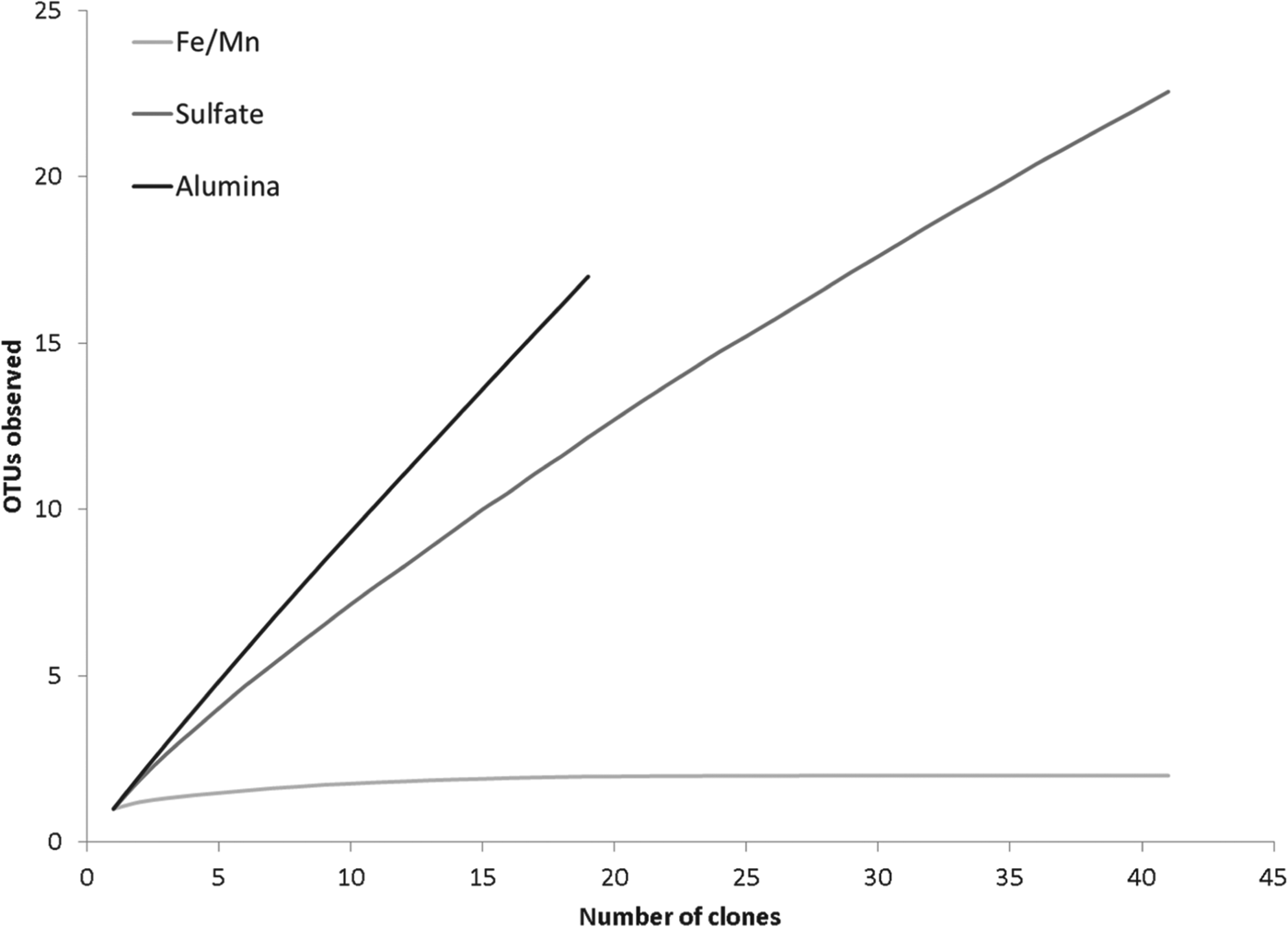
Bacterial 16S rDNA was amplified for samples from sites K, L, V and H. The non-chimaeric sequences from the five coating samples underwent further analysis. Rarefaction curves (Fig. 3) show the expected number of operational taxonomic units (OTUs) observed per number of clones sampled, generating a curve to show the trend of representativeness in terms of diversity each clone library was for each sample. In all, both the community richness estimates and rarefaction curves suggest that communities may be more diverse than found in this initial study.
Rarefaction curves for each rock-coating type. Curves reflect the higher diversity in the aluminium glazes and sulphate crusts.
Aluminium glaze
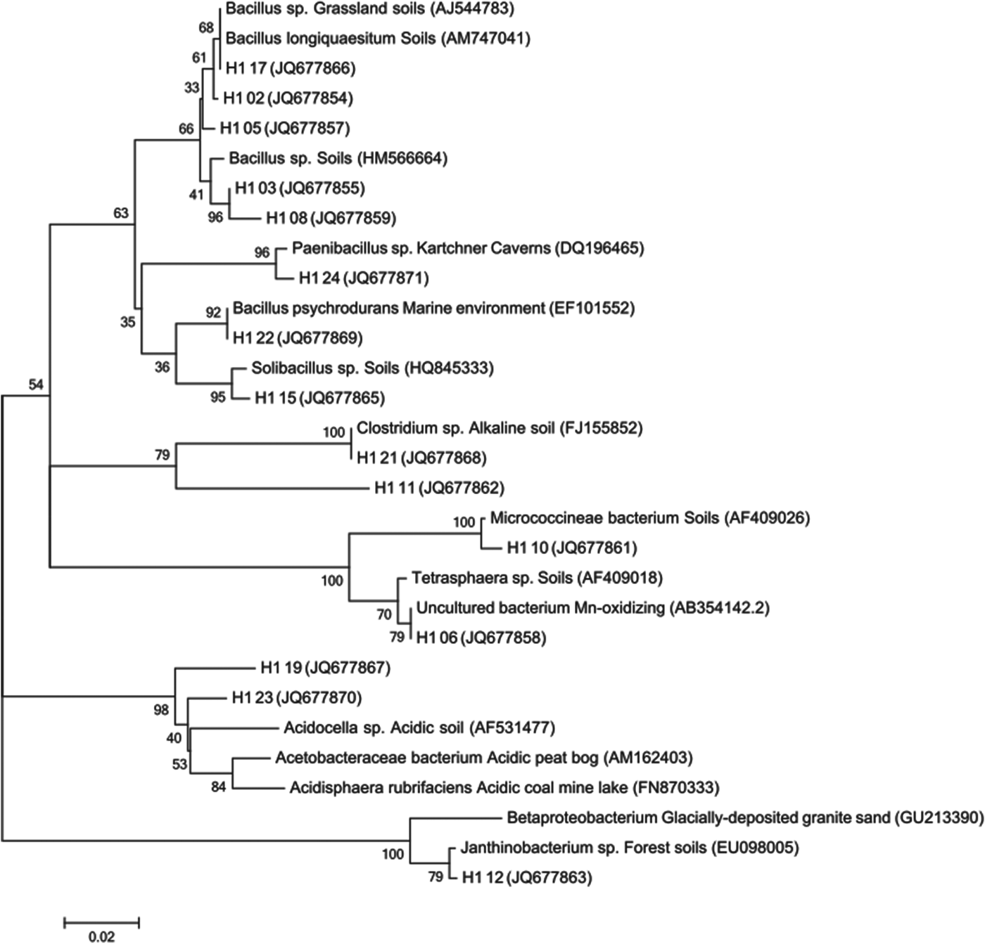
Phylogenetic analyses (Fig. 4) show that the aluminium glaze clones are related primarily to soil bacteria, with relationships to other endoliths, such as those found in tufa and dolomite, and are associated with cold climate representatives in Antarctic soils, tundra and alpine environments. The aluminium glaze contained sequences with nearest neighbours from acid-mine drainage (AMD) environments, including Actinobacteria, Acidisphaera sp. and Acidocella sp. clones. Clones also had nearest neighbours from sites of appreciable uranium concentration, both in mine waste piles and contaminated soil.
Inferred phylogeny for the aluminium glaze (H1) clones with nearest neighbours. Evolutionary histories for Figs. 4 and 5 were inferred using the maximum-likelihood method based on the Jukes–Cantor model (Jukes & Cantor, Reference Jukes and Cantor1969). The percentage of trees in which the associated taxa clustered together is shown next to the branches as a bootstrap value. The trees are drawn to scale, with branch lengths measured as the number of substitutions per site. Accession numbers are shown after each isolate.
Sulphate crust
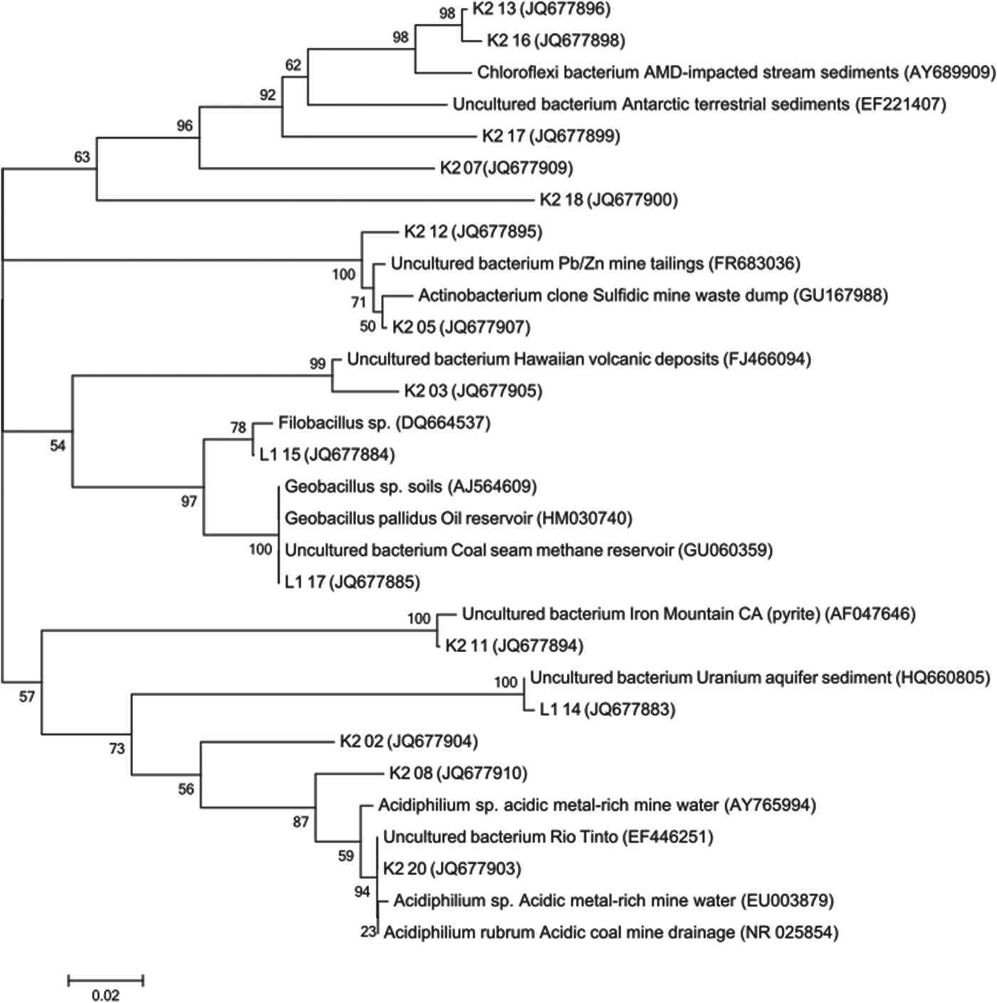
The sulphate crust clones (Fig. 5) display soil, alpine, endolith and AMD relatives. BLAST database searches show a stronger relationship between sulphate crust clones and those found in AMD settings, including sulphur and Pb/Zn mine waste. Sequences from the Actinobacteria and Acidobacteria phyla were found in the K2 site sample, along with Acidiphilum sp. In one jarosite sample, 8 out of 15 clones had nearest BLAST neighbours from coal mine and sulphur-rich mine-tailing sites.
Inferred phylogeny for the sulphate crust (K2, L1) clones with nearest neighbours.
Unlike the other coating types, sulphate crust sequences also have unique nearest neighbours isolated from glacial environments, deep sea sediments and stony corals, and hot springs when compared with the greengenes database.
Fe/Mn film
The Fe/Mn films display the lowest bacterial diversity and exhibit Bacillus spp. as nearest neighbours from a limited number of environments. Because of the low diversity and close relationships of the Fe/Mn film clones, no phylogenetic tree was generated. Fe/Mn films were sampled from both the eastern and western side of the valley in areas of low-to-neutral pH (4.5–7.9) (Campbell et al. Reference Campbell, Dixon, Darmody and Thorn2001). One of the coatings was sampled from a site with mixed bicarbonate/sulphate ion water chemistry and contained predominantly clones with nearest neighbours from acidic environments. It should also be noted that SEM analyses have shown the presence of cocci-type bacteria in the Fe/Mn films, both in this study and as previously reported from Kärkevagge by Dixon et al. (Reference Dixon, Darmody, Schlyter and Thorn1995), whereas only Bacillus sp. (and thus, bacillus morphologies) were identified through 16S rRNA analysis.
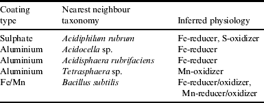
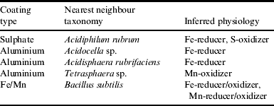
Physiologies of rock-coating bacteria that can be inferred with confidence are shown in Table 1. Other dominant phyla include Actinobacteria, Chloroflexi, Acidobacteria and Firmicutes for the sulphate crusts, Actinobacteria, Firmicutes, α-, β-proteobacteria for the aluminium glazes, and Firmicutes for the Fe/Mn films.
Taxonomy and postulated physiology for those clones with high percent matches within the database and genus-level taxonomy available for nearest neighbours
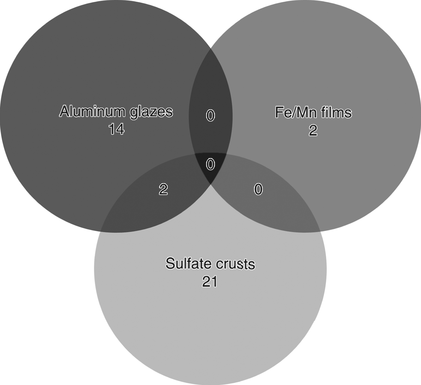
Community structure, when assessed through the number of shared OTUs (Fig. 6), appears to be tied to coating mineralogy. Very little diversity exists between clones from the Fe/Mn films, compared with the aluminium glazes and sulphate crusts. There are two overlapping OTUs between the aluminium glaze and sulphate crust at a pairwise distance of 0.05, and no overlap for any group assuming all sequences represent unique OTUs.
Venn diagram of shared OTUs between rock-coating types at a pairwise distance of 0.05.
Scanning electron microscopy
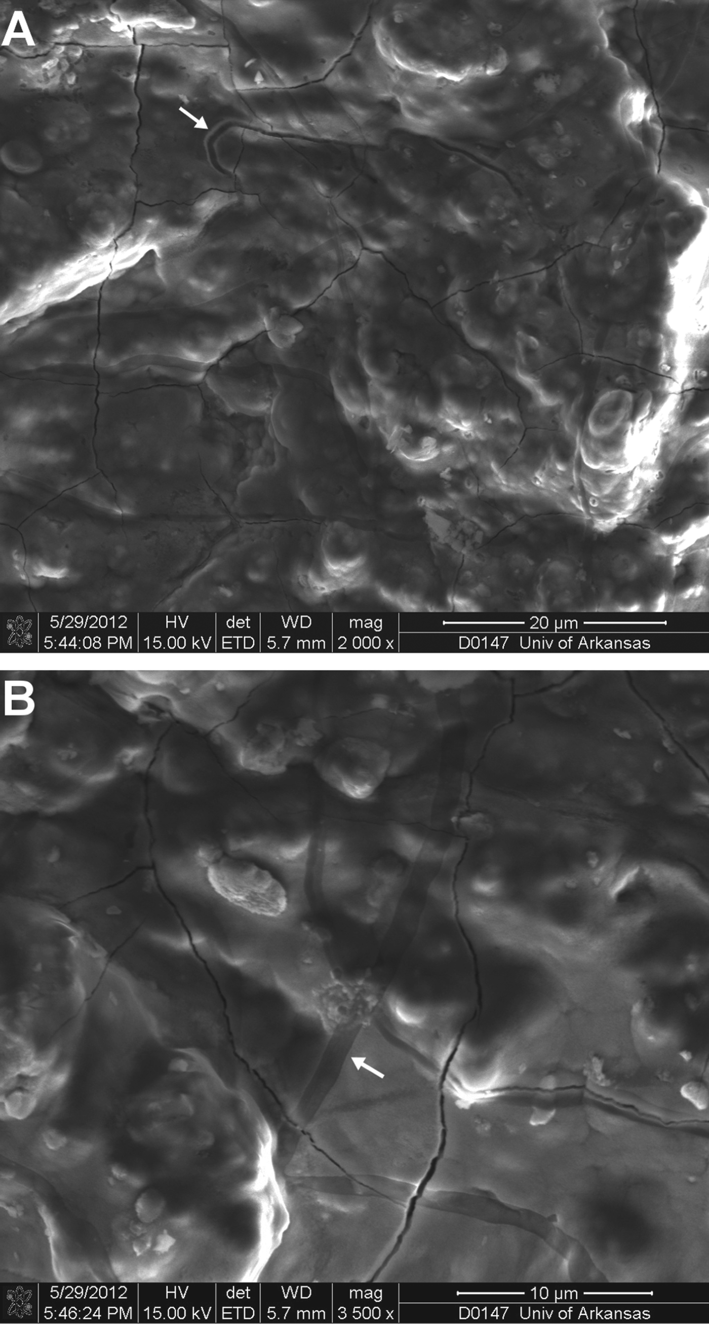
SEM analysis revealed bacterial and fungal morphologies in the three rock-coating types sampled in this study. Fe/Mn films showed strong evidence of iron coating of putative biological forms (Fig. 7). EDX spectra show strong signals for Fe and O, with lesser amounts of Al and S, suggesting a possible iron oxide coating over the biological forms. The aluminium glaze exhibited similar encrustation to the Fe/Mn films. Aluminium glazes also contained multiple dark streaks across the encrusted area (Fig. 8), with similarly sized filaments observed nearby.
SEM micrographs of a Fe/Mn film. (A) Putative cocci-form bacteria and ringed (fungal?) filaments (arrows). (B) Cocci and bacilli-form bacteria covered in a coating highly concentrated in Fe. (C) EDX spectra of the region shown in 7(A).
SEM micrographs of an aluminium glaze. (A) Dark streaks and a deep trench (arrow) indicating possible remnants of fungal filaments. (B) Additional dark streaks (arrow) observed in an area of heavy encrustation. Botryoidal cocci bacteria are visible beneath the smooth alumina coating.
Discussion
Environmental influences on bacterial diversity
All coating-type sequences contained nearest neighbours from cold climates, ranging from glacial ice, deglaciated and tundra soils, Antarctic and Arctic environments, and other environments simply categorized in the databases as ‘cool’ or ‘cold’. Rock coatings are present in other cold environments; in particular, Al-, Si-, SO4- and Fe-rich rock varnishes have been observed in coastal (Victoria Land), inland Antarctica and Tibet (Glasby & Macpherson, Reference Glasby and Macpherson1981; Johnston & Cardile, Reference Johnston and Cardile1984; Dorn et al. Reference Dorn, Krinsley, Liu, Anderson, Clark, Cahill and Gill1992; Ishimaru & Yoshikawa, Reference Ishimaru and Yoshikawa2000; Giorgetti & Baroni, Reference Giorgetti and Baroni2007; Krinsley et al. Reference Krinsley, Dorn and DiGregorio2009).
Summer air temperatures in Kärkevagge range from 6 to 17 °C (Thorn et al. Reference Thorn, Schlyter, Darmody and Dixon1999), moderate enough for mesophilic bacteria to thrive. During the remainder of the year, when air temperatures drop to 1–2 °C and ground surface temperatures to as low as −15 °C, psychrotolerant and psychrophilic bacteria could thrive in the rock coatings at these temperatures (Deming, Reference Deming2002; Rothschild & Mancinelli, Reference Rothschild and Mancinelli2002; Junge et al. Reference Junge, Eicken and Deming2004; Price, Reference Price2007). Alternatively, spore-forming bacteria would withstand the environmental conditions until temperatures were above freezing. Many of the sequences from rock coatings, including the dominant Bacillus sp. in the Fe/Mn films, are related to spore-forming bacteria, capable of producing endospores that could survive dormant in a number of conditions until environmental conditions (e.g. temperature, nutrient availability) were once again conducive to survival and growth (Roszak & Colwell, Reference Roszak and Colwell1987; Vreeland et al. Reference Vreeland, Rosenzweig and Powers2000). Although spore-forming bacteria can be found in any number of environments where conditions do not necessarily promote endospore generation, spore-forming bacteria are often found in cold climates such as Arctic and Antarctic sediments and soils (Vorobyova et al. Reference Vorobyova, Soina, Gorlenko, Minkovskaya, Zalinova, Mamukelashvili, Gilichinsky, Rivkina and Vishnivetskaya1997; Yergeau et al. Reference Yergeau, Newsham, Pearce and Kowalchuk2007), glacial ice and glacial outflow (Miteva & Brenchley, Reference Miteva and Brenchley2005; Mikucki & Priscu, Reference Mikucki and Priscu2007) and permafrost (Vorobyova et al. Reference Vorobyova, Soina, Gorlenko, Minkovskaya, Zalinova, Mamukelashvili, Gilichinsky, Rivkina and Vishnivetskaya1997; Hinsa-Leasure et al. Reference Hinsa-Leasure, Bhavaraju, Rodrigues, Bakermans, Gilichinsky and Tiedje2010).
While arctic and arid environments typically present lower diversity and richness for plants and other higher-order organisms, bacterial diversity and community richness tend not to follow these biogeographic trends (Fierer & Jackson, Reference Fierer and Jackson2006). Desert soils, for example, have shown significantly higher bacterial diversity and species richness than soils in the tropics (Fierer & Jackson, Reference Fierer and Jackson2006). In this case, soil pH plays a pivotal role as an indicator of microbial diversity. Kärkevagge water pH ranges from 4.4 to 8.0, with most values closer to neutral. Soils maintain similar, although more neutral pH values. Comparing local soil and water pH from the rock-coating sample sites shows a relationship between neutral pH and increased bacterial diversity.
Fe(II) oxidation to Fe(III) can occur through abiotic, chemical reactions in neutral or alkaline conditions, whereas Fe(II) remains stable in acidic conditions (Kappler et al. Reference Kappler, Pasquero, Konhauser and Newman2005; Bae & Lee, Reference Bae and Lee2013). Fe/Mn films were sampled from areas with local water chemistry pH ranging from acidic to neutral (Campbell et al. Reference Campbell, Dixon, Darmody and Thorn2001). In more acidic conditions, where Fe(II) would be stable, microbes could be responsible for the oxidation and conversion of pyrite, which is abundant throughout the valley, to the goethite observed in the Fe/Mn films from those sites. In areas of more neutral pH, microbial oxidation of Fe(II) is able to compete with abiotic oxidation (Kappler et al. Reference Kappler, Pasquero, Konhauser and Newman2005; Reference Varnali and EdwardsVarnali & Edwards; Reference Williams, Buck, Soukup and MerklerWilliams et al.; Williamson et al. Reference Williamson, Morris, Shaw, Byrne, Boothman and Lloyd2013).
Lithic microbial communities are found above and below ground, on rock surfaces and within rocks themselves. Desert rock varnish, which differs from Fe/Mn films because of the presence of clay minerals, has been a focus of several of phylogenetic studies. Rock varnish from Black Canyon, New Mexico shares some phyla with those found in the rock coatings in this study, namely Actinobacteria, Ktedobacter, Chloroflexi and Firmicutes (Northup et al. Reference Northup, Snider, Spilde, Porter, van de Kamp, Boston, Nyberg and Bargar2010). Despite the similarities in mineralogy between rock varnish and Fe/Mn films, very few of the isolates from the Black Canyon varnish were from the Firmicutes phylum, whereas the Fe/Mn sequences from Kärkevagge were entirely Firmicutes bacteria, namely Bacillus spp. It should be noted, however, that Bacillus spp. sequences represented some of the only known manganese oxidizers isolated from the varnish samples (Northup et al. Reference Northup, Snider, Spilde, Porter, van de Kamp, Boston, Nyberg and Bargar2010). Inferred physiology, microscopy, culturing and mineralogical analyses of rock varnish suggest a bacterial influence on its generation (Dorn, Reference Dorn1998; Krinsley et al. Reference Krinsley, Dorn and DiGregorio2009; Northup et al. Reference Northup, Snider, Spilde, Porter, van de Kamp, Boston, Nyberg and Bargar2010; Wang et al. Reference Wang, Zeng, Wiens, Schloßmacher, Jochum, Schröder and Müller2011). Sulphate crusts consisting of gypsum and halite also show distinct endolithic bacterial communities (Wierzchos et al. Reference Wierzchos, Ascaso and McKay2006; Stivaletta et al. Reference Stivaletta, López-García, Boihem, Millie and Barbieri2010; Wong et al. Reference Wong, Lacap, Lau, Aitchison, Cowan and Pointing2010). These coating types are unique in that they are translucent enough to allow for photosynthesis, and thus, many of the analyses of these coating types either directly or indirectly suggest a strong presence of photosynthetic bacteria. Sequences in this study contained a small representation of photosynthetic bacteria from the Chloroflexi phylum.
Relationship to AMD
Given its association with AMD, jarosite has frequently been proposed as a possible byproduct of microbial metabolism in these environmental settings (Sharp et al. Reference Sharp, Parkes, Cragg, Fairchild, Lamb and Tranter1999; Baker & Banfield, Reference Baker and Banfield2003; Verplanck, Reference Verplanck2008). The similar geochemistry between AMD sites and that found in Kärkevagge, along with some shared bacterial community compositions, suggests that the rock coatings from Kärkevagge may similarly be generated through bacterial metabolism. The deglaciation of the valley represents the anthropogenic earth-moving processes associated with the development of AMD, which exposes the finely disseminated pyrite found throughout the valley to oxidation (Darmody et al. Reference Darmody, Allen, Thorn and Dixon2001, Reference Darmody, Thorn and Dixon2007). In AMD, acidic waters generated by microbial metabolisms promote the precipitation of sulphate minerals into the environment (Johnson, Reference Johnson1998; Schippers et al. Reference Schippers, Breuker, Blazejak, Bosecker, Kock and Wright2010; Meier et al. Reference Meier, Piva and Fortin2012). Microbes have also been shown to affect the rates of sulphur oxidation (Edwards et al. Reference Edwards, Bond and Banfield2000). Because base metal mining is often associated with pyrite-rich deposits, microbial metabolism is thought to play a role in the acidification of mining sites and the generation of AMD (Baker & Banfield, Reference Baker and Banfield2003). Communities of bacteria found in AMD environments that have physiologies compatible with reactions producing sulphates and other AMD-related minerals. Such taxa include the phyla Nitrospira, Actinobacteria, Firmicutes and Acidobacteria, and the genera Acidocella, Leptospirillum, Acidithiobacillus, Acidisphaera, Leptospirillum, Thiobacillus, Sulfobacillus, Ferroplasma, Sulfobacillus and Acidiphilum (Bond et al. Reference Bond, Smriga and Banfield2000; Baker & Banfield, Reference Baker and Banfield2003; Schippers et al. Reference Schippers, Breuker, Blazejak, Bosecker, Kock and Wright2010). The Actinobacteria, Firmicutes and Acidobacteria phyla and the genera Acidocella, Acidisphaera and Acidiphilium are common sequences isolated from the rock coatings in this study. Similarly, the acidic, sulphur-rich aspects of martian geochemistry have been related to AMD on Earth (Elwood Madden et al. Reference Elwood Madden, Bodnar and Rimstidt2004). This type of geochemical regime is shared between Mars, Kärkevagge and some AMD environments.
The bacterially facilitated formation of Al-rich sulphate minerals in AMD settings has recently been examined in a study by Meier et al. (Reference Meier, Piva and Fortin2012). They identify white–grey aluminium precipitates produced in acidic enrichments by SRB. Equilibrium calculations predict these precipitates to be alunite and gypsum, although this was not supported by the experimental data (Meier et al. Reference Meier, Piva and Fortin2012). Further, Al-sorption to cells has been observed in SRB, with higher absorbance during freezing and thawing of cells and at low pH (Hard et al. Reference Hard, Walther and Babel1999). Although no SRB were identified in the aluminium glaze, it is conceivable that SRB are still present in the coatings, but were not identified in the small clone sample size. A relatively novel species, Acidocella aluminiidurans, has been identified as being Al-tolerant with optimum growth at low-to-neutral (3–7) pH in media containing aluminium sulphate or aluminium chloride (Kimoto et al. Reference Kimoto, Aizawa, Urai, Ve, Suzuki, Nakajima and Sunairi2010). Significantly, the Al-rich crusts in Kärkevagge are dominated by basaluminite (an aluminium oxyhydroxide sulphate), with minor abundances of gypsum and alunite.
Implications for astrobiology on Mars
Kärkevagge represents a strong mineralogical and geochemical, but more limited climatic, analogue for Mars. Goethite and hematite, the primary minerals of the Fe/Mn films in Kärkevagge, have been detected using Mössbauer spectroscopy onboard the MER Spirit at Columbia Hills (Klingelhöfer et al. Reference Klingelhöfer, DeGrave, Morris, Alboom, Resende, Souza, Rodionov, Schröder, Ming and Yen2007). Crystalline hematite mineralization has been observed in Meridiani Planum by the Mars Global Surveyor Thermal Emission Spectrometer, with surface coatings cited as a possible mechanism for the observed coarse-grained hematite (Christensen et al. Reference Christensen, Bandfield, Clark, Edgett, Hamilton, Hoefen, Kieffer, Kuzmin, Lane and Malin2000). Jarosite and gypsum are present in Kärkevagge as dominant minerals in sulphate crusts, and have also been detected on Mars through both observational satellites and MER rovers (Klingelhöfer et al. Reference Klingelhöfer2004; Squyres et al. Reference Squyres2004; Bibring et al. Reference Bibring2005, Reference Bibring2006; Gendrin et al. Reference Gendrin2005; Wang et al. Reference Wang2006). The aluminium glazes in Kärkevagge are predominately basaluminite, and while basaluminite itself has not been detected on Mars, alunite has been detected by the CRISM instrument on board the Mars Reconnaissance Orbiter (Swayze et al. Reference Swayze, Ehlmann, Milliken, Poulet, Wray, Rye, Clark, Desborough, Crowley and Gondet2008). Alunite has been inferred as a subordinate mineral in the aluminium glazes in Kärkevagge (Darmody et al. Reference Darmody, Thorn and Dixon2007). Goethite, hematite, jarosite and gypsum have all been identified as minerals that can be directly and indirectly formed in association with bacteria (Kleinmann et al. Reference Kleinmann, Crerar and Pacelli1981; Clarke et al. Reference Clarke, Konhauser, Thomas and Bottrell1997; Konhauser, Reference Konhauser1997; Dorn, Reference Dorn1998; Banfield et al. Reference Banfield, Welch, Zhang, Ebert and Penn2000; Zachara et al. Reference Zachara, Kukkadapu, Fredrickson, Gorby and Smith2002; Roh et al. Reference Roh, Zhang, Vali, Lauf, Zhou and Phelps2003; Kappler et al. Reference Kappler, Schink and Newman2006; Larese-Casanova et al. Reference Larese-Casanova, Haderlein and Kappler2010; Abreu et al. Reference Abreu2011).
As an environmental analogue, Kärkevagge is a moderately cold, acidic and dry environment, and may represent environmental conditions that existed in the putative early warm and wet Mars (Sagan & Mullen, Reference Sagan and Mullen1972; Pollack et al. Reference Pollack, Kasting, Richardson and Poliakoff1987; Baker et al. Reference Baker, Strom, Gulick, Kargel, Komatsu and Kale1991; Phillips et al. Reference Phillips, Zuber, Solomon, Golombek, Jakosky, Banerdt, Smith, Williams, Hynek and Aharonson2001; Fairén et al. Reference Fairén2011; Manga et al. Reference Manga, Patel, Dufek and Kite2012). Rock coatings are present in extreme Earth environments that are similar to martian conditions, including the Atacama Desert (Wierzchos et al. Reference Wierzchos, Ascaso and McKay2006; DiRuggiero et al. Reference DiRuggiero, Robinson, Souterre, Ravel, Fricke, Ascaso and Wierzchos2012), Antarctica (Weed & Ackert, Reference Weed and Ackert1986; Weed & Norton, Reference Weed and Norton1991; Matsuoka, Reference Matsuoka1995; Wierzchos et al. Reference Wierzchos, Ascaso, Sancho and Green2003) and numerous acidic and sulphur-rich environments (Nordstrom & Alpers, Reference Nordstrom and Alpers1999; Keith et al. Reference Keith, Runnells, Esposito, Chermak, Levy, Hannula, Watts and Hall2001; Jamieson et al. Reference Jamieson, Robinson, Alpers, Nordstrom, Poustovetov and Lowers2005; Fernández-Remolar et al. Reference Fernández-Remolar, Morris, Gruener, Amils and Knoll2005; Fernández-Remolar & Knoll, Reference Fernández-Remolar and Knoll2008).
Terrestrial rock coatings have been proposed as possible analogues for life on the martian surface because of reduced irradiation levels (McKay, Reference McKay1993; Cockell et al. Reference Cockell, Catling, Davis, Snook, Kepner, Lee and McKay2000; Wynn-Williams & Edwards, Reference Wynn-Williams and Edwards2000). Viking demonstrated that surface radiation on Mars can apparently destroy any detectable organics, even those expected from meteoritic delivery (Doi, Reference Doi1973; Dartnell et al. Reference Dartnell, Desorgher, Ward and Coates2007). However, the reduced radiation levels afforded by rock layers could protect organics preserved in the coatings themselves (Wynn-Williams et al. Reference Wynn-Williams, Edwards and Garcia-Pichel1999; Wynn-Williams & Edwards, Reference Wynn-Williams and Edwards2000). Microbes colonizing rock coatings need only be at a depth of 1–2 mm for DNA-affecting radiation to be reduced to levels that the surface of present-day Earth experiences (Friedmann, Reference Friedmann1982; Nienow et al. Reference Nienow, McKay and Friedmann1988; Nienow & Friedmann, Reference Nienow and Friedmann1993; Cockell et al. Reference Cockell, Catling, Davis, Snook, Kepner, Lee and McKay2000).
The ChemCam instrument onboard Mars Science Laboratory (MSL) provides a means of micron-scale depth profiling of rock surfaces and subsequent elemental analysis using laser-induced breakdown spectroscopy (LIBS). A logistical concern described by the ChemCam team was discerning between regolith dust coatings on the surfaces of rocks and the target parent rock. However, this same analysis can be used to collect data on putative rock coatings. Backscatter electron microscopy has shown μm-scale layer sub-structure of rock coatings from Kärkevagge, with especially apparent layers in iron films (Dixon et al. Reference Dixon, Thorn, Darmody and Campbell2002). ChemCam's LIBS instrument provides a means of identifying rock coatings through elemental analysis at depth, thus providing targets for analysis by other MSL instrumentation, such as the Mars Hand Lens Imager (MAHLI) and the Alpha-Particle X-ray Spectrometer (APXS).
Thin rock coatings rich in goethite and hematite have been observed by the Viking Lander on Mars (Clark et al. Reference Clark, Baird, Rose, Toulmin, Keil, Castro, Kelliher, Rowe and Evans1976). These minerals are the primary minerals in the Fe/Mn films from Kärkevagge, and thus, the Fe/Mn films represent terrestrial analogues to the martian coatings observed by Viking. Molecular analyses in this study have identified the presence of Fe- and Mn-concentrating bacteria in the films and SEM has shown the evidence of microbe–mineral interactions and possible biomineralization. This preservation of these features could be applied to future Mars missions in the search for evidence of extinct or extant life on the planet.
Conclusions
Rock coatings on Mars are biologically intriguing targets for potential sites of past or present life. Although the subsurface of the planet may represent environments most hospitable for life as we know it, rock coatings provide a more readily accessible rover and satellite target. As a surface environment, coatings have the added benefit of affording protection from radiation and other stresses that might be harmful for life and its biosignatures. Further, the coating itself can potentially preserve evidence of past life in the form of mineralized filaments and other morphological characteristics. In this study, we have confirmed bacterial colonization of rock coatings in a terrestrial setting and identified taxa that have the capacity to participate in the biomineralization of iron oxides and aluminium and iron sulphates, thus setting the framework for the potential of rock coatings to serve as biosignatures on Mars.
Establishing the biogenicity of formations, which cannot immediately be identified as biological, such as rock coatings, is a persistent challenge in astrobiology. While biofilms and stromatolites have textures and morphologies characteristic of life, biologically mediated mineral accretions such as rock coatings, rock varnish and some cave formations have obfuscated indicators of the presence of micro-organisms. Understanding the community structure of the rock coatings is an essential first step towards the end goal of developing a catalogue of biosignatures that are detectable by the instrumentation aboard current and future Mars missions.
Rock coatings are a unique habitat for microbes with distinct communities and subsequently, a range of metabolic capabilities. Sequences from rock coatings in Kärkevagge possess the capability to participate in the genesis of rock-coating accretions, and possibly are essential to that process. Rarefaction curves and low diversity in the Fe/Mn films suggest that even greater bacterial diversity may be found in these coatings. Our results represent an initial investigation at the bacterial communities present in rock-coating types that have been largely ignored as targets of astrobiological interest, despite their compatibility with martian mineralogy. Thus, we suggest that other rock coatings, in addition to varnish, warrant further study for their potential to form biosignatures.
Acknowledgements
The authors acknowledge Jack Denson, D. Mack Ivey, Tim Kral and Ryan Sheehan for their invaluable assistance in the laboratory. Field work was carried out through funding and logistical support from the Royal Swedish Academy of Sciences and the Abisko Naturvetenskapliga Station (ANS), which the authors gratefully acknowledge. The authors also thank Dr. Mourad Benamara for access to the University of Arkansas imaging laboratory and his expertise. We additionally thank Rasmus Johansson for graphical assistance and an anonymous reviewer for helping improve the quality of this paper.