Recent reports have demonstrated an improved outcome when a conduit is placed from the right ventricle to the pulmonary arteries as the first stage of the sequence of operations used for definitive palliation of hypoplastic left heart syndrome.Reference Sano, Ishino and Kawada1–Reference Mair, Tulzer and Sames3 The adoption of this technique resulted in much improved early postoperative haemodynamic stability, and improved early outcome. Based on this success, and continued poor results in some centres constructing a modified Blalock-Taussig shunt as the first stage of palliation, there has been a rapid adoption of this technique in many centres throughout the world. The potential advantages, including elevated diastolic blood pressure, and thereby stable postoperative haemodynamics, are appealing.Reference Pizarro, Malec and Maher2, Reference Mair, Tulzer and Sames3 It was hoped that adoption of this technique would also decrease the incidence of interstage mortality. There are also concerns, however, with growth of the pulmonary arteries and function of the right ventricle following ventriculotomy. There are also reports that describe stenosis of the conduit, the right and left pulmonary arteries, or their bifurcation.Reference Griselli, McGuirk and Stumper4, Reference Nigro, Bart, Derby, Sklansky and Starnes5 We adopted this technique in November, 2002, and report on our initial experience with special emphasis on problems related to the conduit and their treatment.
Patients and methods
Between November, 2002, and January, 2006, we placed a conduit form the right ventricle to the pulmonary arteries as the first stage of palliation for hypoplasia of the left heart in 32 patients seen at the German Heart Centre, Munich.
In 13 patients the diagnosis was established prenatally. The majority of 28 had the diagnosis of hypoplastic left heart syndrome, with 3 patients having variants, including atrioventricular septal defect with common atrioventricular valve and hypoplastic left ventricle, aortic atresia with hypoplastic aortic arch and ventricular septal defect, and Shone complex. Another patient had a tricuspid atresia with discordant ventricular-arterial connections and a hypoplastic aortic arch.
At preoperative echocardiography the initial size of ascending aorta had a median of 3.0 millimetres, with a range from 1.5 to 7 millimetres. In 28 patients, the function of the systemic ventricle was described as normal, 2 had mildly impaired function, and 2 patients had markedly reduced ventricular function. In 22 patients, there was either no or trivial tricuspid valvar insufficiency, this valve being the systemic atrioventricular valve. Mild regurgitation was observed in 9 patients, and severe regurgitation was present in 1 patient. The operation was performed after initial stabilisation with prostaglandins, and mechanical ventilation and inotropic support when necessary, after a median of 7.5 days, with a range from 1 to 55 days. The median weight at operation was 3.1 kilograms, and median height was 50 centimetres.
Surgical technique
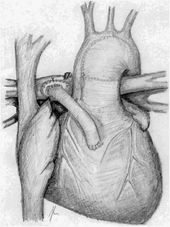
The technique of operation, using a homograft patch to enlarge the aorta, has been described elsewhere.Reference Pizarro, Malec and Maher2, Reference Mair, Tulzer and Sames3 The circulation was arrested at a core temperature of 18 degrees celsius. A single dose of Bretschneider’s solution (Custodiol®; HTK Solution, Dr Franz Koehler Chemie GmBH, Alsbach-Haehnlein, Germany) was delivered through the aortic cannula. The bifurcation of the pulmonary arteries was always closed with a homograft patch. The conduit, made of Goretex, was always positioned to the right of reconstructed neoaorta (Fig. 1) and anastomosed distally to the central part of the patch used to close the bifurcation of the pulmonary arteries. A conduit of 5 millimetres diameter was inserted in all but two patients, who had conduits of 4 and 6 millimetres, respectively. The median time required for extracorporeal circulation time was 139 minutes, with a range from 103 to 360 minutes. The median circulatory arrest time was 52 minutes, with a range from 35 to 71 minutes, and was reduced to a median of 44 minutes in the last 10 patients. Selective antegrade cerebral perfusion was performed at the preference of the surgeon in 2 patients. The sternum was electively left open in all patients. A membrane of Goretex®; Flagstaff; A2 was always attached to the pericardial edges to protect the heart and conduit at the second stage.
The conduit positioned to the right of the neo-aorta.

Results
First stage
Of the patients, 90.6%, or 29 out of 32, survived to be discharged from hospital. Extracorporeal membrane oxygenation was necessary due to myocardial dysfunction in one patient, with the diagnosis of an atrioventricular septal defect and left ventricular hypoplasia. He died on the 4th postoperative day. Death occurred in 2 other patients due to myocardial dysfunction on the 1st and 4th postoperative days, respectively. Delayed sternal closure was performed after a median of 4 days, with a range from 2 to 11 days. The median time to extubation was 8 days, with a range from 5 to 31 days.
Because of distal stenosis, the conduit required revision in 2 patients on the 6th and 7th postoperative days, respectively. At reoperation, the conduit was shortened.
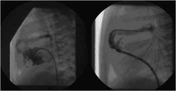
Stents were implanted in 3 patients because of proximal or medial stenosis in the conduit (Fig. 2). An EV3 Primus GPS stent of 5 by 12 millimetres was implanted proximally at the junction of the conduit with the right ventricle in 2 patients 13 and 17 days after the initial procedure. In these two, the second stage was then performed 3 and 3.1 months after placement of the stent.
Proximal stenosis treated with a stent of 5 millimetres.

In another patient, the stent was implanted in the medial portion of the tube 2 months after the initial procedure. Biventricular repair could thereby be postponed for 1 month. In another patient, we attempted placement of a stent 7 days after the initial procedure, and then constructed a bi-directional cavopulmonary anastomosis at the age of 2 months.
In 3 patients, death occurred prior to the second stage of palliation, with 2 of these having a high degree of stenosis noted in the conduit at the last follow-up before admission for a partial cavopulmonary connection. The other patient died suddenly, without any evidence of stenosis in the conduit.
Second stage
So far, 24 out of 26 patients have undergone a bi-directional cavopulmonary anastomosis performed at a mean age of 4.8 plus or minus 1.4 months. In 3 patients the partial cavopulmonary anastomosis was performed semi-urgently because of worsening cyanosis 1.9, 2.9, and 3.09 months after the initial operation, respectively. The mean interval between the first and second stages was 4.3 plus or minus 1.4 months. It was necessary to place a Goretex patch across the proximal pulmonary arteries in 7 patients at the time of the bi-directional cavopulmonary anastomosis. This included either the bifurcation or the proximal right or left pulmonary arteries. In 1 patient, with an ascending aorta measuring 1.5 millimetres, aortic atresia, and ventricular septal defect, we achieved a biventricular repair at the age 3.9 months, closing the ventricular septal defect and placing a homograft of size 14 from the right ventricle to the pulmonary arteries. Death occurred in 1patient 4 months after the second stage after a prolonged and complicated postoperative course in multiorgan failure. One patient required balloon dilation of the proximal left pulmonary artery after construction of the cavopulmonary anastomosis.
Results from perfusion scintigraphy with 40 MBq Tc 99-m were available in 20 out of 24 patients after the second stage operation. The mean ratio of distribution between right and left pulmonary arteries was 1.15 plus or minus 0.48. This information should be interpreted with the obvious limitations of perfusion scintigraphy in mind.
Third stage
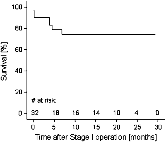
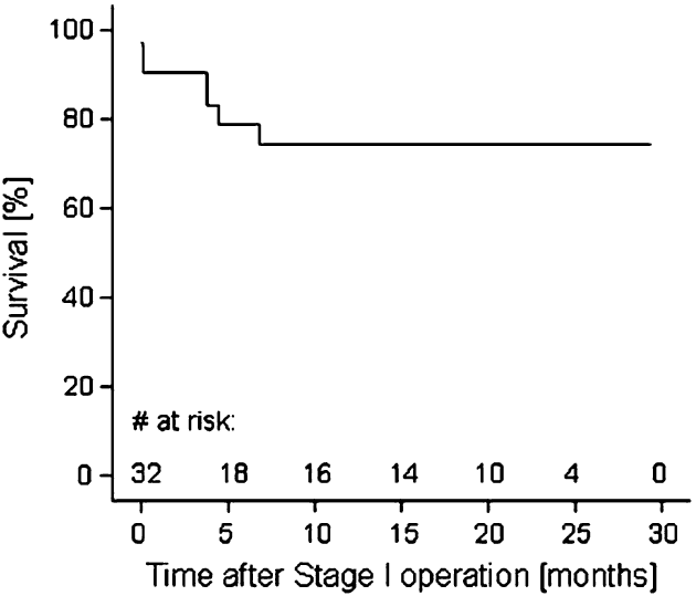
In 8 patients, the Fontan circulation was completed by construction of an extracardiac conduit at a mean age of 19.8 plus or minus 2.2 months. Overall survival of the whole cohort of 32 patients was 78.9 plus or minus 7.8% at 5 months, and 74.3 plus or minus 8.6% up to 25 months. Mean follow-up for the population of 32 patients was 12.2 plus or minus 10.2 months, with a median of 9.3 months, and a range from 0 to 29 months (Fig. 3).
Actuarial survival curve.
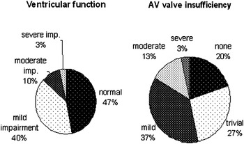
Echocardiographic data on ventricular function and the degree of tricuspid valvar regurgitation either at the last follow-up or postoperatively were available in 30 patients, and are summarised in Fig. 4. There was severe impairment of ventricular function in 1 patient after presumed myocarditis. Another patient had implantation of a mechanical valve after an unsuccessful attempt at repair of the tricuspid valve.
Ventricular function and atrioventricular valvar insufficiency at last follow-up.

Discussion
Placing a conduit from the right ventricle to the pulmonary arteries was associated with improved survival when used at our institution as the first stage of Norwood palliation for hypoplasia of the left heart. Between February, 2000, and November, 2002, only 13 of 25 patients survived to be discharged from hospital after initial palliation (52%). The change of surgical technique is one determinant of improved survival, but several other factors could have contributed. Modifications of perioperative care and surgical management are believed to have improved results for a classic first stage of palliation using a modified Blalock-Taussig shunt, with reported early survival from 79 to 93% in large series of patients.Reference Tweddell, Hoffman and Mussatto6, Reference Stasik, Goldberg, Bove, Devaney and Ohye7
For those using a conduit placed from the right ventricle to the pulmonary arteries, the reported in-hospital survival is up to 94%.Reference Sano, Ishino and Kawada1–Reference Mair, Tulzer and Sames3 In these reports, the postoperative haemodynamic stability associated with this technique is believed to be the main contributor to better results.Reference Maher, Pizarro and Gidding8 A recently published comparative study demonstrated that there is no difference in early survival when the two approached for initial palliation.Reference Tabbutt, Dominguez and Ravishankar9 The retrospective design, and uneven distribution of patients, however, are major drawbacks in this study. Further investigations are required to identify the potential advantages of each technique. The early survival of 90.6% in our patient population is consistent with previous reports.Reference Sano, Ishino and Kawada1–Reference Mair, Tulzer and Sames3
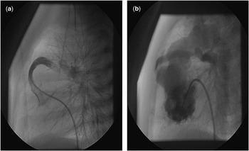
There are concerns regarding the use of the conduit, such as the increased need for revision or reintervention, the development of the pulmonary arteries, and ventricular function. Progressive obstruction of the conduit has already been mentioned in the initial reports by Sano and others.Reference Sano, Ishino and Kawada1, Reference Griselli, McGuirk and Stumper4 In our series, a biphasic manifestation of stenoses was observed. The first occurred early after the operation, and was most likely associated with insufficient resection of right ventricular muscle at the site of ventriculotomy, or kinking of the conduit. The second is, to our belief, a consequence of the conduit being compressed between the heart and sternum, and is an inevitable consequence of the design of the operation (Fig. 5). The proximal part of the conduit, at its anastomosis with the right ventricle, is prone to compression, and thus stenosis, irrespective of whether it is positioned to the left or right to the neo-aorta. We have, therefore, adopted a policy of early routine cardiac catheterisation, at less than 4 months after the initial operation, and early second stage palliation. If obstruction occurs at an early postoperative stage, less than 1 month, and construction of a partial cavopulmonary connection does not seem feasible, we suggest stenting the stenosed shunt.Reference Eicken, Sebening, Genz, Schreiber and Hess10 This avoided a repeated surgical procedure on the shunt, and postponed by a mean of 2.3 months the next stage in 3 of our patients. Hence, the potential negative effects on myocardial function, a key issue and concern in these patients, was prevented.Reference Eicken, Fratz and Gutfried11
A catheterization study in the same patient postoperatively, and before the bidirectional Glenn procedure at the age of 3.6 months. Note the unobstructed conduit at the time of the first study (a), and a small right ventricular aneurysm along with proximal narrowing of the conduit in the second (b).

According to previous reports, use of the conduit placed from the right ventricle to the pulmonary arteries as the initial step in Norwood palliation is associated with better and more evenly distributed growth of the pulmonary arteries.Reference Rumball, McGuirk and Stumper12 It is also associated, nonetheless, with central stenoses, which may require subsequent reconstruction,Reference Maher, Pizarro and Gidding8, Reference Rumball, McGuirk and Stumper12 which was also a frequent finding in our cohort. In our opinion the position of the conduit to the right of the neo-aorta makes them more easily accessible, and if needed, amenable to repair. Thus, we were able to repair the proximal pulmonary arteries by inserting a patch at the second stage in 7 patients.
There are only few publications that comment on ventricular function in patients having either a Blalock-Taussig shunt or a conduit as the initial stage of Norwood palliation.Reference Tanoue, Kado, Shiokawa, Fusazaki and Ishikawa13, Reference Muthurangu, Taylor and Hegde14 The ventriculotomy, and diastolic regurgitation, could possibly affect function, and be a source for late arrhythmias. On the other hand, several other factors, such as the size of a Blalock-Taussig shunt, obstruction to systemic and pulmonary outflows, and the interval to the second and third stages, contribute to ventricular performance. Our intermediate follow-up data show normal or only mildly impaired ventricular function by echocardiographic examination in the majority of our patients. With the obvious limitations of echocardiography for assessing right ventricular function in mind, long term follow-up studies are required to define the impact of ventriculotomy on myocardial function.
The preservation of ventricular function, undistorted pulmonary arteries, and low pulmonary vascular resistance, are all key factors for successful palliation of patients with functionally univentricular hearts. We believe that our concept of aggressive treatment of pulmonary arterial stenosis by intervention or surgery, and early staging so as to unloading the dominant ventricle, contributes to this objective.