Introduction
Bacterial lipopolysaccharide (LPS), commonly known cell-free endotoxin, is the main component of the outer membrane of all Gram negative bacteria (GNB). The endotoxin is shed during proliferation and stationary phases of bacterial growth, and released following cell disintegration and lysis (Hurley, Reference HURLEY1995; Wells and Russell, Reference WELLS and RUSSELL1996). These macromolecules protect bacteria from immunological defence mechanisms produced by the infected host and protect the bacterial membrane from chemical attack (Silhavy et al., Reference SILHAVY, KAHNE and WALKER2010). The endotoxin allows only low molecular weight, hydrophilic molecules to pass through this outer membrane, and so prevents the diffusion of GNB by bile salts, lysozymes, or antimicrobial agents from the digestive tract (Rietschel et al., Reference RIETSCHEL, KIRIKAE, SCHADE, MAMAT, SCHMIDT, LOPPNOW, ULMER, ZAHRINGER, SEYDEL and DI PADOVA1994). The immune system has evolved to recognise such compounds as signals of the presence of harmful bacteria in the gut lumen, and as such, endotoxin is capable of eliciting an immune response in the host once it enters the systemic circulation (Alexander and Rietschel, Reference ALEXANDER and RIETSCHEL2001). Although this initial activation is important for animal's immune system function and health, long-term exposure to endotoxin and subsequent activation of immune system could affect growth and production efficiencies (Mani et al., Reference MANI, WEBER, BAUMGARD and GABLER2012; Khiaosa-ard and Zebeli, Reference KHIAOSA-ARD and ZEBELI2014).
Animals may be exposed to endotoxin via two routes; the first route is systemic or localised GNB infection from an exogenous source (e.g. feed) and the second (which is the most frequent route of endotoxin exposure) is bacterial translocation from the gut (Zebeli and Metzler-Zebeli, Reference ZEBELI and METZLER-ZEBELI2012; Mani et al., Reference MANI, WEBER, BAUMGARD and GABLER2012). GNB are present in the environment, contaminated water and feed, and in the gastrointestinal tract. Thus, the environment of poultry houses can be highly contaminated with endotoxin, since GNB is extremely widespread (Fernandes, Reference FERNANDES2005). Nonetheless, the presence of GNB in the intestinal tract and the use of anti-bacterial drugs can increase poultry exposure to endotoxin. In fact, it has been shown that small amounts of endotoxin are constantly being shed into the portal circulation by the healthy gut and cleared by the liver (Nolan, Reference NOLAN1981; Ruiter et al., Reference RUITER, VAN DER MEULEN, BROUWER, HUMMEL, MAUW, VAN DER PLOEG and WISSE1981). It has been reported that endotoxin is directly recognised by TLR-4 signalling pathway which, in turn, triggers the production of several pro- and anti-inflammatory cytokines that regulate host responses, cause behavioural changes and initiate inflammation (Gardiner et al., Reference GARDINER, HALLIDAY, BARCLAY, MILNE, BROWN, STEPHENS, MAXWELL and ROWLANDS1995). The negative effects of endotoxin such as the reduction of nutrient absorption and increased energy expenditure to support responses to the immune challenge have been reported in chickens (Gardiner et al., Reference GARDINER, HALLIDAY, BARCLAY, MILNE, BROWN, STEPHENS, MAXWELL and ROWLANDS1995; Xie et al., Reference XIE, RATH, HUFF, HUFF and BALOG2000; Hu et al., Reference HU, GUO, LI, YAN, BUN and HUANG2011). This review presents details from studies that have examined and defined the impact of endotoxin release and translocation on the production and gut health of chickens.
Structure and function of LPS
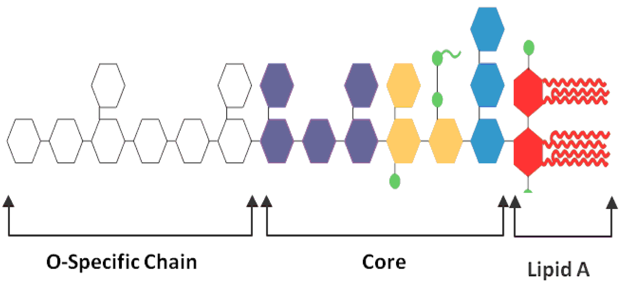
LPS consists of lipid and carbohydrate fractions, and its molecule can be divided into three parts: lipid A, core polysaccharides and O-specific chain (Figure 1). In general, lipid A is responsible for LPS's toxicity (Liebers et al., Reference LIEBERS, RAULF-HEIMSOTH and BRÜNING2008). The basic structure of lipid A consists of a β-(1-6)- linked d-glucosamine disaccharide backbone with two phosphoryl groups linked to the non-reducing glucosaminyl residue and the glycosidic hydroxyl group of the reducing glucosaminyl residue.
Bacterial Lipopolysaccharide (LPS, endotoxin).

These phosphate groups can be substituted with other chemical moieties, such as pyrophosphate, phosphoethanolamine, diphosphoethanolamine, phosphate-esterified l-arabinosamine or l,d-glycero-d-manno-heptose and-galacturonic acid, which brings diversity to natural lipid A (Zähringer et al., Reference ZÄHRINGER, LINDNER and RIETSCHEL1999). The major determinants of the immune-stimulatory capacity of lipid A are the type of disaccharide (d-glucosamine), the length and number of acyl groups, the asymmetry of their distribution, and the presence of two phosphate groups. These structural features are optimal for the induction of a pro-inflammatory response. However, the severity of response is associated with the charge and the three dimensional conformation of the lipid A (Netea et al., Reference NETEA, VAN DEUREN, KULLBERG, CAVAILLON and VAN DER MEER2002).
The polysaccharide portion of LPS consist of a hetero-oligosaccharide that is subdivided into the outer core region that is proximal to the O-specific chain, and an inner-core region that is linked to the lipid A fraction. The outer core contains common hexoses, whereas the inner core consists of 2-keto-3-deoxyoctulosonic acid, and two heptose residues (Rietschel et al., Reference RIETSCHEL, BRADE, HOLST, BRADE, MUELLER-LOENNIES, MAMAT, ZAHRINGER, BECKMANN, SEYDEL, BRANDENBURG, ULMER, MATTERN, HEINE, SCHLETTER, LOPPNOW, SCHONBECK, FLAD, HAUSCHILDT, SCHADE, DI PADOVA, KUSUMOTO and SCHUMANN1996; Sonesson et al., Reference SONESSON, ZÄHRINGER, GRIMMECKE, WESTPHAL and RIETSCHEL1994). The inner core region of LPS of different species of Gram negative bacteria is structurally more uniform than their O-specific chain.
The O-specific chain consists of up to 50 repeating units of oligosaccharide. Rietschel et al. (Reference RIETSCHEL, BRADE, HOLST, BRADE, MUELLER-LOENNIES, MAMAT, ZAHRINGER, BECKMANN, SEYDEL, BRANDENBURG, ULMER, MATTERN, HEINE, SCHLETTER, LOPPNOW, SCHONBECK, FLAD, HAUSCHILDT, SCHADE, DI PADOVA, KUSUMOTO and SCHUMANN1996) showed that the O-specific chain is the least conventional part of LPS due to the variation in the structure of ring form, sequence, substitution, and type of linkage of monosaccharide component. Two phenotypes of LPS are recognised based on structural differences in the O-specific chain; the smooth form of LPS which is characterised by a fully expressed O-specific chain, and the rough form of LPS which has an absent O-specific chain, or missing the outer core section (Sonesson et al., Reference SONESSON, ZÄHRINGER, GRIMMECKE, WESTPHAL and RIETSCHEL1994). The smooth form of LPS is more virulent than the rough form (Todar, Reference TODAR2010). The presence of an intact O-specific chain results has several advantages such as increased adherence to epithelial tissues, higher resistance to phagocytes and protection from damaging reactions with antibodies and complements and increased water-solubilisation for toxic lipid A (Todar, Reference TODAR2010).
Effects on performance and gut morphology
As endotoxin is widely disseminated in poultry houses, some losses in production may be currently disregarded due to a lack of understanding of its impact on broiler chickens. However, studies have shown an effect on bird performance, for example, Xie et al. (Reference XIE, RATH, HUFF, HUFF and BALOG2000) observed a significant reduction in relative body weight gain at 12, 24, and 48 h post injection of endotoxin. The exposure to endotoxin affects broiler biological parameters as well as performance (Rauber et al., Reference RAUBER, PERLIN, FIN, MALLMANN, MIRANDA, GIACOMINI and NASCIMENTO2014). Mireles et al. (Reference MIRELES, KIM and KLASING2005) observed a reduction in body weight and an increase in feed conversion ratio in broilers at 24, 28, 30, 32, or 34 days of age after applying Escherichia coli endotoxin subcutaneously. Available published data indicates that endotoxin decreased feed intake and body-weight gain at different times post injection (Shini et al., Reference SHINI, KAISER, SHINI and BRYDEN2008; Star et al., Reference STAR, KEMP, VAN DEN ANKER and PARMENTIER2008; Yang et al., Reference YANG, GUO, HE, YUAN, YANG and WANG2008; Lai et al., Reference LAI, NIEUWLAND, KEMP, AARNINK and PARMENTIER2009, Zhang et al., Reference ZHANG, ZHAO, CAO, AHMAD, WANG and WANG2013). Hu et al. (Reference HU, GUO, LI, YAN, BUN and HUANG2011) reported that endotoxin challenge (500 µg/ kg of BW) retarded the feed intake and daily gain of growing broilers. This effect of endotoxin on performance may be due to the stimulation of the inflammatory immune response, as it was reported that the increase in the levels of pro-inflammatory cytokines, such as TNF-α and IL-1β, decreased feed consumption (Plata-Salamán et al., Reference PLATA-SALAMÁN, SONTI, BORKOSKI, WILSON and FRENCH-MULLEN1996), weight gain, and FCR (Evock-Clover et al., Reference EVOCK-CLOVER, MYERS and STEELE1997; Steiger et al., Reference STEIGER, SENN, ALTREUTHER, WERLING, SUTTER, KREUZER and LANGHANS1999). Furthermore, multiple immune challenges lead to a cumulative reduction in growth performance (Hanssen et al., Reference HANSSEN, HASSELQUIST, FOLSTAD and ERIKSTAD2004) with evidence indicating a significant decrease in feed intake occurring during the inflammatory challenge.
It is known that activation of immune cells is an energy-consuming process. Khiaosa-ard and Zebeli (Reference KHIAOSA-ARD and ZEBELI2014) reported that the activation of the immune response by endotoxin diverted energy away from production and towards the activation of the immune response. Immune cells rely on oxidative phosphorylation during resting states; however, they switch their metabolic needs to intensive glycolysis during immune activation (Delmastro-Greenwood and Piganelli, Reference DELMASTRO-GREENWOOD and PIGANELLI2013), and this determines the high energy need of the activated immune cells. Supporting this energy diversion, studies have shown that Gram negative bacteria, such as Campylobacter, negatively impacted poultry production with consequences on the well-being of chickens (Awad et al., Reference AWAD, HESS, KHAYAL, ASCHENBACH and HESS2014a; Reference AWAD, ASCHENBACH, GHAREEB, KHAYAL, HESS and HESS2014b; Reference AWAD, MOLNÁR, ASCHENBACH, GHAREEB, KHAYAL, HESS, LIEBHART, DUBLECZ and HESS2015a; Reference AWAD, SMORODCHENKO, HESS, ASCHENBACH, MOLNÁR, DUBLECZ, KHAYAL, POHL and HESS2015b; Ghareeb et al., Reference GHAREEB, AWAD, MOHNL, PORTA, BIARNÉS, BÖHM and SCHATZMAYR2012; Reference GHAREEB, AWAD, MOHNL, BÖHM and SCHATZMAYR2013a). Taken together, the exposure to endotoxin can affect growth performance, and further the Gram negative enteric microbes in commercial poultry may contribute to the losses in productivity, increased mortality, and the contamination of chicken meat.
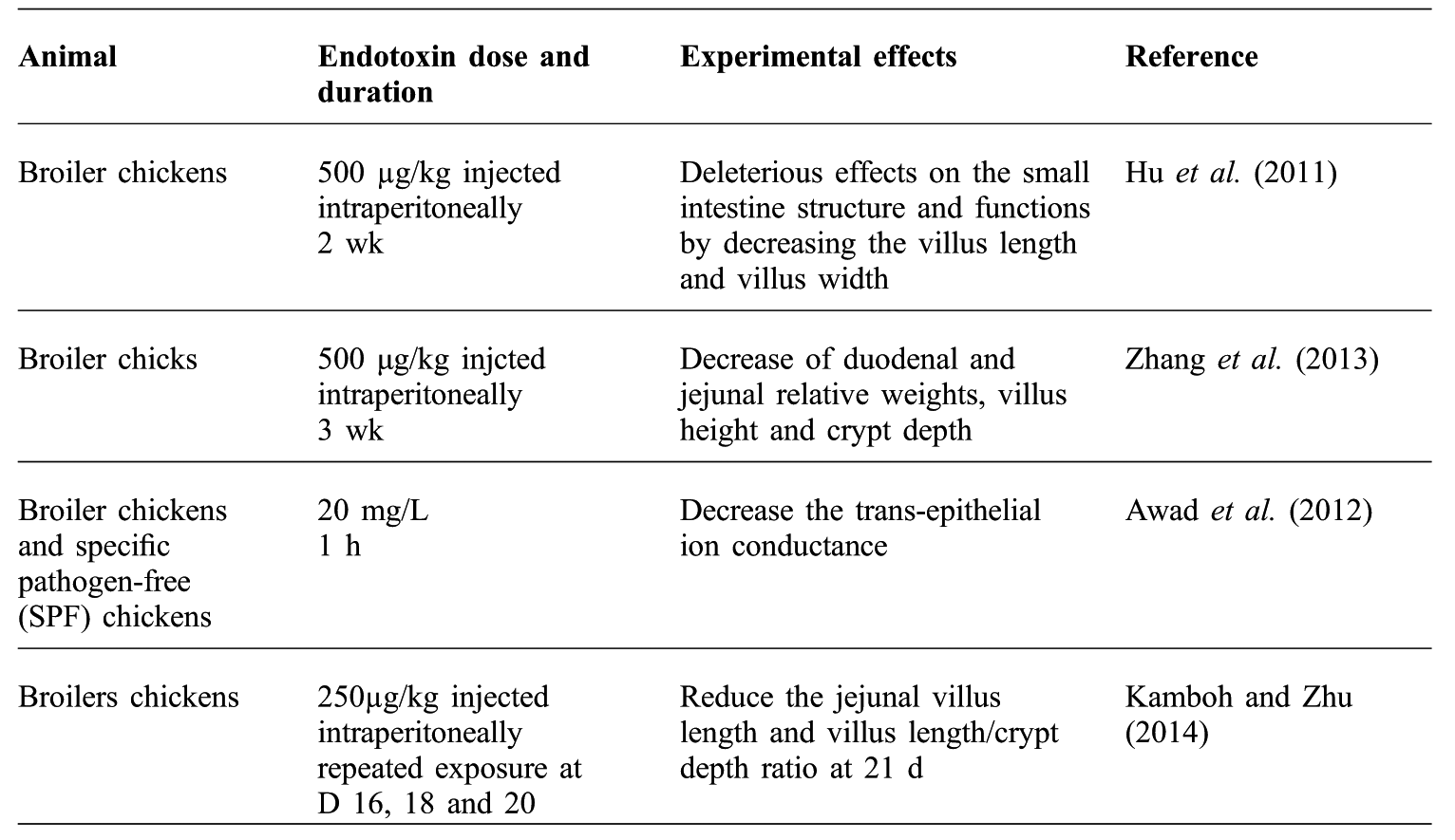
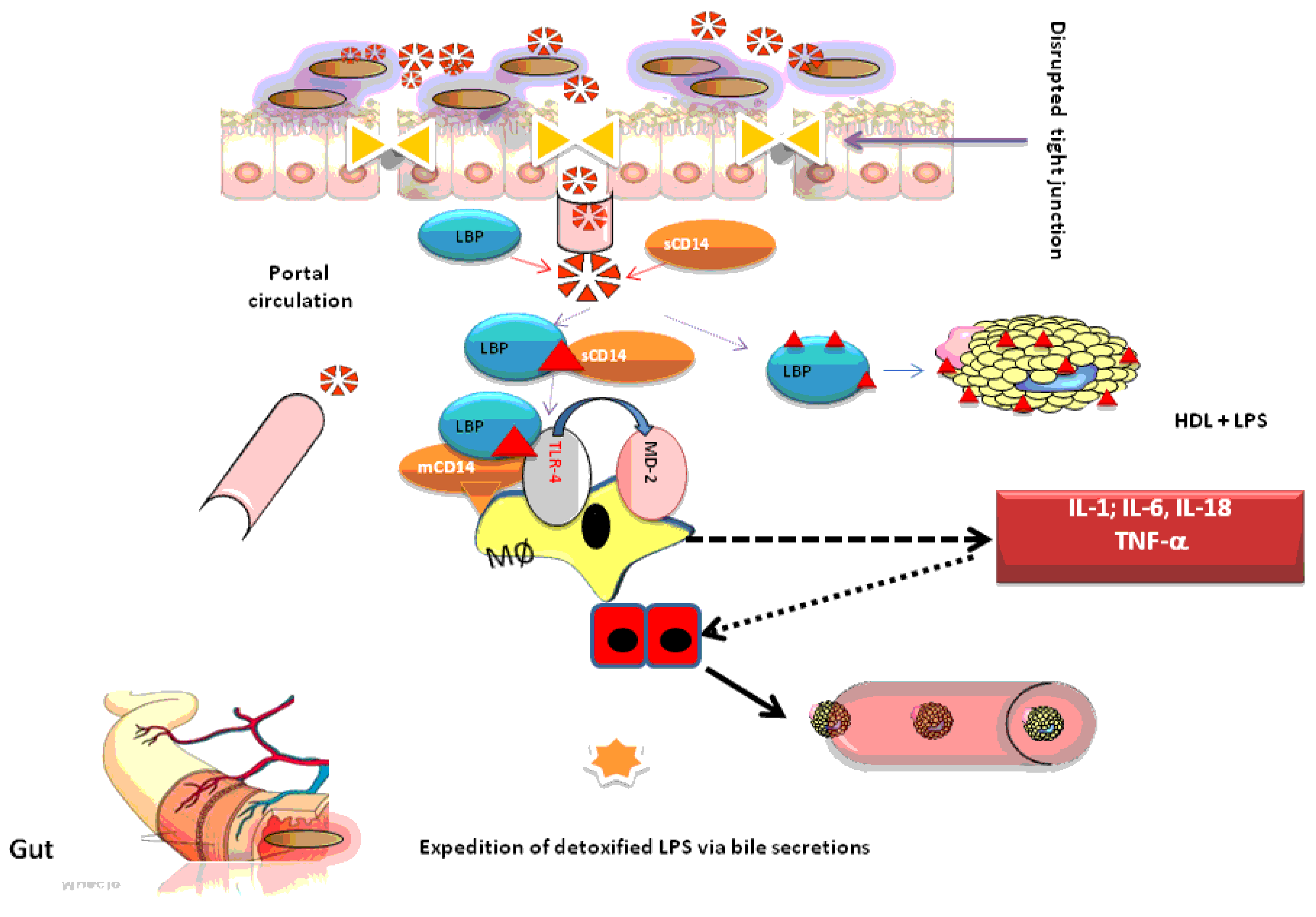
The intestine forms a major physical barrier to prevent pathogens and toxic compounds from crossing the mucosa and entering circulation and then activating the immune system. Thus, intestinal integrity is critical for maintaining a physical barrier between the intestinal lumen and the body (Table 1). Shen et al. (Reference SHEN, WEBER, RALEIGH, YU and TURNER2011) reported that different factors can regulate the permeability properties of the intestinal tight junction barrier. They demonstrated that small quantities of luminal contents including endotoxin, commensals, and pathogens may across the epithelium and enter the circulation through so-called ‘tight junctions’ (TJ), when animals are under stress or have intestinal inflammation. These pathogens can stimulate the localised secretion of pro-inflammatory cytokines from immune and intestinal epithelial cells (Mani et al., Reference MANI, WEBER, BAUMGARD and GABLER2012). Consequently, these inflammatory and stress responses may cause the phosphorylation of the myosin light chain by the kinase enzyme, which results in the contraction and opening of the intestinal epithelial TJ and increases intestinal permeability (Turner, Reference TURNER2009).
Effects of endotoxin exposure on the intestinal integrity and permeability.
Intestinal and systemic diseases are associated with leaky epithelial barriers and increased intestinal permeability to endotoxin. It is known that the reduced trans-epithelial electrical resistance (TEER) is due to compromised TJ, and consequently it is used as a good indicator of gut permeability and epithelial barrier dysfunction as measured by Ussing chambers (Awad et al., Reference AWAD, RAZZAZI-FAZELI, BÖHM and ZENTEK2007, Reference AWAD, RAZZAZI-FAZELI, BÖHM and ZENTEK2008a; Reference AWAD, GHAREEB, NITSCH, PASTEINER, ABDEL-RAHEEM and BÖHM2008b, Reference AWAD, GHAREEB, PAßLACK and ZENTEK2013, Awad and Zentek, Reference AWAD and ZENTEK2015). Albin et al. (Reference ALBIN, WUBBEN, ROWLETT, TAPPENDEN and NOWAK2007) showed that endotoxin increased the intestinal permeability and tissue conductance by decreasing the TEER. In contrast, Awad et al. (Reference AWAD, ASCHENBACH, KHAYAL, HESS and HESS2012) found that the acute exposure to Salmonella spp. endotoxin for one hour decreased the intestinal permeability and had a similar effect to the living bacteria. Accordingly, the compromised barrier function by endotoxin may result in a further translocation of pathogens across the epithelia and consequently have a substantial impact on poultry production (Tomita et al., Reference TOMITA, OHKUBO and HAYASHI2004). Thus, a better understanding of the mechanisms behind the role of endotoxin on gut permeability will allow the development of nutritional- and pharmacological-based strategies to avoid the negative effects of endotoxin.
The structure of the intestinal mucosa is an indicator of a healthy gut. Lew et al. (Reference LEW, BAYNA, MOLLE, DALTON, LAI, BHARGAVA, MENDIOLA, CLOPTON and TANG2013) mentioned that persistent high levels of circulating endotoxin had the ability to cause chronic subclinical diseases and may induce apoptosis by activating the cardiac renin-angiotensin system, and consequently decrease the cell survival. It was reported that endotoxin exposure resulted in a variety of morphological alterations in the digestive tract. Besides the direct effect on the gut, endotoxin may indirectly induce intestinal injury via reduced feed intake (Mitchell and Carlisle, Reference MITCHELL and CARLISLE1992). Kamboh and Zhu (Reference KAMBOH and ZHU2014) demonstrated that endotoxin injection reduced the jejunal villus length and villus length/crypt depth ratio at certain times post injection. Hu et al. (Reference HU, GUO, LI, YAN, BUN and HUANG2011) observed that subclinical level injection of endotoxin could cause deleterious effects on the small intestine structure and function by decreasing the villus length and villus width in broilers (Figure 2).
A summary of the effect of intestinal endotoxin transport and inflammation on gut integrity and function (adapted from Zebeli and Metzler-Zebeli, Reference ZEBELI and METZLER-ZEBELI2012).
Zhang et al. (Reference ZHANG, ZHAO, CAO, AHMAD, WANG and WANG2013) reported a decrease in duodenal and jejunal relative weights, villus height and crypt depth in LPS-challenged chickens, which have been associated with gut stressors such as pathogens and their toxins (Giannenas et al., Reference GIANNENAS, TONTIS, TSALIE, CHRONIS, DOUKAS and KYRIAZAKIS2010; Awad et al., Reference AWAD, VAHJEN, ASCHENBACH and ZENTEK2011a; Reference AWAD, GHAREEB and BÖHM2011b; Reference AWAD, HESS, TWARUŻEK, GRAJEWSKI, KOSICKI, BÖHM and ZENTEK2011c). Epithelial inflammation and villous atrophy occurred at 24 h after an endotoxin challenge (Kanno et al., Reference KANNO, EMIL, KOSI, MONFORTE-MUNOZ and ATKINSON1996). Thus, the present data suggest an endotoxin-induced deterioration of intestinal architecture but a better understanding of the underlying mechanisms of these effects of endotoxin could be helpful in improving our knowledge about intestinal architecture and barrier function.
The nutrient transport function of the gastrointestinal tract (GIT) is be decreased when the intestine is under prolonged inflammatory or metabolic stress. Hu et al. (Reference HU, GUO, LI, YAN, BUN and HUANG2011) found that endotoxin challenge reduced the absorptive function of intestine in broiler chicken. More specifically, Kanno et al. (Reference KANNO, EMIL, KOSI, MONFORTE-MUNOZ and ATKINSON1996) showed that Na and Cl ions, as well as glucose absorption, was decreased even 24 h after parenteral administration of endotoxin. The effects of endotoxin on intestinal villi contribute to a decreased intestinal nutrient absorption and increase co-infection with other pathogens (Parra et al., Reference PARRA, AGUDELO, ORTIZ, RAMÍREZ, RODRÍGUEZ and LÓPEZ2011; Zhang et al., Reference ZHANG, ZHAO, CAO, AHMAD, WANG and WANG2013; Jing et al., Reference JING, MUNYAKA, TACTACAN, RODRIGUEZ-LECOMPTE, O and HOUSE2014). Furthermore, Abad et al. (Reference ABAD, MESONERO, SALVADOR, GARCIA HERRERA and RODRÍGUEZ-YOLDI2001) found that endotoxin decreased the mucosal to serosal transepithelial flux and the transport across brush border membrane vesicles of L-leucine., and inhibited the mucosal Na+-dependent D-galactose intestinal absorption (Amador et al., Reference AMADOR, MARCA, GARCÍA-HERRERA, LOSTAO, GUILLÉN, DE LA OSADA and RODRÍGUEZ-YOLDI2008) and uptake of D-fructose across the rabbit jejunum (García-Herrera et al., Reference GARCÍA-HERRERA, ABAD and RODRÍGUEZ-YOLDI2003). Others reported decreased absorption of various sugars and amino acids (Albin et al., Reference ALBIN, WUBBEN, ROWLETT, TAPPENDEN and NOWAK2007; Flinn et al., Reference FLINN, MANI, JEFFREY, SPENCER and GABLER2010; Meng et al., Reference MENG, CHOUDRY, SOUBA, KARINCH, HUANG, LIN, VARY and PAN2005). These effects may be due to inhibition of Na+-dependent system of transport, as well as a decrease in the Na+/K+ ATPase activity (Abad et al., Reference ABAD, MESONERO, SALVADOR, GARCIA HERRERA and RODRÍGUEZ-YOLDI2001; Amador et al., Reference AMADOR, GARCÍA-HERRERA, MARCA, DE LA OSADA, ACÍN, NAVARRO, SALVADOR, LOSTAO and RODRÍGUEZ-YOLDI2007a; García-Herrera et al., Reference GARCÍA-HERRERA, ABAD and RODRÍGUEZ-YOLDI2003) or pro-inflammatory activity. Amador et al. (Reference AMADOR, GARCÍA-HERRERA, MARCA, DE LA OSADA, ACÍN, NAVARRO, SALVADOR, LOSTAO and RODRÍGUEZ-YOLDI2007b) showed that the pro-inflammatory cytokines such as TNF-α had the ability to decrease the absorption of galactose and the inhibition of the sugar uptake was mediated by the production of TNF- α. Overall, endotoxin mediated inhibition of nutrient absorption seems to be revealed by several signalling cascades including protein kinase C, protein kinase A, and mitogen activated protein kinases, as well as proteasomal degradation (Amador et al., Reference AMADOR, MARCA, GARCÍA-HERRERA, LOSTAO, GUILLÉN, DE LA OSADA and RODRÍGUEZ-YOLDI2008; García-Herrera et al., Reference GARCÍA-HERRERA, MARCA, BROT-LAROCHE, GUILLEN, ACIN, NAVARRO, DE LA OSADA and RODRIGUEZ-YOLDI2008).
Effects on innate immune response
With a well-organised gut associated lymphoid tissue in the submucosal layer to defend against any invading pathogens; the GIT is classified as the largest immune organ in the body (Ghareeb et al., Reference GHAREEB, AWAD, BÖHM and ZEBELI2015; Reference GHAREEB, AWAD, SOODOI, SASGARY, STRASSER and BÖHM2013b). The innate immune response is the first line of defence against infectious diseases and foreign particles (Figure 3).
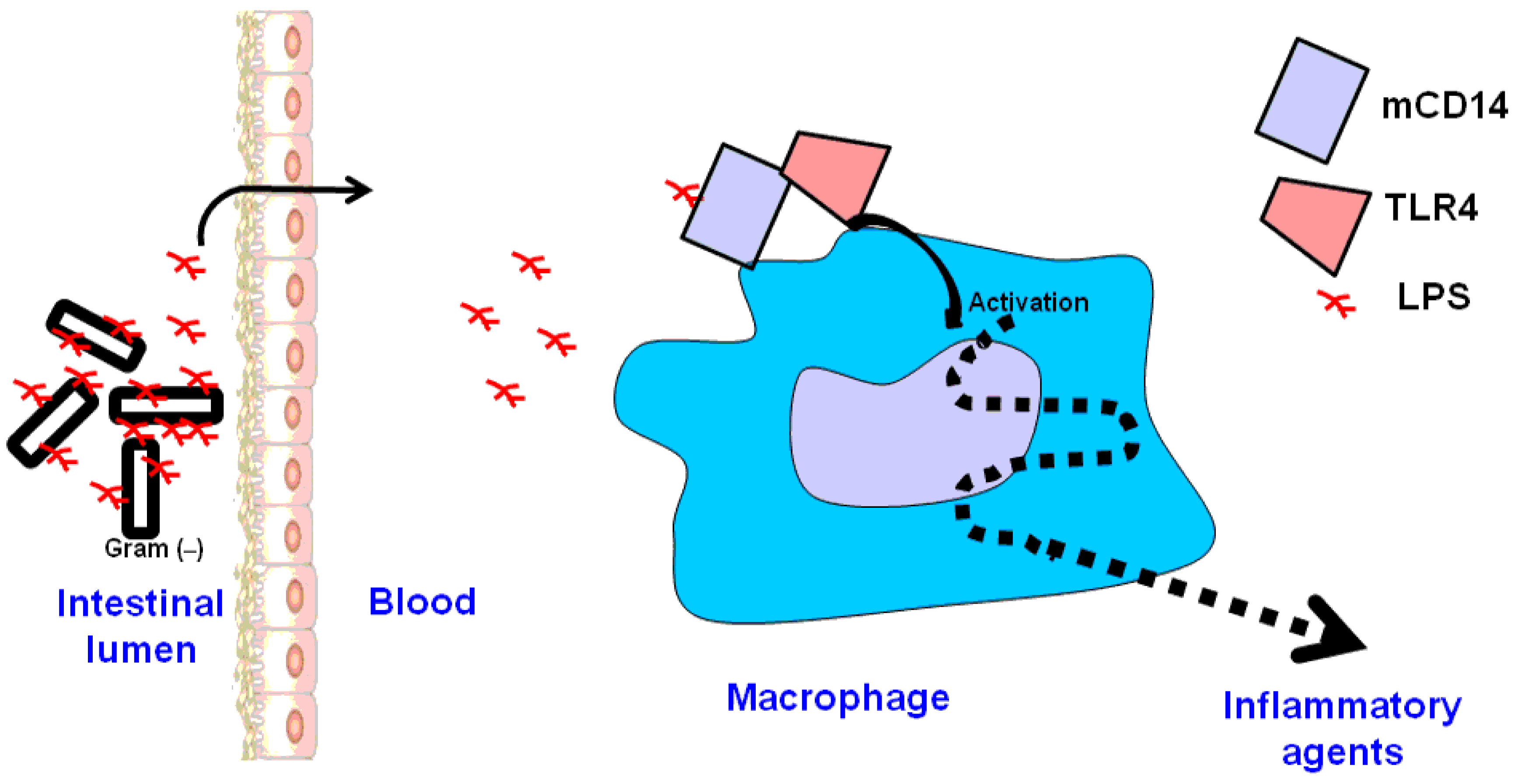
Gram negative bacteria in the intestine releases endotoxin during growth, division, and death.
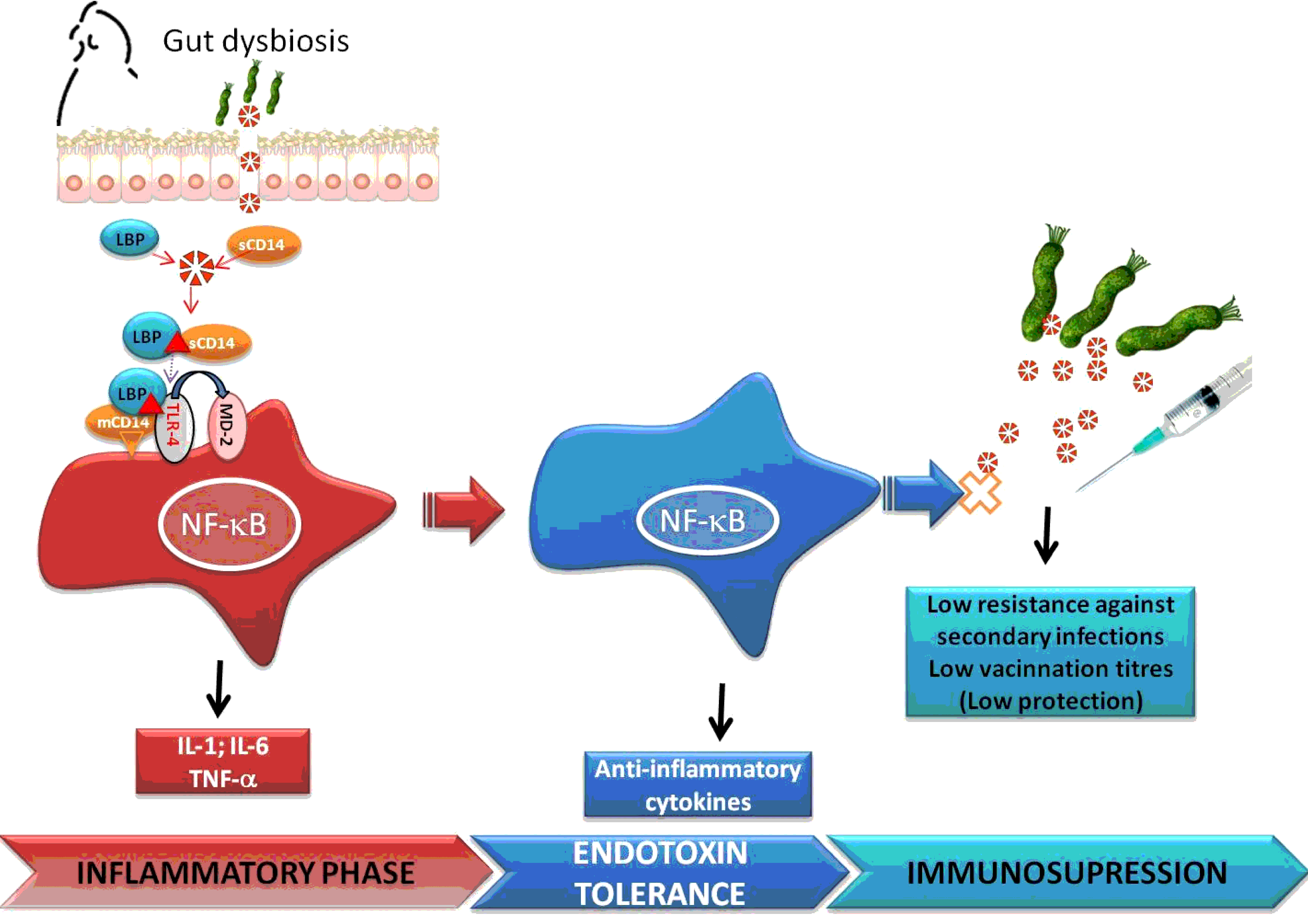
The bacterial endotoxin is one of the most powerful activators of the immune system, leading to a non-specific inflammatory response by increasing the transcription and secretion of cytokines (Mueller et al., Reference MUELLER, LINDNER, KUSUMOTO, FUKASE, SCHROMM and SEYDEL2004; Luyendyk et al., Reference LUYENDYK, SHORES, GANEY and ROTH2002). It has been demonstrated that increased expression of the pro-inflammatory IL-6 in chickens is indicative of inflammatory response induction following endotoxin injection (Yang et al., Reference YANG, GUO, HE, YUAN, YANG and WANG2008; Kaiser et al., Reference KAISER, BEACH, CIRACI and LAMONT2010). These authors reported up-regulation of both avian beta-defensin 10 (AvBD10) and inducible nitric oxide synthase (iNOS), indicating the importance of innate immunity against a bacterial infection. However, repeated exposure to endotoxin resulted in immunosuppression, as indicated by a reduction and/ or a total abolition of the febrile response and as this modulation is part of a temporary relegated immune response to GNB, this phenomenon has been identified as endotoxin tolerance (Roth et al., Reference ROTH, ASLAN, STORR and ZEISBERGER1997; West and Heagy, Reference WEST and HEAGY2002). In support, Maloney and Gray (Reference MALONEY and GRAY1998) reported that the febrile response in Pekin ducks was modulated when they were repeatedly given amounts of endotoxin, and it has been demonstrated that both the amount of endotoxin and the frequency of exposure to endotoxin contributed to the development of tolerance (Marais et al., Reference MARAIS, MALONEY and GRAY2011). Accordingly, endotoxin tolerance can lead to a suppression of the innate immune response (Figure 4) and this renders the host vulnerable to secondary infections (Cavaillon and Adib-Conquy, Reference CAVAILLON and ADIB-CONQUY2006; Biswas and Lopez-Collazo, Reference BISWAS and LOPEZ-COLLAZO2009). This has important implications for poultry industry, as birds are frequently exposed to GNB, and may become more susceptible to common infections and disease.
Suggested model of sustained exposure to endotoxin to induce tolerance and immunosupression in chickens (modified from Biswas and Lopez-Collazo, Reference BISWAS and LOPEZ-COLLAZO2009).
Endotoxin tolerance (ET) is often identified by decreasing the production of pro-inflammatory mediators, mostly TNF-α (Cavaillon et al., Reference CAVAILLON, ADRIE, FITTING and ADIB-CONQUY2003). Greisman and Hornick (Reference GREISMAN and HORNICK1975) found that the ET was characterised by two phases; early and late. The early phase decreased the production of many mediators of inflammation and provided only a short-term refractory response to endotoxin (Greisman and Hornick, Reference GREISMAN and HORNICK1975; Biswas and Lopez-Collazo, Reference BISWAS and LOPEZ-COLLAZO2009). In contrast, late tolerance seemed to rely on the production of anti-endotoxin antibodies such as anti-endotoxin IgA, to block the macrophages responses (Greisman and Hornick, Reference GREISMAN and HORNICK1975). Ametaj et al. (Reference AMETAJ, SIVARAMAN, DUNN and ZEBELI2012) observed that sustained exposure to oral endotoxin induced an oral ET which was associated with increased production of anti-endotoxin IgA.
ET can occur by the down-regulation of proteins that participate in endotoxin signalling and the innate immune response (Fan and Cook, Reference FAN and COOK2004). In both early and late phase, the ET represents a host defence mechanism to prevent tissue damage due to excessive inflammation. However, immunosuppression during ET may be due to a reduced inflammatory response (Cavaillon and Adib-Conquy, Reference CAVAILLON and ADIB-CONQUY2006; Biswas and Lopez-Collazo, Reference BISWAS and LOPEZ-COLLAZO2009), leading to an increased risk of secondary infection and poor vaccination titres (Ploegaert et al., Reference PLOEGAERT, DE VRIES REILINGH, NIEUWLAND, LAMMERS, SAVELKOUL and PARMENTIER2007).
Clearance and detoxification of endotoxin
Endotoxin can be neutralised by the following mechanisms: 1), molecules that bind endotoxin and prevent it from engaging TLR4, 2) enzymes that degrade lipid A to decrease its activity, 3) endotoxin uptake by the liver, as the primary organ involved in the clearance of endotoxin (Braude et al., Reference BRAUDE, CAREY, SUTHERLAND and ZALESKY1955) or 4) target cell adaptations that modify the response to endotoxin. Ulevitch and Johnston (Reference ULEVITCH and JOHNSTON1978) reported that the incubation of endotoxin with plasma makes it less pyrogenic and less inflammatory, suggesting a presence of specific plasma proteins that able to bind endotoxin rendering it inactive and/or detoxify it. Intestinal chylomicrons, which are involved in transporting absorbed fatty acids, appear able to promote the absorption of endotoxin (Ghoshal et al., Reference GHOSHAL, WITTA, ZHONG, DE VILLIERS and ECKHARDT2009). However, it has been demonstrated that chylomicrons can bind the endotoxin and make it inactive via contact with bile (Harris et al., Reference HARRIS, GRUNFELD, FEINGOLD, READ, KANE, JONES, EICHBAUM, BLAND and RAPP1993).
After uptake of endotoxin by hepatocytes and Kupffer cells in the liver, active and inactive forms of endotoxin can be transferred to the bile and excreted into the intestinal lumen (Lóránd, Reference LÓRÁND2004). Vreugdenhil et al. (Reference VREUGDENHIL, ROUSSEAU, HARTUNG, GREVE, VAN'T VEER and BUURMAN2003) found that LBP can enhance the binding of endotoxin to the chylomicrons and consequently reduce its bioactivity. Ulevitch et al. (Reference ULEVITCH, JOHNSTON and WEINSTEIN1979) suggested that endotoxin can be bound by high density lipoprotein (HDL). A major detoxification mechanism for endotoxin is via enzyme modification with acyloxyacyl hydrolase (AOAH). This enzyme selectively removes the secondary fatty acyl chains in the lipid A, producing an endotoxin structure that is capable of binding MD2/TLR4 but does not initiate signalling (Lu et al., Reference LU, ZHANG, TAKASHIMA, WEISS, APICELLA, LI, YUAN and MUNFORD2005). Consequently, many factors can influence plasma endotoxin concentrations through the modification in the detoxification and neutralisation mechanisms.
Conclusions
Published data has shown the effects of luminal and systemic endotoxin on gastrointestinal function and performance in poultry. Even at low concentration, endotoxin is a potent stimulator of pro-inflammatory cytokine production, and this immune activation and associated inflammation makes endotoxin an important factor contributing to an impaired gut health in poultry. However, more research is needed to understand how endotoxin affects gut functions and enters the circulation. Research describing how nutrition and other challenges, such as mycotoxin contamination, modulate endotoxin permeability is warranted (Ghareeb et al., Reference GHAREEB, AWAD, BÖHM and ZEBELI2015). Studies on gut physiology changes associated with subclinical infections is needed in order to improve the understanding of the mechanisms responsible for enteric infection colonisation.