Introduction
Recurrent respiratory papillomatosis is the most common benign neoplasm of the airway in children. The incidence of recurrent respiratory papillomatosis in children is estimated to be 4.3 per 100 000.Reference Derkay1, Reference Cohn, Kos, Taber and Adam2 Of the more than 90 types of human papilloma virus (HPV), HPV types six and 11 are frequently associated with recurrent respiratory papillomatosis.Reference Derkay3 Presentation in a child may range from voice alteration to airway obstruction. The larynx is the most commonly affected site; however, the course and severity of the disease is unpredictable. Extralaryngeal spread of the disease has been reported, with rates varying between 13 and 50 per cent.Reference Derkay1, Reference Armstrong, Derkay and Reeves4–Reference Lindeberg and Elbrond6 Multiple recurrences require frequent excision of papilloma to ensure airway patency. Approximately 11 per cent of patients require a tracheotomy, with a 36 per cent decannulation rate.Reference Armstrong, Derkay and Reeves4
A wide variety of medical and surgical treatment options for recurrent respiratory papillomatosis has been reported; nonetheless, surgery is the mainstay of treatment. The CO2 laser has been the preferred method of removing recurrent respiratory papillomatosis, until the introduction of the microdebrider for laryngeal surgery in 1999. The microdebrider has been suggested as an equivalently safe and lower-cost alternative technique to the laser for removal of recurrent respiratory papilloma.Reference El-Bitar and Zalzal7–Reference Pasquale, Wiatrak, Woolley and Lewis10 Use of a microdebrider for the removal of tracheal papilloma in patients with advanced laryngeal papillomatosis obliterating the glottis has not been previously described. We describe a modified use of a microdebrider with a laryngeal blade to remove tracheal papilloma in children with obliterated larynx and tracheostomy.
Surgical technique
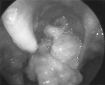
Two patients (boys aged 10 and 11 years) with recurrent respiratory papillomatosis requiring surgical treatment underwent microdebrider removal of tracheal papilloma. The children were diagnosed with laryngeal papillomas at six and 16 months of age and developed tracheobronchial and pulmonary lesions. Along the course of the disease, the children underwent tracheotomy and developed laryngeal papillomas obliterating the laryngeal inlet (Figure 1).
Laryngeal papillomas obliterating the laryngeal inlet.

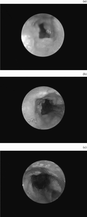
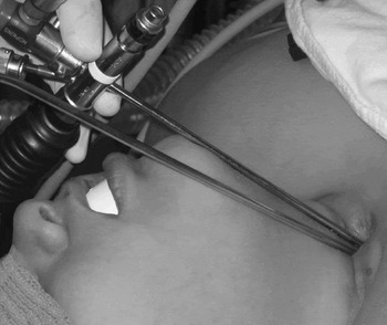
After induction of general anaesthesia via the tracheotomy tube, a 4 mm, flexible fibre-optic scope was passed through the size 6.0 paediatric Shiley tracheotomy tube to examine the trachea and mainstem bronchi, prior to papilloma debridement. Papillomas were observed on the tracheal walls and carina (Figure 2). To remove the tracheal papillomas, a bronchoscope with a ventilating port (size 2.5 and 26 cm long) was introduced into the trachea via the tracheostomy site after removing the tracheotomy tube. A 2 mm, 30° telescope with camera was passed through the bronchoscope, and the tracheal lumen was viewed on a monitor. A microdebrider with an angled tip, 3.5 mm wide and 22.5 cm long, with a Skimmer blade (XOMED Products, Jacksonville, Florida, USA) was used to remove the papillomas. The microdebrider was passed through the stoma site while the ventilating bronchoscope was in place (Figure 3). Suction without irrigation was applied to pull the papillomas away from the underlying tissue, facilitating removal of diseased tissue and preservation of normal tissue. The microdebrider was used in oscillating mode at 1500 rpm. Pledgets saturated with oxymetazoline were applied to achieve haemostasis. Upon removal of visible papillomas, the procedure was terminated, with no complications. The 10-year-old boy required the procedure seven times and the 11-year-old 10 times, between July 2005 and October 2006. No complications (e.g. tracheal stenosis) occurred.
Microdebrider removal of tracheal papillomas; (a) before, (b) during and (c) after removal.

Placement of ventilating bronchoscope and microdebrider via the tracheostomy site.

Discussion
The microdebrider with laryngeal blade has been used to remove laryngeal and tracheal papillomas in children and adults. Earlier studies have documented that the longer microdebrider blades can be used to reach most tracheal lesions in children; however, a ventilating bronchoscope cannot be used simultaneously with the microdebrider.Reference Rees, Tridico and Kirse11 In the presence of an obliterated laryngeal inlet, removal of tracheal papillomas with the microdebrider becomes more challenging due to the lack of passage through the glottis, the length of the instrument and the need for ventilation. In an effort to overcome the above challenges, we describe a surgical technique that allows removal of tracheal papillomas with the microdebrider in children with obstructed laryngeal inlets.
By introducing the microdebrider into the trachea via the tracheostomy site, all lesions located between the carina and the tracheostomy site were accessible for removal with the available lengths of the microdebrider. Due to the limited size of the tracheostomy, a 2.5 mm bronchoscope was chosen for ventilation and direct view to allow passage of both the bronchoscope and the microdebrider. In our paediatric patients, a small ventilating bronchoscope maintained ventilation for a sufficient period to allow papilloma removal. During surgery, when the telescope was being cleaned anaesthesia was maintained through the tracheotomy tube. The microdebrider was used in an oscillating mode of 1500 rpm: however, lower speeds can be chosen for bulky or pedunculated lesions.Reference Patel, Rowe and Tunkel12 The effect of microdebrider use on the number of procedures required to remove tracheal papillomas could not be determined, owing to the short duration of observation.
• Recurrent respiratory papillomatosis is the most common benign neoplasm of the airway in children
• The microdebrider has been suggested as an equivalently safe and lower-cost alternative technique to laser removal of papillomas
• This paper describes a modified use of a microdebrider with laryngeal blade to remove tracheal papilloma in the child with an obliterated larynx and a tracheostomy
• By introducing the microdebrider into the trachea via the tracheostomy site, all lesions located between the carina and the tracheostomy site were accessible for removal with the available lengths of the microdebrider
The microdebrider has the advantages of avoiding thermal injury to adjacent tissue, eliminating the risk of airway fire and requiring a shorter operating time, compared with laser excision of laryngeal papillomas. On the other hand, the microdebrider has the disadvantage of not providing the same level of haemostasis as that achieved with the CO2 laser. However, topical vasoconstriction with oxymetazoline has been effective in achieving adequate control of bleeding during microdebrider removal of paediatric airway lesions. In addition to the well documented use of the microdebrider for removal of papillomas, the microdebrider also has potential for endoscopic treatment of paediatric airway lesions such as cysts, granulation tissue and stenosis.Reference Rees, Tridico and Kirse11 The present study documents the feasibility of microdebrider removal of tracheal papillomas, using the described technique, in the patient with an obliterated larynx.
Acknowledgement
The authors would like to thank Cheryl Langford for her assistance in the preparation of the manuscript.