




No CrossRef data available.
Article contents
Genetic homogeneity within Leishmania (L.) infantum isolated from human and dogs: the relationship with the sandfly fauna distribution in endemic areas of Nueva Esparta State, Venezuela
Published online by Cambridge University Press: 22 February 2005
Abstract
Leishmania infantum has been described as a highly polymorphic group of parasites, responsible for visceral leishmaniasis and cutaneous leishmaniasis. In this paper we report the life-cycle of L. (L.) infantum in an endemic area of visceral leishmaniasis in Venezuela, by using molecular diagnosis and characterization of parasites isolated from dogs, humans with visceral leishmaniasis and sand flies. The molecular characterization was carried out by use of kDNA restriction analysis, dot-blot hybridization with species-specific probes and RFLP of the PCR products. The results demonstrated that L. (L.) infantum is the parasite responsible for VL in the island. The parasites were revealed to be genetically homogeneous with no intra-specific differences between isolates from different individuals. The highest homology of the isolates was with L. (L.) infantum from the Old World rather than with L. (L.) chagasi from the New World. Additionally, we report the geographical distribution of Lutzomyia longipalpis, and the relationship with the transmission of L. (L.) infantum in the studied area.
Information
- Type
- Research Article
- Information
- Copyright
- © 2005 Cambridge University Press
INTRODUCTION
In the New World visceral leishmaniasis is caused by L. (L.) infantum (syn. Chagasi) with the domestic dog as the main reservoir (Mauricio, Stohard & Miles, 2000). It has been reported by Mauricio et al. (1999) that L. (L.) infantum is the species infecting dogs in Latin America but not the only one. Agents of cutaneous leishmaniasis (CL) of the Viannia subgenus such as L. (V.) braziliensis, L. (V.) peruviana and L. (V.) panamensis are frequently found in dogs resulting in non-fatal cutaneous infections. An estimated number of 100000–500000 cases of visceral leishmaniasis occurs annually, with 90% of the cases occurring in India, Sudan and Brazil. In America, the highest incidence in humans has been reported in Brazil with an estimated number of 3000 cases each year.
According to the Ministry of Health, the high endemic area for VL in Venezuela is found in Sucre state, where 398 cases have been recorded in the last 10 years. Other states with low incidence are Yaracuy (21 cases) and Nueva Esparta with 54 cases from 1998 to 2002. However, in recent years, there has been an increase of the disease in humans throughout the country, with the re-emergence in rural communities (Delgado et al. 1998; Feliciangeli et al. 1998) and the appearance in urban and suburban communities (Aguilar et al. 1998). Recently, an epidemiological evaluation of the situation of VL in Eastern Venezuela, revealed an increase in the number of human cases and domestic dogs (Zulueta et al. 1999), the same situation has been observed in the Nueva Esparta State where the visceral leishmaniasis in humans has increased from 8 cases between 1990 and 1997 (1·25 cases/year) to an average of 10·4 cases per year in the past 5 years. From the total cases reported in Nueva Esparta, 85% occurred in children aged from 1 to 2 years. This increment of VL in humans in that part of the country, has motivated an epidemiological study to evaluate the risk to the population. The study included clinical, serological, and parasitological evaluations of humans and dogs in different localities, where human cases have been reported during the last 5 years, as well as an entomological survey. In a previous study (Zerpa et al. 2000) the presence of a very significant number of infected dogs was demonstrated by serological testing in different communities of the State. Regarding sand fly vectors, Lutzomyia longipalpis has long been recognized as the sole vector of the AVL. However, after Mangabeira (1969) drew attention to the morphological differences between males of Lu. longipalpis from Pará and Ceará, North Eastern Brazil, the study of the genetic variability led to the conclusion that this taxon is a complex of sibling species (Lanzaro et al. 1993). Outside Venezuela this is composed of at least 3 species (Uribe, 1999; Mutebi et al. 2002). In Venezuela, 2 species were identified by isoenzyme diagnostic markers (Lampo et al. 1999; Arrivillaga et al. 2000). In this study we report the natural cycle of L. (L.) infantum in the study area with the molecular characterization of the parasites from humans, dogs and sand flies, which resulted in a homogeneous group of parasites, not previously reported. We discuss the possibilities of an imported leishmaniasis in the island as well as the VL case distribution in relation to the sand fly fauna in different ecological situations.
MATERIALS AND METHODS
Study area
This study was carried out in 2 small villages in Nueva Esparta State, Venezuela. The island of Margarita, Venezuela (10°51′50″–11°11′06N and 63°46′06″–64°24′32″W), has an extension of 1085 km2 and a population of 373851 inhabitants (INE, 2001). Climate on the island is characterized by an average temperature of 26–28 °C and a bimodal pattern of annual precipitation (400–1100 mm) with 2 peaks (June–August and December–February).
So far, cases of visceral leishmaniasis have only been reported in the eastern area of the island, concentrated in dry zones at low altitude, while no cases have been recorded in the life-zone described as humid pre-mountain forest at the foothills of the massif El Copey (910 m a.s.l).
Sample collection and parasite isolation
Parasites were isolated from human cases and serologically positive dogs by inoculation of bone marrow, blood or lymph node infected samples in blood agar culture medium (DIFCO) containing 10% defibrinated rabbit blood and 1000 units of penicillin/ml. For parasitological studies biopsies were taken from dogs under anaesthetic conditions. Twenty-five cultures were isolated from infected dogs from 56 clinically suspicious or serologically positive animals, but only 2 positive cultures were isolated from patients in the studied endemic areas. Twelve isolates from dogs grew vigorously in the culture medium and were mass cultured in blood/agar plates at 26 °C for genetic studies. Sand fly sampling was carried out in localities where AVL cases have been detected. Additionally, where AVL had not been reported it was also screened for sand fly fauna. Collections were carried out using CDC traps placed between 19 p.m. and 7 a.m. in the main bedroom and outdoors in chicken houses or close to the dogs' resting houses.
Total DNA and kDNA isolation
Parasites were collected in the stationary phase by centrifuging at 5000 g for 10 min, washed 3 times with phosphate buffer pH 7·2 and lysed in buffer (0·05 M EDTA, 0·2 M NaCl, 10% SDS and 10 μg/ml of proteinase K) for 1 h at 56 °C. kDNA was obtained as previously described (Barrios et al. 1994). Briefly, after phenol/chloroform extraction, the aqueous phase was collected in a clean tube and centrifuged at 30800 g for 45 min at 4 °C. Finally, the kDNA was resuspended in 100 μl of TE buffer (10 mMTris HCl, 1 mM EDTA, pH 8) and storage at −20 °C.
Total DNA was obtained from 1×10⁹ promastigotes as previously described (Rodriguez et al. 1997).
Chromosomal DNA preparation and PFG electrophoresis
For chromosome analysis, promastigotes were washed once in phosphate-buffered saline, pH 7·0. Chromosomal DNA was prepared as previously described (Rodriguez et al. 2000). Agarose plugs, containing approximately 1×10⁷ promastigotes were placed in the well of a 1% (w/v) agarose gel (100 ml), poured on a 14 cm (5·2″) wide × 13 cm (5″) long surface. The gel was run in a Chef-DR II Pulsed Field Electrophoresis System (Bio-Rad), using TBE buffer 0·5×(45 mM Tris, 45 mM boric acid and 1·0 mM EDTA, pH 8) at a constant voltage of 3·5 v/cm and a ramp of 110–120 s pulse frequency for 80 h. After running, the gel was stained with ethidium bromide (0·5 μl/ml) and photographed with Polaroid film 665.
DNA digestion and hybridization
Five μg of kDNA from each isolate were digested with 10 Units of Msp1 endonuclease according to the manufacturer's instructions (New England Biolabs, Beverly, MA and Bethesda Research Laboratories, Gaithersburg, MD). The kDNA-digested products were separated by electrophoresis in 4·5–10% linear-gradient polyacrylamide gels, run at 7 mA overnight. The gels were stained with silver nitrate and photographed. Dot blot hybridization was carried out using a kDNA probe labelled with digoxigenin, using a non-radioactive labelling kit (Boehringer–Mannheim). Southern blot hybridization was carried out using Gp63 probes labelled with dCT32P (Amersham) 3000 Ci/mmol in hybridization buffer (5×SSC, 1% SDS, 0·5% Denhardt's and 50 μg/ml salmon sperm) at 42 °C, washed at 65 °C 3 times with 2×SSC, 0·5% SDS and finally exposed to photographic film (GRI Ltd) overnight at −80 °C.
Polymerase Chain Reaction (PCR)
For PCR the DNA was extracted from bone marrow aspirates obtained from patients, dogs, and from the digestive tract of Lu. longipalpis. Samples were collected in TE modified buffer (10 mM Tris pH 8, 10 mM EDTA), DNA was extracted as we previously described (Rodriguez et al. 2002). The polymerase chain reaction was carried out with AJS-31 and DBY primers, 5′GGGGTTGGTGTAAAATAG and 5′CCAGTTTCCCGCCCCGGA derived from kDNAs of L. infantum which are specific for the L. donovani complex (Lambson, Smyth & Barker, 1999, 2000). The PCR reaction mix containing 5 μl (10 ng) of total DNA, 10 mM Tris-HCl (pH 8), 1·5 M MgCl2, 0·01% gelatin, 0·2 mM dNTP mix, 100 ng of each primer, 10 μl of water, and 2·0 units of Taq polymerase (Promega) in a total volume of 25 μl, and overlayed with 20 μl of mineral oil. Amplification took place during 35 cycles, the annealing temperature was at 67 °C for 1 min, extension at 72 °C for 1 min and denaturation at 95 °C for 1min, except the first cycle of denaturation which was for 5 min; finally there was an extension cycle for 10 min. The programme was run on a thermocycler (MJ Research PT 100). Ten μl of the PCR product was run in a 1% agarose gel and the gel stained with ethidium bromide (10 mg/ml). DNA was purified from PCR products by ethanol precipitation, and finally digested with 10 Units of HaeIII restriction enzyme according to the instructions of the manufactures.
RESULTS
Sandfly collection and abundance
In Table 1 we give the results of the entomological sampling in the 11 localities surveyed, located in different life-zones in the western area of the Margarita Island. Because of a variable effort of capture, we give the total numbers of males and females collected and the relative numbers per trap used. We also give the results of the natural search for Leishmania spp. that was more than 1% in the 2 samples of females dissected, one from Achipano, the suburb of Porlamar, the capital of the Island, and the other from the village La Vecindad.
Table 1. Geographical distribution of sandfly fauna (Phlebotomine sandflies caught indoors (*) and outdoors in localities of the Island of Margarita where VL cases have been reported ([bull ]) or not (○), situated in different life zones (Ttw=Tropical thorn woodland, Tvdf=Tropical very dry forest, P-mdf=Pre-mountain dry forest, P-mmf=Pre-mountain moist forest).)

Lutzomyia longiplapis predominated in the driest areas, tropical thorn woodland, the tropical very dry forest and the pre-mountain dry forest, while Lu. gomezi and, to a lesser extent Lu. evansi, were found in the pre-mountain moist forest, which is limited to Cerro Copey and the area at the foothills.
Diagnosis and culturing of the parasites
In Fig. 1 we show (i) endemic localities where human cases have been reported and the parasites have been isolated from humans, dogs and sand flies and (ii) non-endemic localities where entomological surveys were also carried out.

Fig. 1. Map of Nueva Esparta State, showing the localities where the entomological and clinical studies were carried out.
Parasites were diagnosed in 56 biopsies from different tissues by PCR, using primers DBY and AJS-1. Fig. 2 is an example of a PCR reaction. The diagnostic band of 805 bp was obtained in biopsies from bone marrow isolated from dogs and humans, as well as samples obtained from the digestive tract of Lu. longipalpis captured in the endemic area. The isolate from L. evansi used in this experiment was previously described as L. chagasi by Feliciangeli et al. (1999), this isolate gave a band of 780 bp similar to L. (L.) chagasi (PP75, international reference strain).

Fig. 2. Gel electrophoresis of PCR products obtained after 35 amplification cycles using DNA extracted from human, dogs and sandfly. Lane 1, human DNA from bone marrow; lanes 2 and 3, dog DNA from bone marrow; lane 4, Lu. longipalpis midgut; lane 5, DNA from parasites isolated from Lu. evansi and previously identify as L. chagasi; lane 6, L. (L.) infantum (MHOM/TN/80/IPT1; lane 7, L. (L.) donovani (MHOM/IN/80/DD8); lane 8, L. (L.) chagasi (MHOM/BR/74/PP75).
Leishmania parasites were isolated from 12/24 samples obtained from dogs (Table 2), from 2 human samples but no parasites were isolated from sand flies. Nine isolates were adapted to artificial culture medium. Massive culture of parasites was obtained in blood agar base artificial medium for molecular characterization purposes.

Molecular characterization of parasites
The isolated parasites were characterized by different molecular techniques. Fig. 3 shows the restriction pattern after digestion of the PCR product with HaeIII restriction enzyme. The new isolates run in the gel (lanes 2–8) showed at least 4 bands under 1018 bp; corresponding to the pattern obtained for L. (L.) infantum (MHOM/TN/80/IPT1, lane 11), but different from the pattern obtained for L. (L.) chagasi (MHOM/BR/74/PP75, lane 12) and L. (L.) donovani (MHOM/IN/80/DD8, lane 13).

Fig. 3. Gel electrophoresis in 1% agarose of the PCR products digested with HaeIII restriction enzyme. Lane 1, Leishmania (V.) braziliensis (LTB300); lane 2, human bone marrow; lane 3, Lu. longipalpis; lanes 4–9, dog bone marrow; lane 10, L. (L.) infantum (IPT1); lane 11, L. (L.) chagasi (PP75); lane 12, L. (L.) donovani (DD8); lane 13, L. (L.) mexicana (Bel21); lane 14, molecular weight markers (1 kb ladder, Gibco-BRL).
Fig. 4 shows the results after digestion of the kDNA with the restriction enzyme MspI, this enzyme cuts the kDNA and produces 4 fragments in the majority of the new isolates (lanes 1–9), the restriction fragments share homologies with the same size of 3 fragments obtained for L. (L.) infantum (lane 12) and with 2 fragments of L. (L.) chagasi (lane 13), other enzymes such as EcoRI and PstI gave the same homologies between the dog isolates and L. (L.) infantum (results not shown). Dot blot hybridization of the kDNA to kDNA probe from L. (L.) infantum, (IPT1), gave a high hybridization signal (Fig. 5A) with the kDNA–PCR samples from dogs, humans and sand flies, which is similar to the hybridization signal obtained when the L. (L.) donovani (DD8) probe was used (Fig. 5B). However, a very low hybridization signal was obtained when the hybridization was to L. (L.) chagasi (PP75) kDNA probe (Fig. 5C).

Fig. 4. Polyacrylamide gel electrophoresis of total kDNA digested with MspI restriction enzyme. Lane 1, molecular weight markers (1 kb ladder, Gibco-BRL); lanes 2 and 3, kDNA from human isolate; lanes 4–10, kDNA from dogs isolates; lane 11, L. (L.) donovani (DD8); lane 12, L. (L.) infantum (IPT1); lane 13, L. (L.) chagasi (PP75).

Fig. 5. Dot blot of kDNA isolated from dog isolates hybridized to total kDNA probes from international reference strains. (A) Leishmania (L.) donovani kDNA probe; (B) L. (L.) infantum kDNA probe; (C) L. (L.) chagasi kDNA probe.
Southern blotted chromosomal DNA separated by pulse field electrophoresis was hybridized to GP63 probe labelled with dCT32P. GP63 probe is a 2 kb EcoRI/Xba fragment representing the coding region of the gene in L. infantum. The results in Fig. 6 show a high hybridization signal corresponding to a chromosomal band of 945 kb in samples of new isolates from dogs (lanes 1–5), which is the same signal obtained for L. (L.) infantum (lane 8) an additional hybridization band over 945 Mb was observed in L. (V.) braziliensis (lane 6). The signal is different for L. (L.) chagasi with 2 hybridization signals of 825 and 945 kbp and L. (L.) donovani with 2 bands of 1020 and 945 kbp.

Fig. 6. Pulsed field electrophoresis hybridized to GP63 (coding region) probe. Lanes 1 and 2, human isolates; lanes 3–6, dog isolates; lane 7, Leishmania (L.) donovani (DD8); lane 8, L. (L.) infantum (IPT1); lane 9, L. (L.) chagasi (PP75).
DISCUSSION
This article describes for the first time, to our knowledge, the epidemiological cycle of L. infantum in an endemic area of visceral leishmaniasis in the island of Margarita, Nueva Esparta State in the North of Venezuela, where cutaneous leishmaniasis has not been reported. Human cases, infected dogs and infected sand flies were diagnosed by conventional methods (smear, culture, ELISA) and PCR using AJS and DBY primers (Lambson et al. 1999, 2000). The PCR product revealed the diagnostic expected band of 805 bp in human, sandflies and dog samples, and the amplified product was the same as that obtained for the Old World reference strains; L. (L.) infantum (MHOM/TN/80/IPT1) and L. (L.) donovani (MHOM/IN/80/DD8), but was different from L. (L.) chagasi (MHOM/BR/74/PP75), with a size band of 780 bp. The digested PCR products, also demonstrated high homology within the field isolates and L. (L.) infantum, (IPT1) rather than with L. (L.) chagasi (PP75).
The highest homology within the studied isolates with Old World international reference strains was corroborated by dot blot hybridization of total kDNA and the Southern blot hybridization of chromosomal DNA to GP63 probe (Victoir et al. 1995), the results indicated that the Gp63 gene is located in a chromosome of approximately 975 kb which is in the range of that previously described for L. (L.) infantum from the Mediterranean Basin (Guerbouj et al. 2001). The results presented here also demonstrated the presence of L. (L.) infantum in a specimen of Lu. longipalpis, in 23 females captured, no parasites were observed in other species captured in AVL endemic and non-endemic areas. The presence of promastigotes in the digestive tract of Lu. longipalpis captured on the island was previously reported by Feliciangeli et al. (1998), but the parasite was not identified until the present work. The above-mentioned species has been described as a vector for L. (L.) chagasi that is considered to be the aetiological agent of American visceral leishmaniasis (Grimaldi, Tesh & McMahon-Prat, 1989; Cupolillo, Grimaldi & Momen, 1994; Tesh, 1995). However, this species of sand fly has a wide geographical distribution in the New World, occurring in tropical areas from Mexico to Argentina (Lainson et al. 1985; Ward, 1985; Young & Duncan, 1994). A recent report from foci of cutaneous leishmaniasis in Colombia demonstrates the role of this species in the transmission of cutaneous leishmaniasis, but the association with visceral leishmaniasis was not proven (Alexander & Young, 1982; Lopez et al. 1996). Cutaneous leishmaniasis has not been reported on the island of Margarita. However, visceral leishmaniasis was reported earlier by Pifano & Romero (1964) but the parasite was not identified and Lu. evansi was incriminated as the vector in the absence of Lu. longipalpis. More recently, 2 isolates from the Island were identified as L. (L.) infantum zymodeme MON-1 (Zerpa, 2001) which is in agreement with the early results reported by Moreno et al. (1990) in Trujillo isolates from humans and dogs. Our results are in concordance with the previous report about the relationship of L. (L.) infantum with the outcome of visceral leishmaniasis in Venezuela; however, Leishmania infantum, zymodeme MON-1 is highly polymorphic, (Hide, Bañuls & Tibayrenc, 2001). The results shown in this paper suggest that the group of studied isolates is genetically homogeneous, and even there was no difference in isolates from different foci in the same or different municipalities. As far as we know, this is the first report where the molecular characterization of the parasites revealed the natural epidemiological cycle of L. (L.) infantum in an area of visceral leishmaniasis in the country, and the genetic homogeneity of isolates from human, with no HIV history, dogs and sand fly vector. This result is not in agreement with a previous report about the heterogeneity of L. infantum described by Guerbouj et al. (2001). The genetic characterization of the isolates compare with the international reference strains (IPT1), provide evidence for the existence of L. (L.) infantum on the island of Margarita, genetically homologous to L. (L.) infantum from the Old World but with minor homology to L. (L.) chagasi (PP75, international reference strain) from the New World. This finding, together with the clinical manifestations of the disease in infants aged from 1 to 2 years (Zerpa et al. 2002), suggests that this disease will be imported in dogs coming with tourists, visiting the island from the Mediterranean Basin and have been adapted to the ecological conditions of the island.
On the island of Margarita the parasite found a good vector, Lu. longipalpis s.l. In fact, the entomological study showed that, in all the localities where VL cases have been recorded, Lu. longipalpis s.l. was present. Additionally, this species was found naturally infected and the parasites were indistinguishable from those of dogs and humans, which proves its role as possible vector of L. infantum in the area. As in other foci in Colombia, (Travi et al. 1990) and Venezuela (Feliciangeli et al. 1998), Lu. evansi seems to predominate in most humid areas that do not seem to be endemic for VL on the island of Margarita. However, the potential to be incriminated as a possible vector has not to be discarded. On the other hand, it is also interesting to notice that in the eastern area of the island, which is almost completely covered by tropical thorn woodland, no VL cases have been recorded, but anti-Leishmania antibodies have been detected in the population (Zerpa et al. 2002).
A vector control trial targeted to L. longipalpis s.l., has recently been carried out on the island, using indoor spraying of λ-cyhalotrin. The population density of Lu. longipalpis was significantly reduced in Santa Ana in comparison to the population density in a control village, Las Cabreras (Feliciangeli et al. 2003). However, it is necessary to evaluate the impact of this measure on the incidence of the disease, and further studies are needed to fully understand the transmission dynamics of the disease in order to design opportune strategies of prevention and control on the island of Margarita, which is the most important tourist area in Venezuela. The high incidence of serologically positive dogs (43% in 35 households) and co-habitants, together with the low incidence of sand fly vectors, have motivated an epidemiological surveillance and other studies concerning the transmission of visceral leishmaniasis in that area.
We wish to thank Mrs Irene Gamundi for the critical reading of the manuscript. This work received financial support from the Iniciativa Cientifica Millenium, Grant 4572-VE.
References
REFERENCES
AGUILAR, C. M.,
FERNANDEZ, E.,
FERNANDEZ, R.,
CANNOVA, D. C.,
FERRER, E.,
CABRERA, Z.,
SOUZA, W. J. S. &
COUTINHO, S. G. (
1998).
Urban visceral leishmaniasis in Venezuela.
Memorias do Instituto Oswaldo Cruz
93,
15–16.CrossRefGoogle Scholar
ALEXANDER, B. &
YOUNG, D. G. (
1992).
Dispersal of Phlebotomine sandflies (Diptera: Psychodidae) in a colombian focus of Leishmania (Viannia) braziliensis.
Memorias do Instituto Oswaldo Cruz
87,
397–403.CrossRefGoogle Scholar
ARRIVILLAGA, J. C.,
RANGEL, Y.,
OVIEDO, M. &
FELICIANGELI, M. D. (
2000).
Genetic diversity among Venezuelan populations of Lutzmoyia longipalpis (Diptera: Psychodidae: Phlebotominae).
Journal of Medical Entomology
37,
325–330.CrossRefGoogle Scholar
BARRIOS, M. A.,
RODRIGUEZ, N.,
FELICIANGELI, M. D.,
ULRICH, M.,
TELLES, S.,
PINARDI, M. E. &
CONVIT, J. (
1994).
Coexistence of two species of Leishmania in the digestive tract of the vector Lutzomyia ovallesi.
American Journal of Tropical Medicine and Hygiene
51,
669–675.CrossRefGoogle Scholar
CUPOLILLO, E.,
GRIMALDI, G. &
MOMEN, H. (
1994).
A general classification of New World Leishmania using numerical zymotaxonomy.
American Journal of Tropical Medicine and Hygiene
57,
296–311.CrossRefGoogle Scholar
DELGADO, O.,
FELICIANGELI, M. D.,
GOMEZ, B.,
ALVARADO, J.,
GARCÍA, L. &
BELLO, C. (
1998).
The re-emergence of American visceral leishmaniasis in an old focus in Venezuela: human and canine infection.
Parasite
5,
317–323.CrossRefGoogle Scholar
FELICIANGELI, M. D.,
ZERPA, O.,
RODRIGUEZ, N.,
BRAVO, A.,
GALINDO, W. &
CONVIT, J. (
1998).
Hallazgo de Lutzomyia longipalpis (Diptera: Psychodidae) naturalmente infectada con promastigotes en un foco endémico de leishmaniasis visceral en la isla de margarita, Venezuela.
Boletin de la Division de Malariologia y Saneamiento Ambiental
38,
73–75.Google Scholar
FELICIANGELI, M. D.,
RODRIGUEZ, N. M.,
DE GUGLIELMO, Z. &
RODRIGUEZ, A. (
1999).
The reemergence of American visceral leishmaniasis in an old focus in Venezuela. II. Vectors and parasites.
Parasite
6,
113–120.Google Scholar
FELICIANGELI, M. D.,
MAZZARI, M. B.,
SAN BLAS, S. &
ZERPA, O. (
2003).
Control trial of Lutzomyia longipalpis s.l in the island of Margarita, Venezuela.
Tropical Medicine and International Health
8,
1131–1136.CrossRefGoogle Scholar
GRIMALDI, G.,
TESH, R. &
McMAHON-PRAT, D. (
1989).
A review of geographic distribution and epidemiology of leishmaniasis in the New World.
American Journal of Tropical Medicine and Hygiene
41,
687–725.CrossRefGoogle Scholar
GUERBOUJ, S.,
GUISANI, I.,
SPEYBROECK, N.,
LE RAY, D. &
DUJARDIN, J. C. (
2001).
Genomic polymorphism of Leishmania infantum: a relationship with clinical pleomorphism? Infection.
Genetic and Evolution
1,
49–59.CrossRefGoogle Scholar
HIDE, M,
BAÑULS, A. L. &
TIBAYRENC, M. (
2001).
Genetic heterogeneity and phylogenetic status of Leishmania (Leishmania) infantum Zymodeme MON-1: epidemiological implications.
Parasitology
123,
425–432.CrossRefGoogle Scholar
LAMBSON, B.,
SMYTH, A. &
BARKER, D. (
1999).
Sequence homology within a minicircle class of the Leishmania donovani complex.
Molecular and Biochemical Parasitology
101,
229–232.CrossRefGoogle Scholar
LAMBSON, B.,
SMYTH, A. &
BARKER, D. (
2000).
Development and characterization of a kinetoplast DNA probe and its use in the detection of parasites.
Experimental Parasitology
94,
15–22.CrossRefGoogle Scholar
LAMPO, M.,
TORGERSON, D.,
MARQUEZ, L. M.,
RINALDI, M.,
GARCÍA, C. Z. &
ARAB, A. (
1999).
Occurrence of sibling species of Lutzomyia longipalpis (Diptera: Psychodidae) in Venezuela: first evidence from reproductively isolated sympatric populations.
American Journal of Tropical Medicine and Hygiene
64,
1004–1009.CrossRefGoogle Scholar
LANZARO, G. C.,
OSTROVSKA, K.,
HERRERO, M.,
LAWYER, P. G. &
WARBURG, A. (
1993).
Lutzomyia longipalpis is a species complex: Genetic divergence and interspecific hybrid sterility among three populations.
American Journal of Tropical Medicine and Hygiene
48,
839–847.CrossRefGoogle Scholar
LOPEZ, Y.,
OSORIO, L.,
ALVAREZ, G.,
ROJAS, J.,
JIMÉNEZ, F.,
GOMEZ, C. &
FERRO, C. (
1996).
Sandfly Lutzomyia longipalpis in a cutaneous leishmaniasis focus in Central Colombia.
Memorias do Instituto Oswaldo Cruz
97,
415–419.CrossRefGoogle Scholar
MANGABEIRA, O. (
1969).
Sobre a sistematica e biologia dos Phlebotomus do Ceara.
Revista Brasileira de Malariologia e Doenças Tropicais
21,
3–26.Google Scholar
MAURICIO, I. L.,
HOWARD, M. K.,
STOHARD, J. R. &
MILES, M. A. (
1999).
Genomic diversity in Leishmania donovani complex.
Parasitology
119,
237–246.CrossRefGoogle Scholar
MAURICIO, I. L.,
STOHARD, J. R. &
MILES, M. A. (
2000).
The strange case of Leishmania chagasi.
Parasitology Today
16,
188–189.CrossRefGoogle Scholar
MUTEBI, J. P.,
TRIPET, F.,
ALEXANDER, J. B. &
LANZARO, G. C. (
2002).
Genetic differentiation among populations of Lutzomyia longipalpis (Diptera: Psychodidae) in Central and South America.
Annals of the Entomological Society of America
95,
740–752.CrossRefGoogle Scholar
MORENO, G.,
RIOUX, J. A.,
LANOTTE, G.,
PRATLONG, F. &
SERRES, E. (
1986).
Le complexe Leishmania donovani s.l. Analyse enzimatique et traitment numerique individualisation du complexe Leishmania infantum. Corollaires biogeographiques et phyletiques. A propos de 146 souches originaires de l'Ancien et du Nouveau Monde. In coll Int CNRS/INSERM 1984, Leishmania Taxonomie et Phylogenese Applications eco-epidemiologiques,
IMEEE.
Montpellier, pp.
105–117.
MORENO, G.,
SCORZA, J. V. &
AÑEZ, N. (
1990).
Leishmania infantum en el estado Trujillo, Venezuela.
Acta Científica Venezolana
41,
27.Google Scholar
LAINSON, R.,
SHAW, J. J.,
RYAN, L.,
RIBEIRO, R. S. &
SILVEIRA, F. T. (
1985).
Leishmaniasis in Brazil XXI. Visceral leishmaniasis in the Amazonan region and further observations on role of Lutzomyia longipalpis (Lutz and Neiva, 1912) as the vector.
Transactions of the Royal Society of Tropical Medicine and Hygiene
79,
223–226.Google Scholar
RODRÍGUEZ, N.,
DE LIMA, H.,
RODRÍGUEZ, A.,
BREWSTER, S. &
BARKER, D. C. (
1997).
Genomic DNA repeat from Leishmania (Viannia) braziliensis (Venezuelan strain) containing repeats and microsatellites.
Parasitology
5,
349–358.CrossRefGoogle Scholar
RODRIGUEZ, N.,
RODRIGUEZ, A.,
CARDONA, M.,
BARRIOS, M. A.,
McCANN, S. H. &
BARKER, D. C. (
2000):
Leishmania (Viannia) guyanensis: A new minicircle class exclusive to this species isolated from a DNA cosmid library useful for taxonomic purposes.
Experimental Parasitology
94,
143–149.CrossRefGoogle Scholar
RODRIGUEZ, N.,
DE LIMA, H.,
AGUILAR, C. M.,
RODRIGUEZ, A.,
BARKER, D. C. &
CONVIT, J. (
2002).
Molecular epidemiology of cutaneous leishmaniasis in Venezuela.
Transactions of the Royal Society of Tropical Medicine and Hygiene
96,
105–109.CrossRefGoogle Scholar
TESH, R. B. (
1995).
Control of zoonotic visceral leishmaniasis: Is it to change strategies?
American Journal of Tropical Medicine and Hygiene
52,
287–292.Google Scholar
TRAVI, B. L.,
VELEZ, J. D.,
BRUTUS, L.,
SEGURA, I.,
JARAMILLO, C. &
MONTOYA, J. (
1990).
Lutzomyia evansi, an alternate vector of Leishmania chagasi in a Colombian focus of visceral leishmaniasis.
Transactions of the Royal Society of Tropical Medicine and Hygiene
84,
676–677.CrossRefGoogle Scholar
URIBE, B. (
1999).
The status of the Lutzomyia longipalpis species complex and possible implications for Leishmania transmission.
Memorias do Instituto Oswaldo Cruz
94,
729–734.CrossRefGoogle Scholar
VICTOIR, K.,
DUJARDIN, J. C.,
DE DONKER, S.,
BARKER, D. C.,
AREVALO, J.,
HAMERS, R. &
LERAY, D. (
1995).
Plasticity of GP63 gene organization in Leishmania (V) braziliensis and Leishmania (V) peruviana.
Parasitology
111,
265–273.CrossRefGoogle Scholar
WARD, R. D. (
1985).
Vector biology and control. In
Leishmaniasis
(ed. Chang, K. P. & Bray, R.), pp.
199–212.
Elsevier Science Publishers,
Amsterdam, New York and Oxford.
YOUNG, D. G. &
DUNCAN, M. A. (
1994).
Guide to the identification and geographic distribution of Lutzomyia sandflies in Mexico, the West Indies, Central and South America. (Diptera: Psychodidae).
Memoirs of the American Entomologist Institute
54,
1–881.
Associated Publishers, Gainsville, FL.Google Scholar
ZERPA, O.,
ULRICH, M.,
NEGRON, E.,
RODRÍGUEZ, N.,
CENTENO, M.,
RODRÍGUEZ, V.,
BARRIOS, R. M.,
BELISARIO, D.,
REED, S. &
CONVIT, J. (
2000).
Canine visceral leishmaniasis in Margarita Island (Nueva Esparta, Venezuela).
Transactions of the Royal Society of Tropical Medicine and Hygiene
94,
484–487.CrossRefGoogle Scholar
ZERPA, O.,
PRATLONG, F.,
ULRICH, M. &
CONVIT, J. (
2001).
Isolation of Leishmania infantum MON-1 from canine and human visceral leishmaniasis on Margarita Island, Venezuela.
Memorias Instituto Oswaldo Cruz
96,
901–902.CrossRefGoogle Scholar
ZERPA, O.,
ULRICH, M.,
BENITEZ, M.,
AVILA, C.,
RODRÍGUEZ, V.,
CENTENO, M.,
BELISARIO, D.,
REED, S. &
CONVIT, J. (
2002).
Epidemiological and immunological aspects of human visceral leishmaniasis on Margarita island, Venezuela.
Memorias Instituto Oswaldo Cruz
97,
1079–1083.CrossRefGoogle Scholar
ZULUETA, A. M.,
VILLARROEL, E.,
RODRÍGUEZ, N.,
FELICIANGELI, M. D.,
MASSARRI, M.,
REYES, O.,
RODRÍGUEZ, V.,
CENTENO, M.,
BARRIOS, R. M. &
ULRICH, M. (
1999).
Epidemiologic aspects of American visceral leishmaniasis in an endemic focus in Eastern Venezuela.
American Journal of Tropical Medicine and Hygiene
61,
945–950.CrossRefGoogle Scholar
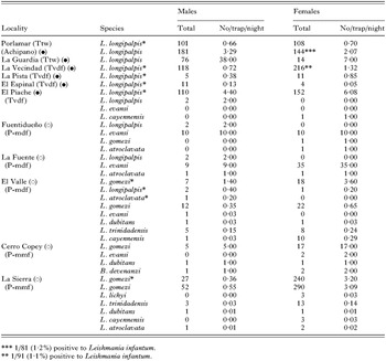
Table 1. Geographical distribution of sandfly fauna
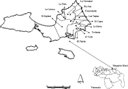
Fig. 1. Map of Nueva Esparta State, showing the localities where the entomological and clinical studies were carried out.
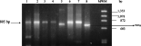
Fig. 2. Gel electrophoresis of PCR products obtained after 35 amplification cycles using DNA extracted from human, dogs and sandfly. Lane 1, human DNA from bone marrow; lanes 2 and 3, dog DNA from bone marrow; lane 4, Lu. longipalpis midgut; lane 5, DNA from parasites isolated from Lu. evansi and previously identify as L. chagasi; lane 6, L. (L.) infantum (MHOM/TN/80/IPT1; lane 7, L. (L.) donovani (MHOM/IN/80/DD8); lane 8, L. (L.) chagasi (MHOM/BR/74/PP75).
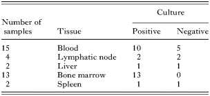
Table 2. Number of cultures isolated from different tissues
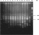
Fig. 3. Gel electrophoresis in 1% agarose of the PCR products digested with HaeIII restriction enzyme. Lane 1, Leishmania (V.) braziliensis (LTB300); lane 2, human bone marrow; lane 3, Lu. longipalpis; lanes 4–9, dog bone marrow; lane 10, L. (L.) infantum (IPT1); lane 11, L. (L.) chagasi (PP75); lane 12, L. (L.) donovani (DD8); lane 13, L. (L.) mexicana (Bel21); lane 14, molecular weight markers (1 kb ladder, Gibco-BRL).
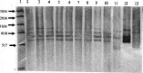
Fig. 4. Polyacrylamide gel electrophoresis of total kDNA digested with MspI restriction enzyme. Lane 1, molecular weight markers (1 kb ladder, Gibco-BRL); lanes 2 and 3, kDNA from human isolate; lanes 4–10, kDNA from dogs isolates; lane 11, L. (L.) donovani (DD8); lane 12, L. (L.) infantum (IPT1); lane 13, L. (L.) chagasi (PP75).
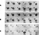
Fig. 5. Dot blot of kDNA isolated from dog isolates hybridized to total kDNA probes from international reference strains. (A) Leishmania (L.) donovani kDNA probe; (B) L. (L.) infantum kDNA probe; (C) L. (L.) chagasi kDNA probe.
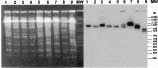
Fig. 6. Pulsed field electrophoresis hybridized to GP63 (coding region) probe. Lanes 1 and 2, human isolates; lanes 3–6, dog isolates; lane 7, Leishmania (L.) donovani (DD8); lane 8, L. (L.) infantum (IPT1); lane 9, L. (L.) chagasi (PP75).
 You have
Access
- 6
- Cited by