Introduction
Transgender persons still experience a considerable amount of social rejection, exclusion, discrimination, and hate-motivated harassment even after they have begun living according to their gender identity (EU Agency for Fundamental Rights, 2014). Limited self-report data of transgender persons suggest elevated feelings of isolation, emotional deprivation, and an urge to meet others’ needs (Simon et al. Reference Simon, Zsolt, Fogd and Czobor2011). However, studies of social rejection in transgender persons in the laboratory are missing to characterize the neural correlates of rejection sensitivity or regulation of these feelings. Knowledge of the impact of ostracism on the brain may aid in the understanding of social-cognitive effects on the underlying neurobiology, quality of life, and mental health in socially marginalized groups (Meyer-Lindenberg & Tost, Reference Meyer-Lindenberg and Tost2012).
In the laboratory, one frequently used paradigm to elicit social rejection is the cyberball task (Williams et al. Reference Williams, Cheung and Choi2000; Eisenberger et al. Reference Eisenberger, Lieberman and Williams2003), in which the participant plays a game of toss-the-ball with two other, virtual players. After a short period, the participant is excluded from the game whilst the two virtual players only pass the ball amongst each other, thus eliciting feelings of exclusion. In the standard variant, the participant is made to believe they are playing against two (or three) other real people who are connected to them (for example through the internet) (Williams & Jarvis, Reference Williams and Jarvis2006). From a clinical perspective, initial sensitivity to social rejection in the cyberball task predicts therapeutic outcome in depressed patients (Mueller et al. Reference Mueller, De Rubeis, Lange, Pawelzik and Sutterlin2016), supporting a strong role for social exclusion in mental health (Meyer-Lindenberg & Tost, Reference Meyer-Lindenberg and Tost2012).
In the original study by Eisenberger et al. (Reference Eisenberger, Lieberman and Williams2003), social distress was related to activity in the ventro-lateral prefrontal cortex (vlPFC) and the dorsal anterior cingulate cortex (dACC). These authors proposed commonalities between ostracism-induced social distress and the experience of physical/social pain, apparent in ACC activation, and its supposed inhibition through the vlPFC. A between-group study then not only documented reduced frontal activation in people with mental health problems, which may indicate reduced ability to regulate this ostracism experience, but also reduced functional connectivity between the vlPFC and the ACC (Maurage et al. Reference Maurage, Joassin, Philippot, Heeren, Vermeulen and Mahau2012). Interestingly, these researchers (Maurage et al. Reference Maurage, Joassin, Philippot, Heeren, Vermeulen and Mahau2012) additionally used a re-inclusion condition after the exclusion to examine how easily participants would be able to regulate their social rejection feelings in order to reintegrate into the social game. Such re-inclusion evoked larger activation in patients relative to comparisons in the ACC, which was hypothesized to reflect persistent feelings of exclusion. Of note, while the study by Eisenberger et al. (Reference Eisenberger, Lieberman and Williams2003) reported dorsal ACC activation, Maurage et al. (Reference Maurage, Joassin, Philippot, Heeren, Vermeulen and Mahau2012) reported a more ventral focus, i.e. pregenual ACC. A recent meta-analysis of ACC activation during the cyberball task (Rotge et al. Reference Rotge, Lemogne, Hinfray, Huguet, Grynszpan and Tartour2015) notes both regions of the ACC to be active during social exclusion. Thus, one immediate question is to what extent transgender persons, a group strongly stigmatized in society, would experience social exclusion, how they would regulate this experience, and how easily they would feel ready to re-integrate.
The present study addressed this serious issue. Transgender men, transgender women (TW), cisgender men (CM) and cisgender women (CW) completed the cyberball task during fMRI using both social exclusion and re-inclusion conditions. Based on available self-report data in transgender persons (Simon et al. Reference Simon, Zsolt, Fogd and Czobor2011; EU Agency for Fundamental Rights, 2014), the importance of social inclusion for mental health in marginalized groups (Meyer-Lindenberg & Tost, Reference Meyer-Lindenberg and Tost2012), and the involvement of the ACC in social exclusion (Rotge et al. Reference Rotge, Lemogne, Hinfray, Huguet, Grynszpan and Tartour2015), we hypothesized; (1) larger neural activation in the dorsal/ventral ACC but reduced vlPFC activation during an experience of ostracism in transgender persons relative to comparisons. Because previous research has shown that frontal regions are connected with the ACC to regulate social rejection feelings (Eisenberger et al. Reference Eisenberger, Lieberman and Williams2003; Maurage et al. Reference Maurage, Joassin, Philippot, Heeren, Vermeulen and Mahau2012), we expected this inverse relationship to reflect (2) less regulatory (negative) functional connectivity between the ventral ACC and vlPFC in trans persons relative to cisgender persons. Finally, we anticipated these feelings to last longer in trans persons as reflected by (3) larger (persisting) activation in the ACC during social re-inclusion in transgender persons relative to comparisons, indicating greater difficulty to reconnect after being socially excluded.
Materials and methods
Participants
Twenty transgender men (TM, female-to-male) (age = 36.80 years, s.d. = 8.36 years), 19 TW (male-to-female) (age = 40.53 years, s.d. = 8.55 years) and 20 CM (age = 32.50 years, s.d. = 10.13 years), and 20 CW (age = 34.50 years, s.d. = 11.20 years) participated. Participants did not differ significantly in age (F(3,75) = 2.48, p = .068). Because of this trending effect, however, age was included as a covariate of no interest in all analyses. Transgender persons (TM, TW) were recruited through the Department of Endocrinology at Ghent University Hospital. TM and TW were on at least 2 years of cross-sex hormone therapy and at least 1 year after sex-affirming surgery. They were thus living for at least 3 years in the new gender role. Comparison (cisgender) participants (CM, CW) were recruited by word-of-mouth. Given recent associations of depression with social exclusion sensitivity in the cyberball task (Mueller et al. Reference Mueller, De Rubeis, Lange, Pawelzik and Sutterlin2016) and a high prevalence of depression in transgender persons (Heylens et al. Reference Heylens, Elaut, Kreukels, Paap, Cerwenka and Richter-Appelt2014), all participants completed the Beck Depression Inventory (BDI, Beck et al. Reference Beck, Steer and Garbin1988) as well as the Spielberger State/Trait Anxiety Inventory (STAI, Spielberger et al. Reference Spielberger, Gorsuch and Lushene1970). Although a one-way ANOVA indicated a statistically significant group difference in depression scores [F(3,69) = 4.07, p = .010], follow-up post-hoc tests showed that TW had only marginally higher BDI scores than CM (p = .054), with no differences between any other groups (all ps > 0.289) (cf. Table 1). No group differences emerged for state [F(3,73) = 0.72, p = .543] or trait [F(3,73) = 0.07, p = .976] anxiety. The study was approved by the ethical committee of Ghent University Hospital. The authors assert that all procedures contributing to this work comply with the ethical standards of the relevant national and institutional committees on human experimentation and with the Helsinki Declaration of 1975, as revised in 2008. Participants received 35 EUR for compensation. Prior to the study, all participants signed an informed consent form.
Demographic information of the four groups including depression (BDI) and anxiety (STAI) scores as well as hormone levels

Task and procedure
Exactly the same fMRI version of the cyberball as described in Maurage et al. (Reference Maurage, Joassin, Philippot, Heeren, Vermeulen and Mahau2012) was used. Participants were made to believe that they would be playing against two other players via the intranet (two medical students, always one male, one female, both with common first names who were located in a different hospital building). In case of technical difficulties or delays, and when it seemed that participants would not believe or no longer believed the cover story, the experimenter would tell them that one of the players had to leave for an appointment and that they would now be playing against someone else. This change was then reinforced by visibly changing the player's name on the screen and/or a fake phone call that was observable to the participant. The experiment began with participants seeing the intranet connection status (‘you are not connected’ for the first condition and ‘you are connected’ for the three other conditions) on the top of the screen. As in the regular version of the game, participants played a round of bass-the-ball with the two other players. Once participants received the ball from one of the other players, they had 2.5 s to pass the ball back to the same player or the other player using a two-button response pad (Lumina response box). If the player did not throw within that time the ball was automatically passed to another player. Participants were encouraged to pass the ball to other players as quickly as possible without thinking too much about it and just enjoy the game. The experiment progressed in a fixed order of four conditions:
(1) Implicit social exclusion (ISE). Participants saw the ‘not connected’ status, which we told participants would reflect the fact that we were still establishing a connection and getting the MRI equipment to run while the other two players could already be observed playing. (2) First inclusion condition (INCL1) in which participants were connected and could freely play with the other players. (3) Explicit social exclusion (ESE) in which participants first received five throws but were then excluded for the rest of the time. Here, the other players (i.e. the computer) only passed the ball amongst each other. (4) In the second inclusion condition (INCL2) participants were re-included in the game. The rationale for this fourth condition was both to explore the brain correlates of re-inclusion but also to allow for a friendly and non-frustrating end of the game for all participants. As noted above, whilst participants knew they were excluded in condition 1 due to technical issues, they believed they were included for conditions 2–4. Apart from the change in connection status from conditions 1 to 2 (ISE>INCL1), participants were not told about the transition to the other remaining conditions, and the task appeared as continuous.
Each condition had a fixed duration of 125 s (50 volumes). Computer players’ speed varied randomly between 500 ms and 2 s, and was adapted to obtain 100 throws per condition. To avoid overlap between the activations associated with each condition during the transition phase, we excluded the first 10 volumes of each condition from analyses (thus leaving a total of 40 volumes per condition). Participants viewed the task on a screen behind the magnetic bore through a back mirror mounted on the coil.
fMRI data acquisition
Functional MRI data were acquired in a single run of 8 : 49 min on a 3 T Siemens Trio MRI scanner (Erlangen, Germany) and an eight channel phased array head coil (Phillips Medical Systems) as a series of blood-oxygen-sensitive T2*-weighted echo-planar image volumes. Acquisition parameters were TE = 32 ms, TR = 2500 ms, flip angle = 90deg, FOV = 220 mm2, slice thickness = 3.5 mm with no interslice gap, distance factor = 0%. Each volume comprised 36 axial slices acquired in ascending interleaved sequence. Recording comprised one run of 208 volumes (50 volumes per condition, interleaved by 2 volumes transition periods).
Prior to this, a high-resolution T1-weighted MPRAGE anatomical image was also acquired (duration = 5 : 14 min) in ascending order with a FOV = 256 mm2, slice thickness 1 × 1 × 1 mm2, TR = 2250 ms, TE = 2.52 ms, flip angle = 9°.
fMRI data processing and statistical analysis
Data were pre-processed using Statistical Parametric Mapping (SPM8, Welcome Department of Cognitive Neurology, UK), implemented in Matlab R2012b (The Mathworks, Natick, MA, USA). Functional images were preprocessed and first realigned to the first scan to correct within- and between-run motion; coregistered with the anatomical scan; normalized to the MNI template using an affine fourth-degree b -spline interpolation formation to a voxel size of 3 × 3 × 3 mm3 and spatially smoothed using a 8 mm FWHM Gaussian kernel. Condition-related changes in regional brain activity were estimated for each participant by a general linear model in which the responses evoked by each condition were modeled by a standard hemodynamic response function. The six motion parameters resulting from the realignment step were added as covariates of no interest to all contrasts. Similar to Maurage et al. (Reference Maurage, Joassin, Philippot, Heeren, Vermeulen and Mahau2012), we were interested in two main experimental contrasts. First, the ESE–ISE contrast (social exclusion) would isolate the neural activity associated with feelings of social exclusion since ESE and ISE were identical (the participant never received the ball) and only differed in the fact that in the ISE condition people knew there was a reason for exclusion (technical problems) while in the ESE condition the social exclusion experience was modeled. Second, the INCL2– INCL1 contrast (re-inclusion) would examine to what extent participants were able to re-integrate after social exclusion given that in both conditions participants were actively participating and that the INCL2 condition directly followed the social exclusion condition.
In order to balance the small sample size of each group without compromising statistical rigor, we opted to examine three a priori regions of interest (ROIs). Specifically, we were interested in between-group differences in the dACC and ventral ACC (vACC), and the vlPFC. For the two ACC ROIs, we selected two coordinates from the (ACC-focused) meta-analysis by Rotge et al. (Reference Rotge, Lemogne, Hinfray, Huguet, Grynszpan and Tartour2015), one in the dorsal ACC [MNI xyz: 8 24 24] and one in the ventral (pregenual) ACC [10 32-2]. The vlPFC ROI consisted of right Brodmann area 45, which has been reported in prior work (Masten et al. Reference Masten, Eisenberger, Borofsky, Pfeifer, McNealy and Mazziotta2009; Maurage et al. Reference Maurage, Joassin, Philippot, Heeren, Vermeulen and Mahau2012). ROIs were created using the WFU Pickatlas toolbox (Maldjian et al. Reference Maldjian, Laurienti, Burdette and RA2003): the dACC and vACC by drawing a 6 mm sphere around the aforementioned main coordinates and the vlPFC by selecting the appropriate Brodmann area. Mean parameter estimates for each of the three ROIs were extracted using the REX toolbox for SPM and analyzed in SPSS (p ⩽ 0.05, two-tailed). The main (within-group) effects were examined using one-sample t tests (with activity being different from 0) specifically testing for increases in activation. Between-group comparisons were conducted using an ANCOVA with four groups (CM, CW, TM, and TW) and covariates of no interest of age and TBV. Age was entered as a covariate because of the trending difference in age between groups. TBV was included because we wanted to remove any variance possibly associated with subtle anatomical variations, as we had found small group differences (in different regions than the ones studied here) previously in this cohort (Mueller et al. Reference Mueller, Landre, Wierckx and T'Sjoen2017b). When the data were reanalyzed without TBV as a covariate, the findings did not substantially change. Cohen's d and partial eta squared were taken as measures of effect size, as appropriate. Finally, to assess whether the trending significant group effect in depression could have driven any significant findings, the influence of depression (BDI scores) was assessed repeating the model but with depressive symptoms added as a covariate.
Finally, although we did not have any a priori hypotheses regarding circulating hormone levels, because transgender persons were taking cross-sex hormones long-term, for sake of completeness and to assess any potential hormonal effects on the data post-hoc, these data were additionally assessed (supplementary materials). In brief, hormones were unlikely to influence the present findings and are not further considered.
Functional connectivity (psychophysiological interaction, PPI)
Also akin to prior studies (Maurage et al. Reference Maurage, Joassin, Philippot, Heeren, Vermeulen and Mahau2012), PPI analyses (Friston, Reference Friston2004) were conducted for the vACC ROI during the ESE–ISE contrast, i.e. to examine PPIs during social exclusion. However, extending that prior work, we additionally computed PPI analyses for the same vACC ROI for the re-inclusion condition [INLC2–INLC1] given that no prior fMRI study of social exclusion has examined functional connectivity during social re-inclusion. We thus extracted the deconvolved activity time course centered around the vACC coordinates from the main analysis [MNI xyz: 10 32-2] and a 6 mm sphere drawn around it from each participant and corrected for the effect of interest. The product of this activation time course was calculated with a condition-specific regressor probing social exclusion (ESE–ISE) and re-inclusion (ISI2–ISI1), respectively, to create PPI terms. The social exclusion and re-inclusion PPI analyses were conducted separately. PPI analyses were conducted for each participant and entered into a random-effects analysis with one-sample t tests. Between-group comparisons were conducted using an ANCOVA with four groups (CM, CW, TM, and TW) and covariates of no interest of age and TBV. Because the PPI analyses aimed to examine whole brain connections of the vACC ROI, corrections for multiple comparisons were applied using a combined voxelwise and cluster size thresholding approach with a voxelwise threshold of p = 0.001 and a corrected alpha of p = 0.01 (calculated with 3dClustSim in Afni), which resulted in a minimum cluster size of 37 contiguous voxels at the whole brain level. The estimated connectivity was represented as a t- value with the positive t-value representing a positive interaction with vACC and a negative t-value a negative interaction.
Procedure
After participants arrived at the MRI center, they were informed about the task and signed the informed consent form. Then a structural MRI was taken, which was followed by the cyberball task. At the end of the experiment, participants were asked how much they believed the cover story, and were fully debriefed regarding the deception. No participants showed any adverse reactions or were upset. They were then thanked and reimbursed for their time and participation.
Results
Task-related effects
Within-group effects
Exclusion: explicit exclusion - implicit exclusion [ESE-ISE]
No significant effects emerged.
Re-inclusion: inclusion 2 – inclusion 1 [ISI2–ISI1]
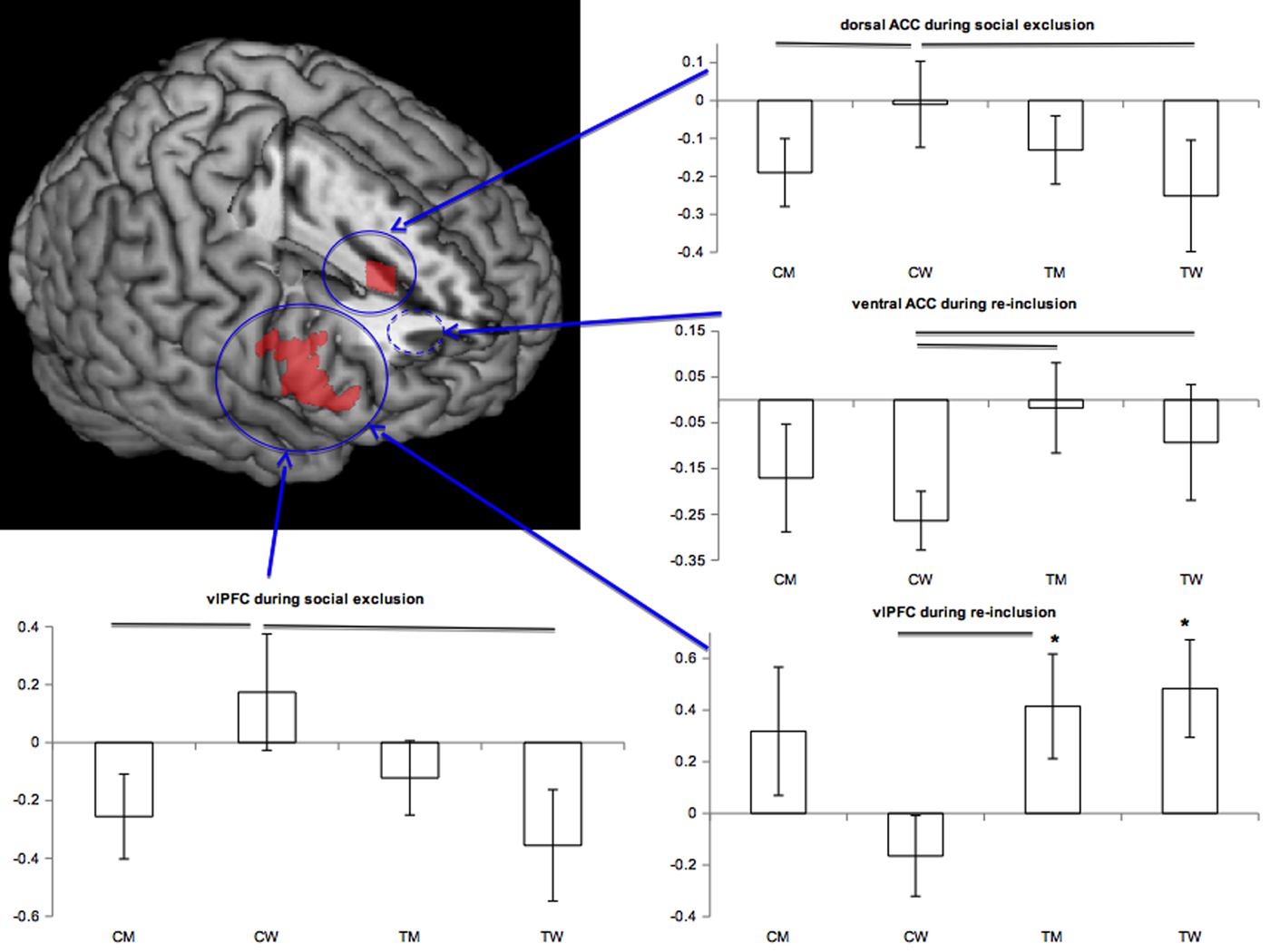
During re-inclusion, TW (t(18) = 2.56, p = 0.020, d = 0.59) and TM (t(19) = 2.05, p = .055, d = 0.46) showed increased vlPFC activation relative to baseline with medium effect sizes (Fig. 1, asterisks).
Figure displays significant average region-of-interest activations for the social exclusion and re-inclusion contrasts in the dACC (upper blue circle), vACC (dashed blue circle), and vlPFC/BA 45 (lower blue circle). The individual group activations (y-axis = mean parameter estimates) are displayed for cisgender men (CM), cisgender women (CW), trans men (TM), and trans women (TW) on the right and below. Solid black lines indicate statistically significant differences between groups, whereas asterisks indicate significant main effects within groups. Error bars denote SEM. The vACC circle is dashed because the precise anatomical location could not be displayed on the same slice as it lies anterior/inferior to the dACC site (cf. yellow circle Fig. 2). The rendering was done using MRICron.

Between-group comparisons
Exclusion: [ESE–ISE]
During the ostracism experience, CW exhibited significantly larger activation in dACC [F(1,36) = 9.09, p = .005,  $\eta _p^2 $ = 0.20] and vlPFC [F(1,36) = 5.54, p = .024,
 $\eta _p^2 $ = 0.13] than CM with medium-to-large and medium effect sizes, respectively. Similarly, CW showed the same pattern relative to TW significantly in the vlPFC [F(1,35) = 5.86, p = .021,
 $\eta _p^2 $ = 0.14] and at trend-level in the dACC [F(1,35) = 3.39, p = 0.074,
 $\eta _p^2 \; \; $ = 0.09] (small-to-medium effect) (Fig. 1).
Re-inclusion: [ISI2–ISI1]
During re-inclusion, both transgender groups exhibited more activation than CW. TW had relatively more activation than CW in the vACC [F(1,35) = 4.06 p = 0.052,  $\eta _p^2 $ = 0.10] while TM had relatively larger activations than CW in the vACC [F(1,36) = 6.13, p = 0.018,
 $\eta _p^2 \; $ = 0.15] and vlPFC [F(1,35) = 5.59, p = 0.024,
 $\eta _p^2 \; $ = 0.13] with medium effect sizes. The effect in the dACC was trending [F(1,36) = 3.30, p = 0.078,
 $\eta _p^2 $ = 0.08] (small-to-medium effect) (Fig. 1).
Functional connectivity (PPI analyses)
Within-group effects
Exclusion: [ESE-ISE]
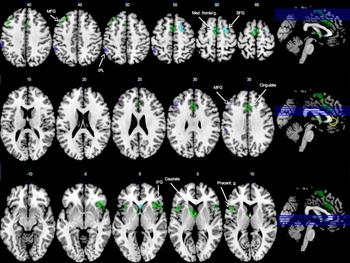
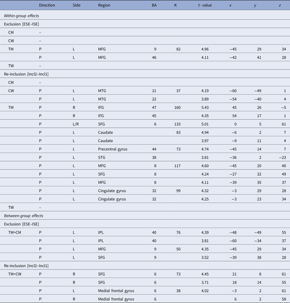
Functional connectivity as revealed by PPI analyses showed a positive connectivity between the vACC and middle frontal gyrus in TM only (see Table 2, Fig. 2).
Between-group effects from the functional connectivity (PPI) analyses during social exclusion (whole brain, thresholded at p < 0.001 for illustration) indicating stronger positive connectivity of the vACC ROI (yellow circle) with areas involved in down-regulation in lateral PFC in TM relative to CM, CW, and TW. TM>CM (in blue); TM>CW (in cyan); TM only during exclusion (in violet); TM only during re-inclusion (green). White numbers denote z coordinate. SFG, superior frontal gyrus; IFG, inferior frontal gyrus; MFG, middle frontal gyrus; IPL, inferior parietal lobule; Med. frontal g., medial frontal gyrus.

Table indicates the within and between group effects of the functional connectivity analyses, corrected for multiple comparisons at voxelwise threshold p < 0.001, and clusterwise threshold of p < 0.01

CM, Control men; CW, control women,;TM, Trans men; TW, Trans women; Direction P, positive; Side R, right; L, left; BA, Brodman area; SFG, superior frontal gyrus; MFG, middle frontal gyrus; IFG, inferior frontal gyrus; STG, superior temporal gyrus; MTG, middle temporal gyrus; IPL, inferior parietal gyrus, Coordinates [x y z] are in MNI space.
Re-inclusion: [ISI2–ISI1]
During re-inclusion, CW showed positive connectivity between the vACC and the left middle temporal gyrus. TM showed widespread positive connectivity of the vACC with all parts of the lateral prefrontal cortex, i.e. the left superior and middle and, crucially, the right inferior frontal gyrus (IFG). Additional connections emerged with the left cingulate and precentral gyri, the left superior temporal gyrus and left caudate (Table 2, Fig. 2).
Correlation of brain activity with mood scores
Given the marginally higher levels of depressive symptoms in TW relative to CM, we assessed associations between neural activity in the exclusion and re-inclusion contrasts and depression (BDI) scores for each group separately. Only a negative correlation emerged for CW in the dACC ROI [r(19) = −0.54, p = 0.017] during re-inclusion. This correlation suggested that higher depressive scores were associated with less activation in this region. However, the range of BDI scores in the majority of CW was in the minimal range (0–9, 83.3%) with three women scoring in the mild depression range (10–18, 16.7%) and the correlation thus has to be interpreted with caution. No other correlations were significant.
Discussion
This study identified the neural correlates of ostracism in transgender persons already living according to their (preferred) gender identity. Based on pan-European data on discrimination and stigmatization (EU Agency for Fundamental Rights, 2014), it was hypothesized that transgender persons would exhibit greater neural activations during social exclusion relative to CM and women and that they would also experience more difficulty reintegrating. Several critical findings emerged. First, during social exclusion, CW exhibited greater activation than CM or trans women in the dACC and vlPFC. Second, during re-inclusion, both transgender groups showed more activation in the vACC than CW. Third, these data were nicely corroborated by functional connectivity profiles, which showed aberrant functional connectivity between the vACC and vlPFC in TM.
During a lab-based ostracism experience, task-based effects were present in socially relevant brain structures. Although no effects within groups emerged during exclusion, between-group contrasts revealed larger neural activity in CW relative to CM or trans women in two areas hypothesized to be central to social pain and its regulation, the ACC and vlPFC (Eisenberger et al. Reference Eisenberger, Lieberman and Williams2003; Masten et al. Reference Masten, Eisenberger, Borofsky, Pfeifer, McNealy and Mazziotta2009; Rotge et al. Reference Rotge, Lemogne, Hinfray, Huguet, Grynszpan and Tartour2015). VlPFC modulation has been documented in previous cyberball studies (Eisenberger et al. Reference Eisenberger, Lieberman and Williams2003; Sebastian et al. Reference Sebastian, Tan, Roiser, Viding, Dumontheil and Blakemore2011; Maurage et al. Reference Maurage, Joassin, Philippot, Heeren, Vermeulen and Mahau2012; Moor et al. Reference Moor, Guroglu, Op de Macks, Rombouts, Van der Molen and Crone2012) and meta-analyses report right IFG modulation/activation during psychosocial as well as psychophysiological stress (Kogler et al. Reference Kogler, Muller, Chang, Eickhoff, Fox and Gur2015). In particular, this vlPFC activity is hypothesized to counteract (via top-down control) vACC activity that is elicited during the experience of social exclusion (Eisenberger et al. Reference Eisenberger, Lieberman and Williams2003; Maurage et al. Reference Maurage, Joassin, Philippot, Heeren, Vermeulen and Mahau2012). In line with these interpretations, the current data would suggest that CW experienced psychosocial stress during exclusion, but also engaged brain areas involved in the regulatory effort of this experience. Moreover, the neural pattern in trans women appeared to resemble that of their sex assigned at birth. However, during re-inclusion a different pattern emerged.
As hypothesized, during re-inclusion in the game, both trans groups showed strong activations in both the within and the between-group contrasts in the vACC and vlPFC. Particularly, TM and TW indicated larger activity in the vACC relative to CW when being re-included. Transgender persons experience more social isolation and are more vulnerable to harm (Simon et al. Reference Simon, Zsolt, Fogd and Czobor2011; EU Agency for Fundamental Rights, 2014). Interestingly, theories on minority stress propose that minority groups such as trans persons learn to anticipate social rejection and discrimination, but also develop vigilance in interaction with dominant group members (Meyer, Reference Meyer2003). Thus, within this context, it is conceivable that vulnerability to exclusion might also make it harder to forgive and re-engage in the social process or that this vulnerability is associated with less trust in the other based on the experience. Consequently, while CW might experience higher social distress during exclusion with higher concurrent activation of regulatory areas (vlPFC), transgender persons might experience lingering feelings of exclusion during attempts at re-inclusion similar to the pattern observed by Maurage et al. (Reference Maurage, Joassin, Philippot, Heeren, Vermeulen and Mahau2012). However, these conjectures are tentative at this stage and the precise socio-cognitive processes underlying these neural effects remain to be determined.
In any case, the functional connectivity analyses (PPI) in transgender persons further corroborated these conjectures. As noted above, a negative relationship between the vACC and the vlPFC could counteract social distress and aid top-down regulatory efforts (Eisenberger et al. Reference Eisenberger, Lieberman and Williams2003; Maurage et al. Reference Maurage, Joassin, Philippot, Heeren, Vermeulen and Mahau2012). In the present study, TM in particular possessed an aberrant positive (rather than negative) connectivity between the vACC and a lateral frontal network indicating potential further vulnerability. By contrast, TW did not show such changes in functional connectivity. Interestingly, the fact that TM and women differed from one another in this respect might additionally have gender-specific implications regarding social inclusion/exclusion in society (Saito et al. Reference Saito, Kondo, Kondo, Ojima and Hirai2012; Benenson et al. Reference Benenson, Markovits, Hultgren, Nguyen, Bullock and Wrangham2013).
Some strengths and limitations require discussion. A major strength is that the present study is currently one of the largest fMRI studies of the cyberball task. Secondly, it included not only the regular exclusion contrast, but also the re-inclusion contrast and the respective functional connectivity profiles of both conditions. A limitation, however, is that the study focused on social rejection sensitivity in trans persons after adopting their gender identity. Consequently, we cannot disentangle longitudinal aspects of rejection sensitivity prior to transitioning. A second limitation is regrettably the omission to collect a self-report scale of distress against which the neural findings could have been evaluated (correlated). Nonetheless, the vACC and IFG findings in particular, and their functional connectivity, are consistent with meta-analytic reports of activations in this region during psychosocial stress (Kogler et al. Reference Kogler, Muller, Chang, Eickhoff, Fox and Gur2015) and prior fMRI work using the cyberball task (Eisenberger et al. Reference Eisenberger, Lieberman and Williams2003; Sebastian et al. Reference Sebastian, Tan, Roiser, Viding, Dumontheil and Blakemore2011; Maurage et al. Reference Maurage, Joassin, Philippot, Heeren, Vermeulen and Mahau2012; Moor et al. Reference Moor, Guroglu, Op de Macks, Rombouts, Van der Molen and Crone2012).
In conclusion, the present study identifies neural correlates of social exclusion and re-inclusion in a socially-stigmatized and often ostracized group, that of transgender persons. The ventral ACC in particular was sensitive to social exclusion in transgender persons. This finding was further supported by reduced functional connectivity with regions hypothesized in down-regulation of such experience (lateral PFC), particularly in TM. Future work should more specifically examine neural subcomponents of social exclusion (e.g. exclusion v.. re-integration) and their associated role in risk and resilience for mental health problems and quality of life in marginalized groups (Meyer-Lindenberg & Tost, Reference Meyer-Lindenberg and Tost2012; Mueller et al. Reference Mueller, De Cuypere and T'Sjoen2017a).
Supplementary material
The supplementary material for this article can be found at https://doi.org/10.1017/S0033291717003828
Acknowledgements
We would like to thank Pierre Maurage for helpful comments on an earlier draft of this article and Anna Hudson for grammar and language editing. This project was supported by the MRP of Ghent University (Multidisciplinary Research Partnership ‘The integrative neuroscience of behavioural control’ to SCM).
Conflict of Interest
None of the authors has a conflict of interest to declare.