Bovine mastitis, inflammation of a cow's mammary glands, is a significant problem in dairy farming because the disease lowers productivity and milk quality, resulting in higher production costs (Reshi et al. Reference Reshi, Husain, Bhat, Rehman, Razak, Bilal and Mir2015). Mastitis is caused mostly by microbial infection but also by chemical, physical, or traumatic impacts that result in an oxidative stress response in the mammary gland (Sordillo & Aitken, Reference Sordillo and Aitken2009; Poławska et al. Reference Poławska, Bagnicka, Niemczuk and Lipińska2012). A high milk yield is also known to be associated with clinical mastitis in dairy cows (Chassagne et al. Reference Chassagne, Barnouin and Chacornac1998; O'Reilly et al. Reference O'Reilly, Green, Peeler, Fitzpatrick and Green2006). For example, in an investigation of risk factors for clinical mastitis in British dairy herds, the risk of clinical mastitis was significantly increased when the dairy herds produced an average annual milk yield of more than 7000 l per cow (O'Reilly et al. Reference O'Reilly, Green, Peeler, Fitzpatrick and Green2006).
In cells, reactive oxygen species (ROS) are normally produced via aerobic metabolism in the mitochondrial electron transport chain (Decoursey & Ligeti, Reference Decoursey and Ligeti2005). To maintain cellular homeostasis, host antioxidant defence systems are balanced with ROS production. In contrast, large amounts of ROS production that exceed host antioxidant capabilities can cause oxidative stress and consequent inflammatory reactions and diseases (Poławska et al. Reference Poławska, Bagnicka, Niemczuk and Lipińska2012; Wiegman et al. Reference Wiegman, Michaeloudes, Haji, Narang, Clarke, Russell, Bao, Pavlidis, Barnes and Kanerva2015; Jin et al. Reference Jin, Yan, Shi, Sheng, Shi, Zhao and Li2016a; Shi et al. Reference Shi, Yan, Jin, Shi and Guo2016). Since the bovine mammary gland is metabolically highly active during lactation, dairy cows with high-yielding mammary glands may be exposed to a considerable amount of ROS (Jozwik et al. Reference Jozwik, Krzyzewski, Strzalkowska, Polawska, Bagnicka, Wierzbicka, Niemczuk, Lipinska and Horbanczuk2012). Also, it was suggested that intensive growth of the mammary gland and the synthesis of milk are causes of oxidative stress and inflammation in the mammary gland (Jozwik et al. Reference Jozwik, Krzyzewski, Strzalkowska, Polawska, Bagnicka, Wierzbicka, Niemczuk, Lipinska and Horbanczuk2012). Previous reports also indicated that lactation can contribute to cellular oxidative stress in the mammary gland (Sordillo & Aitken, Reference Sordillo and Aitken2009; Jin et al. Reference Jin, Wang, Liu, Hu, Zhao and Liu2016b). For example, both lipid peroxidation and total amount of antioxidants have been found to increase in lactating dairy cows (Castillo et al. Reference Castillo, Hernandez, Valverde, Pereira, Sotillo, Alonso and Benedito2006). Furthermore, the level of hydroperoxides in low-density lipoprotein lipids was shown to be proportional to milk yields of dairy cows (Lohrke et al. Reference Lohrke, Viergutz, Kanitz, Gollnitz, Becker, Hurtienne and Schweigert2004). These data suggest that oxidative stress is increased during milk production in the mammary gland.
In cells, oxidative stress activates multiple intracellular signalling molecules, including mitogen-activated protein kinases (MAPK; e.g., extracellular signal receptor-activated kinase 1/2 [ERK1/2], p38, and c-Jun N-terminal kinase), Akt, and nuclear factor-kappa B (NF-κB) (Cargnello & Roux, Reference Cargnello and Roux2011; Liu et al. Reference Liu, Sun, Sun, Ma and Cheng2012; Ma, Reference Ma2013). Both MAPKs and Akt mediate cellular inflammatory responses by activation of NF-κB (Liu et al. Reference Liu, Lin, Liu, Bian, Zhang, Gao and Li2015). NF-κB is a major transcription factor for transcriptional regulation of cyclooxygenase-2 (COX-2) (Kim et al. Reference Kim, Kim, Kim, Lee, Kundu, Na, Cha and Surh2014a). Previous studies have shown that there is increased expression of COX-2 in inflammatory diseases, including bovine mastitis (Corl et al. Reference Corl, Robinson, Contreras, Holcombe, Cook and Sordillo2010; Kang et al. Reference Kang, Lee, Lee, Petriello, Kim, Do, Lim, Lee and Han2016). When oxidative stress or inflammation is occurring in cells, antioxidant systems are normally reinforced for a host defensive mechanism. Haeme oxygenase-1 (HO-1) is an antioxidant enzyme that catalyses the conversion of haeme into biliverdin, carbon monoxide, and free iron (Wang et al. Reference Wang, Stavchansky, Zhao, Bynum, Kerwin and Bowman2008). HO-1 and the haeme-degradation byproducts play protective roles against cellular oxidative injury and inflammation (Loboda et al. Reference Loboda, Damulewicz, Pyza, Jozkowicz and Dulak2016). Expression of HO-1 is regulated by oxidative stress in cells (Loboda et al. Reference Loboda, Damulewicz, Pyza, Jozkowicz and Dulak2016). In addition to the cellular antioxidant system, many antioxidant substances have been identified. N-acetylcysteine (NAC) is an antioxidant reagent that serves as an ROS scavenger, a precursor of cellular cysteine and glutathione (GSH), and an inhibitor of NF-κB nuclear translocation (Sun, Reference Sun2010; Halasi et al. Reference Halasi, Wang, Chavan, Gaponenko, Hay and Gartel2013). NAC also protects cells from apoptosis by scavenging ROS and restoring GSH levels (Sun, Reference Sun2010).
Since high milk production can produce oxidative stress and is known to be associated with bovine mastitis, in the current study we investigated the hypothesis that oxidative stress increases inflammatory responses in bovine mammary cells. We intentionally produced ROS (e.g., superoxide anion) in bovine mammary alveolar cells (MAC-T) and investigated resulting cellular inflammatory responses (e.g., alterations of COX-2, HO-1, ERK1/2, Akt and NF-κB). To ensure the role of ROS in the inflammatory responses, cells were treated with an antioxidant, NAC.
Materials & methods
Materials
Dulbecco's modified Eagle's medium (high glucose) was purchased from Lonza (Basel, Switzerland). Foetal bovine serum (FBS) was obtained from PAN (Aidenbach, Germany). Phosphate buffered saline (PBS) and trypsin were purchased from Gibco (Grand Island, NY). Penicillin/streptomycin was purchased from Welgene (Daegu, Korea). Progesterone, insulin, and menadione (2-methyl-1,4-naphthoquinone (MD), vitamin K3) were obtained from Sigma Aldrich (St. Louis, MO). Trypan blue dye exclusion solution and NAC were purchased from Amresco (Solon, OH). Dihydroethidium (DHE) was purchased from Invitrogen (Carlsbad, CA). ProLong Gold Antifade reagent containing 4,6-diamidino-2-phenylindole (DAPI) was purchased from Life Technologies (Carlsbad, CA). PD98059 and LY294002 inhibitors were purchased from Santa Cruz Biotechnology (Santa Cruz, CA), and BAY11-7082 was purchased from Calbiochem (Darmstadt, Germany). Antibodies for COX-2, p-Akt, HO-1, NF-κB p65, glyceraldehyde 3-phosphate dehydrogenase (GAPDH), proliferating cell nuclear antigen (PCNA), and goat anti-rabbit IgG-HRP were purchased from Santa Cruz Biotechnology (Santa Cruz, CA). Antibodies for p-ERK1/2, ERK1/2, and Akt were purchased from Cell Signaling Technology (Danvers, MA).
Cell culture and treatments
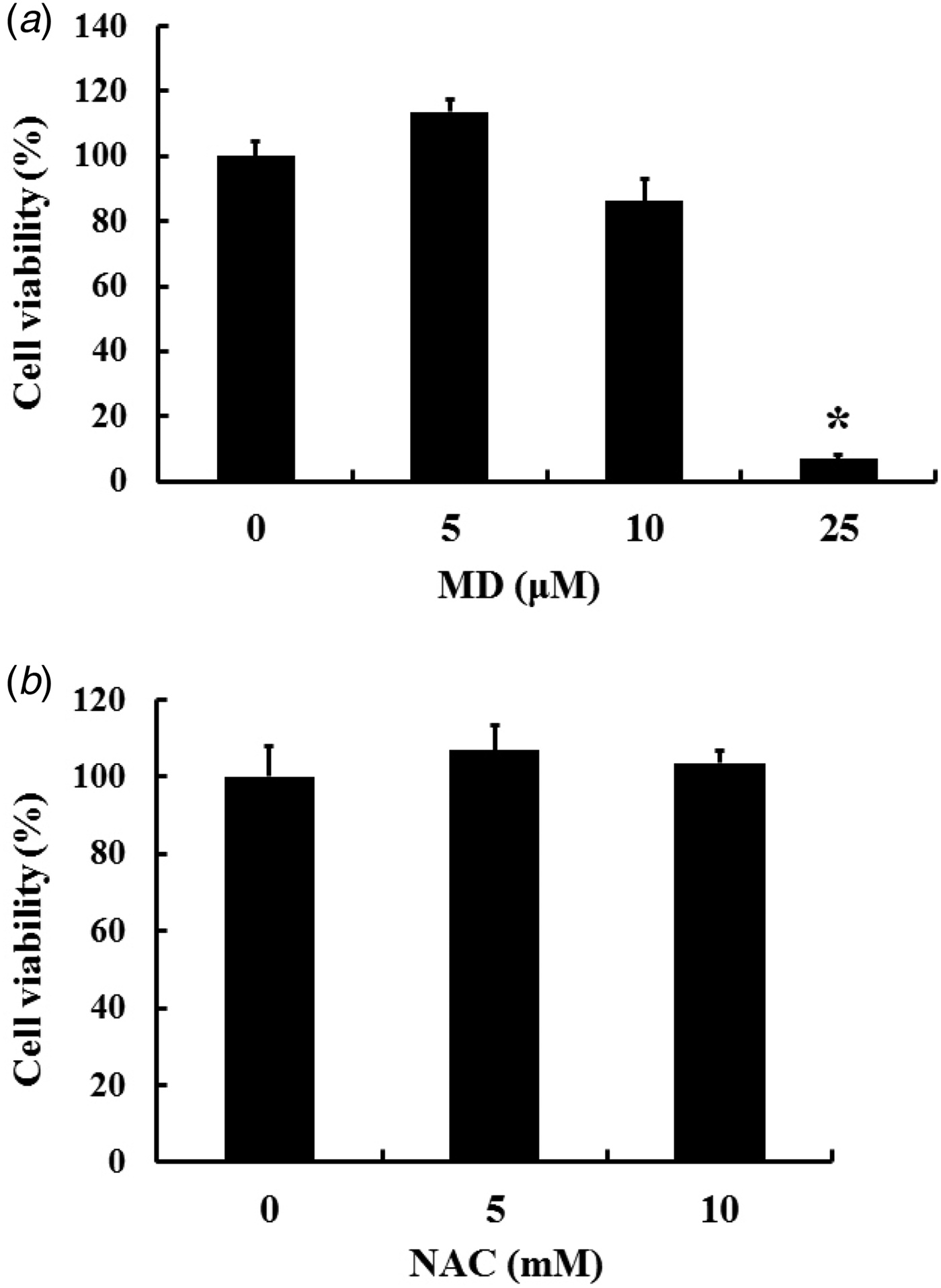
Bovine mammary alveolar epithelial cells (MAC-T) were maintained in Dulbecco's modified Eagle's medium supplemented with 10% FBS, penicillin/streptomycin, 5 µg/ml insulin, and 1 µg/ml progesterone at 37 °C in a humidified atmosphere containing 5% CO2. Cells were grown to approximately 90% confluency and synchronised overnight in medium containing 1% FBS before initiation of cell treatments. Cells were treated with MD at concentrations of 5 and 10 µm for 6 h. To observe the effects of antioxidants, cells were pretreated with NAC at concentrations of 5 and 10 mm for 1 h. Concentrations of NAC and MD were chosen based on the cell viability data presented in this study (Fig. 1). To investigate the effects of pharmacological inhibitors, cells were pretreated with the designated inhibitor (PD98059, LY294002, or BAY11-7082) at various concentrations for 1 h, followed by treatment with MD for 6 h. A lower concentration of MD (5 µm) was used because co-exposure of pharmacological inhibitors and MD affected cell viability.
Cytotoxicity of MD and NAC in MAC-T cells. Cells were treated with various concentrations of (a) MD (0–25 µm, 6 h) and (b) NAC (0–10 mm, 24 h). Trypan blue dye exclusion assay was used to determine the viability of cells. Experiments were repeated four times. Data represent the mean ± se. * Significantly different compared to control (P < 0·05).

Cell viability assay
Trypan blue dye exclusion assay was implemented to determine cell viability. Cells grown in 6-well plates were treated with MD (0, 5, 10, or 25 µm) for 6 h and NAC (0, 5, or 10 mm) for 24 h. After treatments, cells were trypsinised and centrifuged. The cell pellets were resuspended in warm PBS. The number of viable cells was determined by trypan blue dye exclusion and counted manually using a haemocytometer.
Cell lysate preparation
To prepare protein samples, cells were harvested in RIPA-buffer containing 50 mm Tris (pH 8·0), 150 mm NaCl, 1% triton X-100, 0·5% sodium deoxycholate, 0·1% SDS, and protease inhibitors (2 µg/ml aprotinin, 10 µg/ml leupeptin, 1 µg/ml pepstatin A, 1 mm PMSF, 5 mm EDTA, 1 mm EGTA, 10 mm sodium fluoride, and 1 mm sodium orthovanadate). Lysed cells were centrifuged at 18 000 g for 20 min at 4 °C. The supernatants were collected and concentrations of protein samples were quantitated using the Bradford protein assay (Sigma-Aldrich, St. Louis, MO) at 595 nm using a spectrophotometer (Biotek, Winoski, VT). The protein samples were stored at −80 °C until use.
Assessment of intracellular superoxide levels
Cellular superoxide production was measured as described previously (Kang et al. Reference Kang, Lee, Lee, Petriello, Kim, Do, Lim, Lee and Han2016). Briefly, MAC-T cells were grown to confluence on a cover glass in 6-well plates. Cells were treated with NAC (5 or 10 mm) for 1 h, followed by MD (10 µm) for 6 h. Following these treatments, cells were incubated with a final concentration of 1 µm DHE in a 5% CO2 incubator for 30 min. Cells were washed 3 times with PBS, fixed with 4% formaldehyde and then washed with PBS. Slides were mounted with ProLong Gold Antifade reagent containing DAPI (Invitrogen, Carlsbad, CA) to visualise the nuclei. Slides were estimated under an Olympus IX71 fluorescence microscope and the images were captured digitally by an Olympus DP71 camera and DP controller software (Olympus Optical Co. Ltd, Tokyo, Japan). DHE is oxidised in the presence of ROS, especially superoxide anion, and the reaction stains cells a bright red.
Measurement of glutathione levels
GSH levels were measured as per Ellman's method using a GSH assay kit (Bioassay Systems, Hayward, CA). Briefly, cells were collected by centrifugation at 18 000 g for 5 min at 4 °C, lysed by sonication in 1 ml cold PBS containing 1 mm EDTA, and finally, centrifuged at 10 000 g for 15 min at 4 °C. The supernatant was used for the assay according to the manufacturer's instructions.
SDS-PAGE and western blot analysis
To investigate the level of protein expression, quantitated protein samples were separated using 10% SDS-PAGE and subsequently were transferred onto nitrocellulose membranes using a semi-dry gel transfer system (Bio-Rad, Hercules, CA). The membranes were blocked with 3% non-fat milk buffer for 2 h and incubated overnight at 4 °C with primary antibodies. After washing with TBST, the membranes were incubated with appropriate secondary antibodies conjugated with horseradish peroxidase at room temperature for 1·5 h and visualised using ECL detection reagents (Lugen, Seoul, Korea). Bands were quantified using ImageJ software and normalised to housekeeping proteins.
Nuclear fractionation
Cells grown in 10-cm plates were treated with NAC (5 and 10 mm) for 1 h or MD (10 µm) for 6 h. After treatment, cells were harvested and lysed with hypotonic buffer solution (20 mm Tris [pH 7·4], 10 mm NaCl, and 3 mm MgCl2) containing protease inhibitor cocktail. Triton-X 100 (10%) was added to cell lysates, which were then centrifuged at 650 g for 10 min at 4 °C. The supernatant was collected as the cytosolic fraction and the pellet was resuspended in cell extraction buffer (100 mm Tris [pH 7·4], 100 mm NaCl, 1% Triton X-100, 10% glycerol, and 0·1% SDS) containing protease inhibitor cocktail. The homogenates were centrifuged at 14 000 g for 30 min at 4 °C and the supernatant was collected as the nuclear fraction. The cytosolic and nuclear fractions were stored at −80 °C until use. The nuclear fraction was analysed by SDS-PAGE and western blot for NF-κB p65 nuclear translocation. PCNA, a nuclear fraction-specific protein, was used as a loading control.
Statistical analysis
Data were expressed as mean ± standard error of the mean (sem). Statistical significance was determined with SPSS-PASW statistics software version 18·0 for Windows (SPSS, Chicago, IL). One- or two-way ANOVA was performed and treatment groups were compared using Tukey post-hoc test. A probability value of P < 0·05 was considered statistically significant.
Results
Effect of MD and NAC on cell viability
In the present study, cells were treated with MD to produce oxidative stress in MAC-T cells. Trypan blue dye exclusion assay was used to determine the appropriate concentrations of MD and NAC. MAC-T cells were treated with various concentrations of MD (0–25 µm for 6 h) and NAC (0–10 mm for 24 h). MD is a quinone that generates intracellular ROS, especially superoxide anion, through a redox cycle. We treated cells with MD to produce cellular oxidative stress, a compound used by several other studies to generate cellular oxidative stress in various epithelial cells (Torres-Gonzalez et al. Reference Torres-Gonzalez, Gawlowski, Kocalis, Scott and Dillmann2014; Deferme et al. Reference Deferme, Briede, Claessen, Cavill and Kleinjans2015). When MAC-T cells were treated with MD, there were no significant cytotoxic effects at concentrations up to 10 µm (Fig. 1a). A significant increase in cell death was observed at 25 µm MD (Fig. 1a). NAC did not affect the cell viability at all concentrations tested (Fig. 1b). Based on these data, cells were treated with up to 10 µm MD and 10 mm NAC in the subsequent experiments.
MD-induced oxidative stress in MAC-T cells and protective effects of NAC
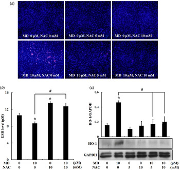
To investigate whether MD can produce oxidative stress in MAC-T cells, intracellular production of superoxide anion was determined. Substantial amount of superoxide (red fluorescence) was observed after treatment of cells with MD (10 µm for 6 h) (Fig. 2a). Pretreatment of cells with NAC (5 and 10 mm for 1 h) prevented MD-induced superoxide production (Fig. 2a). These data suggest that MD is an appropriate agent for producing superoxide anion in MAC-T cells. To determine whether other mechanisms are involved in MD-induced oxidative stress, cellular GSH levels were measured. Treatment of cells with MD decreased cellular GSH levels while supplementation of NAC restored cellular GSH levels (Fig. 2b). These data suggest that MD may produce oxidative stress through generation of superoxide and depletion of GSH, whereas NAC may prevent oxidative stress in cells by decreasing superoxide and restoring cellular GSH. HO-1 is an enzyme involved in cellular antioxidant and cytoprotective mechanisms. Generally, HO-1 is upregulated in oxidative stress environments (Loboda et al. Reference Loboda, Damulewicz, Pyza, Jozkowicz and Dulak2016). Our results show that treatment of cells with MD (10 µm for 6 h) increased HO-1 expression and pretreatment of NAC (5 and 10 mm for 1 h) attenuated HO-1 expression (Fig. 2c). These data suggest that oxidative stress produced by MD can initiate cellular antioxidant mechanisms and NAC can prevent this defensive mechanism by removing oxidative stress.
MD-induced intracellular oxidative stress in MAC-T cells. (a) Production of superoxide in MAC-T cells. Cells were pretreated with NAC (5 and 10 mm, 1 h), followed by incubation with MD (10 µm, 6 h). Cells were stained with DHE to detect superoxide production. The intensity of red fluorescence (superoxide production) was assessed using a fluorescence microscope. The nuclei were stained with DAPI. The images shown here are representative of three independent experiments. (b) MD-induced intracellular GSH changes in MAC-T cells. Cells were pretreated with NAC (10 mm, 1 h), followed by treatment of MD (10 µm, 2 h). Intracellular expression of GSH was measured with a GSH assay kit. Results represent the mean ± se. Experiments were repeated three times. (c) HO-1 expression in MAC-T cells. Cells were pretreated with NAC (5 and 10 mm, 1 h), followed by treatment with MD (10 µm, 6 h). Western blot analysis was used to measure protein expression in whole cell lysates. Results represent the mean ± se. Experiments were repeated three times. *Indicates a significant difference vs. control (P < 0·05). #Indicates a significant difference vs. MD only (P < 0·05).

MD-induced COX-2 expression and the protective role of NAC
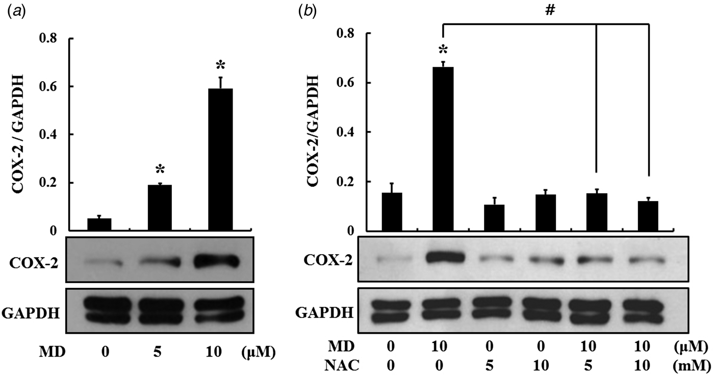
Oxidative stress is known to trigger inflammatory responses in cells. To determine whether MD-induced oxidative stress can induce an inflammatory response, MAC-T cells were treated with MD (5–10 µm) for 6 h. Since NF-κB, a redox sensitive transcription factor, is known to regulate COX-2 genes, protein expression of COX-2 was observed as an indicator of oxidative stress-induced inflammatory responses. Treatment of cells with MD markedly increased COX-2 expression in a dose-dependent manner (Fig. 3a). Next, in order to identify if antioxidant substances can modulate COX-2 expression, cells were treated with NAC (5 and 10 mm) for 1 h prior to MD treatment (10 µm for 6 h). We found that NAC inhibited MD-induced COX-2 expression (Fig. 3b). These results show that NAC can protect MAC-T cells against inflammatory responses caused by oxidative stress.
MD-induced COX-2 expression in MAC-T cells. (a) Cells were treated with MD (5 and 10 µm, 6 h). (b) Cells were pretreated with NAC (5 and 10 mm, 1 h), followed by treatment with MD (10 µm, 6 h). Western blot analysis was used to measure protein expression in whole cell lysates. Results represent the mean ± se. Experiments were repeated three times. *Indicates a significant difference vs. control (P < 0·05). #Indicates a significant difference vs. MD only (P < 0·05).

MD-induced activation of ERK1/2, Akt, and NF-κB, and the protective role of NAC
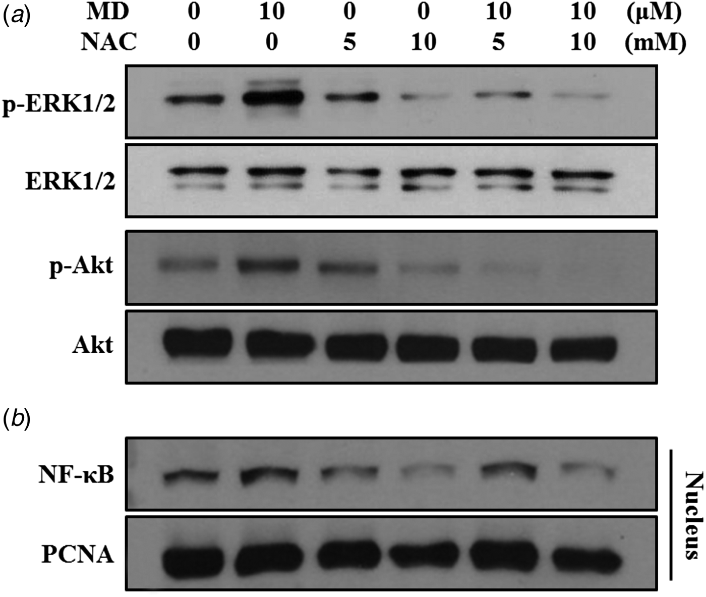
ROS, such as superoxide anion, can activate several intracellular signalling molecules, including ERK1/2, Akt, and NF-κB. Previous studies have reported that these signalling molecules are associated with inflammatory responses, including COX-2 expression (Kang et al. Reference Kang, Lee, Lee, Petriello, Kim, Do, Lim, Lee and Han2016). To identify the mediators of MD-induced COX-2 expression and the role of NAC, activation of cell signalling molecules (e.g., ERK1/2, Akt, and NF-κB) were examined in cells. Cells were treated with NAC for 1 h, followed by MD treatment for 1 h. Results showed that MD significantly increased phosphorylation ERK1/2 and Akt, while pretreatment with NAC diminished phosphorylation of these proteins (Fig. 4a). NF-κB is a transcription factor that is linked to activation of RNA transcription of many inflammatory response genes, including COX-2. Activation of NF-κB was determined by measuring the amount of NF-κB p65 in the nuclear fraction. MD significantly increased nuclear translocation of NF-κB, whereas NAC attenuated the observed nuclear translocation (Fig. 4b). These results suggest that NAC can decrease oxidative stress-induced activation of ERK1/2, Akt, and NF-κB, and these intracellular molecules are highly associated with inflammatory responses in MAC-T cells.
MD-induced activation of ERK1/2, Akt, and NF-κB in MAC-T cells. (a) Cells were pretreated with NAC (5 and 10 mm, 1 h), followed by treatment with MD (10 µm, 1 h). Western blot analysis was used to measure phosphorylation of the signalling molecules. (b) Cells were pretreated with NAC (5 and 10 mm, 1 h), followed by MD treatment (10 µm, 1 h). Nuclear translocation of NF-κB was determined in the nuclear fraction. PCNA was used as the loading control. The western blots shown here are representative images of three independent experiments.

Pharmacological inhibition of ERK1/2, Akt, and NF-κB, and COX-2 expression
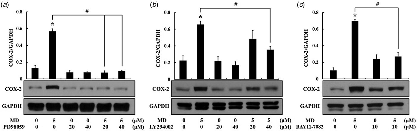
In order to confirm the association of ERK1/2, Akt, and NF-κB in MD-induced COX-2 expression, cells were treated with pharmacological inhibitors for ERK1/2 (PD98059), Akt (LY294002), and NF-κB (BAY11-7082). MD-induced COX-2 expression was significantly decreased when cells were treated with PD98059, LY294002, and BAY11-7082 (Fig. 5a–c). These results demonstrate that ERK1/2, Akt, and NF-κB can mediate MD-induced inflammatory responses, such as COX-2 expression.
Pharmacological inhibition of signalling molecules results in the decrease of COX-2 expression. MAC-T cells were pretreated with inhibitors for (a) ERK1/2 (PD98059), (b) Akt (LY294002), and (c) NF-κB (BAY11-7082) for 1 h, followed by incubation with MD (5 µm, 6 h). COX-2 expression was measured in whole cell lysates using western blot analysis. Results represent the mean ± se. Experiments were repeated three times. *Indicates a significant difference vs. control (P < 0·05). #Indicates a significant difference vs. MD only (P < 0·05).

Discussion
Since the production of a high milk yield is known to produce oxidative stress and may result in mastitis in dairy cows (O'Reilly et al. Reference O'Reilly, Green, Peeler, Fitzpatrick and Green2006; Sordillo & Aitken, Reference Sordillo and Aitken2009), we hypothesised that cellular oxidative stress increases inflammatory responses in bovine mammary cells. Oxidative stress-induced inflammatory responses have not been fully identified in bovine mammary alveolar cells. To examine the hypothesis, we generated ROS (i.e., superoxide anion) and investigated resulting inflammatory responses in bovine mammary alveolar cells (MAC-T). Our results support our hypothesis that oxidative stress can increase inflammatory responses in MAC-T cells and a decrease of oxidative stress can attenuate such responses.
In the present study, cells were treated with MD to produce oxidative stress through the generation of superoxide anion, a compound that has been widely used to generate intracellular ROS in past studies (Torres-Gonzalez et al. Reference Torres-Gonzalez, Gawlowski, Kocalis, Scott and Dillmann2014). Consistent with previous studies, we found that MD generated a great amount of cellular ROS. Pretreatment of cells with the antioxidant NAC eliminated MD-induced ROS in cells. Similar results have been reported in past studies where NAC reduced high glucose-induced ROS and MD-induced ROS production in cultured cells (Kim et al. Reference Kim, Shin, Sohn and Lee2014b). Such antioxidant effects of NAC are due to its role as a precursor of cellular cysteine and GSH as well as its ROS scavenging effects (Sadowska et al. Reference Sadowska, Manuel-Y-Keenoy and De Backer2007). Therefore, NAC has been used as an antidote for GSH deficiency and a general antioxidant (Jeong et al. Reference Jeong, Seok, Petriello and Han2017). In the present study, intracellular GSH levels increased in cells treated with NAC. In addition, pretreating cells with NAC reversed MD-induced reduction of GSH levels. To further test the effects of oxidative stress in MD-treated cells, the modulation of antioxidant defensive systems was investigated. Oxidative stress often stimulates cells to initiate their antioxidant defence systems (Loboda et al. Reference Loboda, Damulewicz, Pyza, Jozkowicz and Dulak2016). HO-1 is a representative antioxidant protein that counteracts many environmental insults, including oxidative stress and inflammatory responses (Ryter et al. Reference Ryter, Alam and Choi2006). Our results demonstrate that expression of HO-1 increased in cells treated with MD. Therefore, cellular defence machinery was activated to adapt to oxidative stress. ROS production that exceeds host antioxidant capacity induces oxidative stress and inflammatory responses (Poławska et al. Reference Poławska, Bagnicka, Niemczuk and Lipińska2012; Wiegman et al. Reference Wiegman, Michaeloudes, Haji, Narang, Clarke, Russell, Bao, Pavlidis, Barnes and Kanerva2015). COX-2 is an inflammatory mediator that converts arachidonic acid into prostaglandin E, a protein involved in inflammatory responses. Upregulation of COX-2 expression is found in cellular inflammatory responses (Ricciotti & FitzGerald, Reference Ricciotti and FitzGerald2011). Furthermore, overexpression of COX-2 was also observed in cells stimulated by LPS and mastitis-inducing bacteria such as Staphylococcus aureus (De & Mukherjee, Reference De and Mukherjee2014; Kang et al. Reference Kang, Lee, Lee, Petriello, Kim, Do, Lim, Lee and Han2016). Thus, COX-2 activation is a reliable end-point in cells suffering from inflammation. Our data also showed that COX-2 expression increased in cells treated with 5–10 µm MD in a dose-dependent manner. This suggests that inflammatory responses can be increased by oxidative stress. In contrast, decrease of oxidative stress by pretreating cells with NAC decreased MD-induced COX-2 expression. Similarly, a previous report demonstrated that ROS produced by Propionibacterium acnes increased expression of COX-2, and NAC reduced COX-2 expression in cells (Tsai et al. Reference Tsai, Lee, Wang, Cheng, Chen and Shen2013). Expression of COX-2 is generally mediated by the NF-κB pathway (Zha et al. Reference Zha, Chen, Sun, Mao, Chu, Deng, Cai, Li, Liu and Cao2014). As shown in the results of our study, NF-κB was significantly activated by MD, which upregulated expression of COX-2. MD-induced superoxide production and the subsequent cellular oxidative stress is closely associated with the activation of NF-κB in MAC-T cells. NF-κB, a redox-sensitive transcription factor, is regulated by intracellular GSH level (Rahman & MacNee, Reference Rahman and MacNee2000). Deficiency of intracellular GSH can activate NF-κB, while sufficient intracellular GSH can inhibit activation of NF-κB (Rahman & MacNee, Reference Rahman and MacNee2000). Thus, in the present study, the inactivation of NF-κB is mainly due to providing NAC, a precursor of intracellular GSH in MAC-T cells.
Further, intracellular signalling mechanism linked to oxidative stress-induced inflammation was investigated in MAC-T cells. Phosphorylation of cell signalling molecules such as ERK1/2 and Akt are known to be linked to the activation of NF-κB (Chung et al. Reference Chung, Kim, Na, Kim, Kim, Haegeman and Surh2014; Kim et al. Reference Kim, Kim, Kim, Lee, Kundu, Na, Cha and Surh2014a). Previously, it was shown that COX-2 expression was suppressed in mammary epithelial cells when blocking ERK1/2 or NF-κB pathways (Chung et al. Reference Chung, Kim, Na, Kim, Kim, Haegeman and Surh2014). According to our previous reports, inhibition of pathways for Akt and ERK1/2 attenuated LPS-induced COX-2 expression in MAC-T cells (Kang et al. Reference Kang, Lee, Lee, Petriello, Kim, Do, Lim, Lee and Han2016). In the same context, our results also showed that phosphorylation of ERK1/2 and Akt was increased in cells treated with MD and these signalling molecules were responsible for COX-2 expression. These results were subsequently confirmed in cells treated with pharmacological inhibitors for ERK1/2, Akt, and NF-κB, where inhibition of these molecules significantly decreased MD-induced COX-2 expression. The overall intracellular mechanisms occurred by oxidative stress in MAC-T cells were illustrated in Supplementary Figure S1.
In conclusion, our results demonstrate that oxidative stress (e.g., superoxide production) can activate inflammatory signalling cascades such as ERK1/2, Akt, and NF-κB, and ultimately upregulate COX-2 expression in bovine mammary alveolar epithelial cells. COX-2 is a sensitive biomarker in oxidative stress-induced inflammatory responses in this cell type. In addition, our research elucidated antioxidant and anti-inflammatory effects of NAC in bovine mammary alveolar cells. These results suggest that decrease of oxidative stress may result in reduced risk of inflammation in the udder of lactating cows. The study may be relevant to mastitis in high producing cows from which no pathogen can be isolated.
Supplementary material
The supplementary material for this article can be found at https://doi.org/10.1017/S002202991700067X.
This paper was supported by Konkuk University in 2017.
Disclosure
The authors declare no conflict of interest.