INTRODUCTION
Several authors consider Doris verrucosa a highly widespread species, with the geographic range including the Atlantic coast of Europe, the Mediterranean Sea to the Canary Islands, and the Western Atlantic (e.g. Marcus, Reference Marcus1955; Marcus & Marcus, Reference Marcus and Marcus1967; Schmekel, Reference Schmekel1968; Schmekel & Portmann, Reference Schmekel and Portmann1982; Thompson & Brown, Reference Thompson and Brown1984; Just & Edmunds, Reference Edmunds and Just1985; Valdés, Reference Valdés2002; Valdés et al., Reference Valdés, Hamann, Behrens and Dupont2006; Garcia et al., Reference Garcia, Álvares and Troncoso2008).
Two distinct species reported from Brazil are related to Doris L, 1758. To the south, in Santa Catarina, Morretes (Reference Morretes1949) reported Doridigitata derelicta Fischer, Reference Fischer1867, a species described from Arcachon Basin (France). Earlier, Bergh (Reference Bergh1878) described Staurodoris januarii from Rio de Janeiro. However, von Ihering (Reference von Ihering1886, Reference von Ihering1915) considered the latter as a synonym of D. verrucosa, and this opinion was accepted by Bergh (Reference Bergh1904) (Marcus, Reference Marucs1957) and by subsequent authors (Bouchet & Valdés, Reference Bouchet and Valdés2000; Valdés, Reference Valdés2002).
Bouchet & Valdés (Reference Bouchet and Valdés2000) reviewed the nomenclature of D. verrucosa, from the original description by Linnaeus (Reference Linnaeus1758) up to recent papers. They proposed the conservation of the generic and specific names, and designated a neotype, maintaining the species with an Atlantic/European distribution. Recent field guides (Valdés et al., Reference Valdés, Hamann, Behrens and Dupont2006; Garcia et al., Reference Garcia, Álvares and Troncoso2008; Rios, Reference Rios2009) considered D. verrucosa an amphi-Atlantic species.
In this paper we perform a detailed anatomical study of what has been considered Doris verrucosa, including specimens collected on the Atlantic coast of Spain, Mediterranean and Brazil. The identity of another previously described species from Brazil (type locality in Rio de Janeiro), Doris januarii (Bergh, Reference Bergh1878), is also investigated in order to solve the systematics of the Doris verrucosa species complex in the Atlantic and Mediterranean. This paper is the first step of a larger project dealing with the revision of Doris in the Western Atlantic.
MATERIALS AND METHODS
All specimens were preserved in 70% ethanol. Dissections were performed under a stereomicroscope by standard techniques, with the specimens immersed in fixative (Simone, Reference Simone2004, Reference Simone2011). The initial steps of the anatomical investigation were done through a longitudinal cut on the tissue covering the dorsal visceral mass. Digestive, circulatory, excretory, reproductive and central nervous systems were investigated in detail. The terminology used for odontophore muscles was based on Ponder et al. (Reference Ponder, Colgan, Healy, Nützel, Simone, Strong, Ponder and Lindberg2008) and Simone (Reference Simone2011). Digital photographs were taken at each dissection step. Drawings were done with the aid of a camera lucida. Scanning electron microscopy (SEM) was used to examine details of the radula at the Laboratório de Microscopia Eletrônica of Museu de Zoologia da Universidade de São Paulo (MZSP). Several specimens were also photographed prior to sacrifice.
The following abbreviations are used in the figures: aa: anterior aorta; ab: afferent branchial vein; ag: albumen gland; am: ampulla; ap: posterior aorta; ar: afferent branchial ring; au: auricle; at: aortic trunk; av: auricle vein; bc: bursa copulatrix; bg: blood gland; bs: buccal sphincter; bt: branchial tubercle; cb: buccal commissure; ce: cerebral ganglia; cg: connective buccal ganglia; cp: pedal commissure; cs: circular sinus; cu: caecum; cv: ctenidial vein; dd: duct of digestive gland; dg: digestive gland; ef: oesophageal folds; er: efferent branchial ring; es: oesophagus; ev: efferent branchial vein; ey: eye; fg: female gland; ft: foot; gb: buccal ganglia; gc: gill circle; ge: gonopore; gf: gill filament; gg: gastro-oesophageal ganglia; gp: pedal ganglia; hd: hermaphrodite duct; il: inner lip; in: intestine; le: lateral extension; mo: mouth; m2 – m10: odontophore muscles; mp: penis's muscle; ms: medial sinus; mt: oral tube muscle; oc: odontophore cartilage; ol: outer lip; ot: oral tube; ov: oviduct; pa: papilla; pc: pericardium; pe: penis; pl: pleural ganglia; pr: prostate; ra: radula; rc: renal chamber; rg: rhinophoral ganglia; ri: rhinophore; rm: retractor muscle gill; rp: reproductive system; rs: radular sac; rt: rhinophoral tubercle; rv: renal vesicle; sg: salivary gland. st: stomach; sn: nervous system; sr: seminal receptacle; ud: uterine duct; va: vagina; vb: buccal vein; vd: vas deferent; ve: ventricle; vp: reproductive system vein; vn: nervous system vein: vs: radular sac vein; vv: auricoventricular valve.
Institutional abbreviations
MNHN – Muséum National d'Histoire Naturelle, Paris, France.
MNRJ – Museu Nacional do Rio de Janeiro, Brazil.
MZSP – Museu de Zoologia da Universidade de São Paulo, São Paulo, Brazil.
ZMUC – Zoologisk Museum, Københavns Universitet, Copenhagen, Denmark.
SYSTEMATICS
Genus Doris Linnaeus, Reference Linnaeus1758
Type species: Doris verrucosa Linnaeus, Reference Linnaeus1758
Doris verrucosa Linnaeus, Reference Linnaeus1758
(Figures
1–8)
Neotype of Doris verrucosa (MNHN 24625), fixated specimen. (A) Dorsal view. (B) Ventral view. (C) Posterior region, gill circle detailed, dorsal view. Photos provided by MNHN, Paris.

Doris verrucosa (MNRJ 2416), fixated specimen. (A) Dorsal view. (B) Ventral view. (C). Anterior portion with detail for elongate tubercles around rhinophoral cavity, dorsal view. (D). Posterior portion, detail of gill circle, dorsal view. Scales: 2 mm.
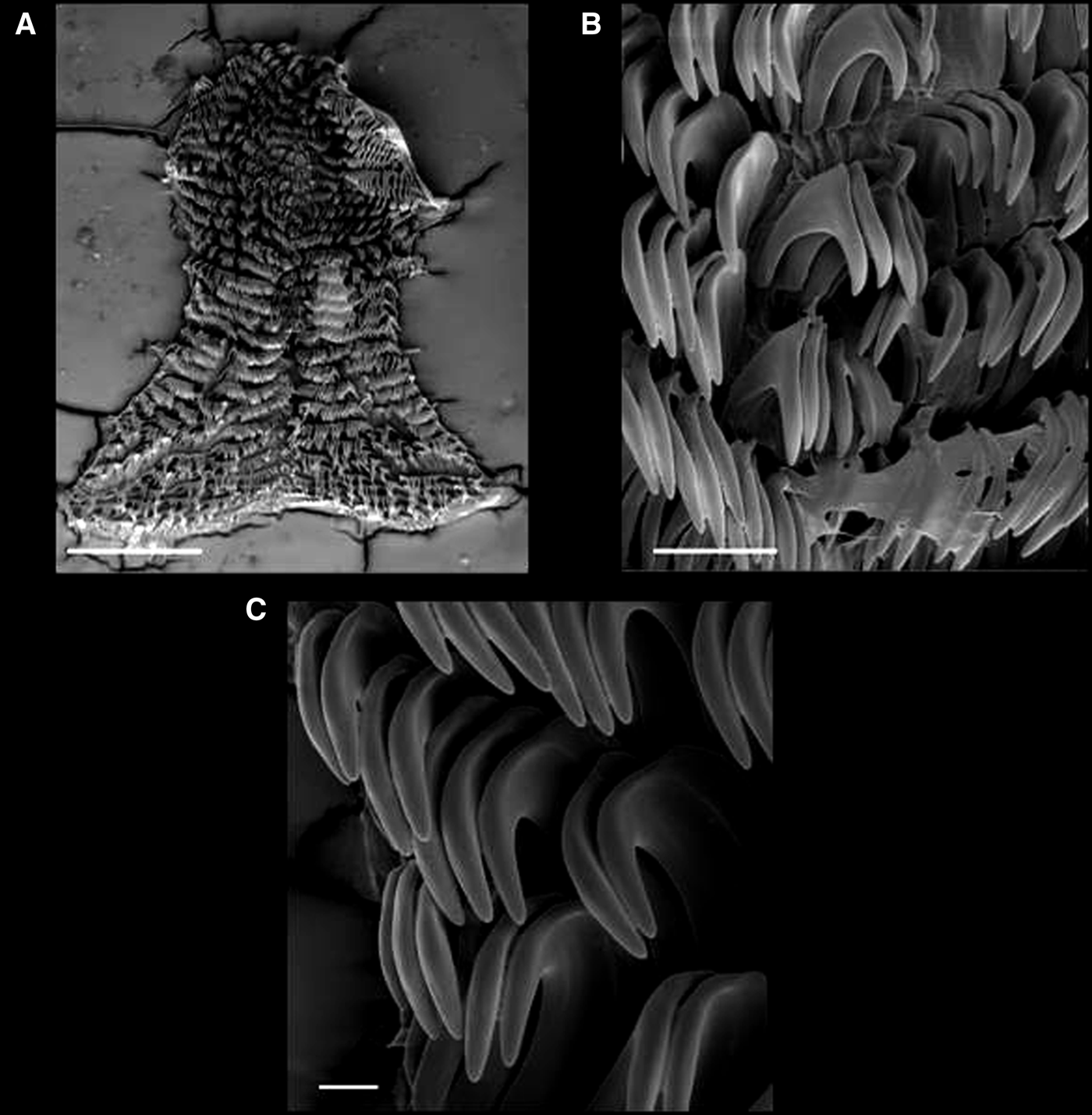
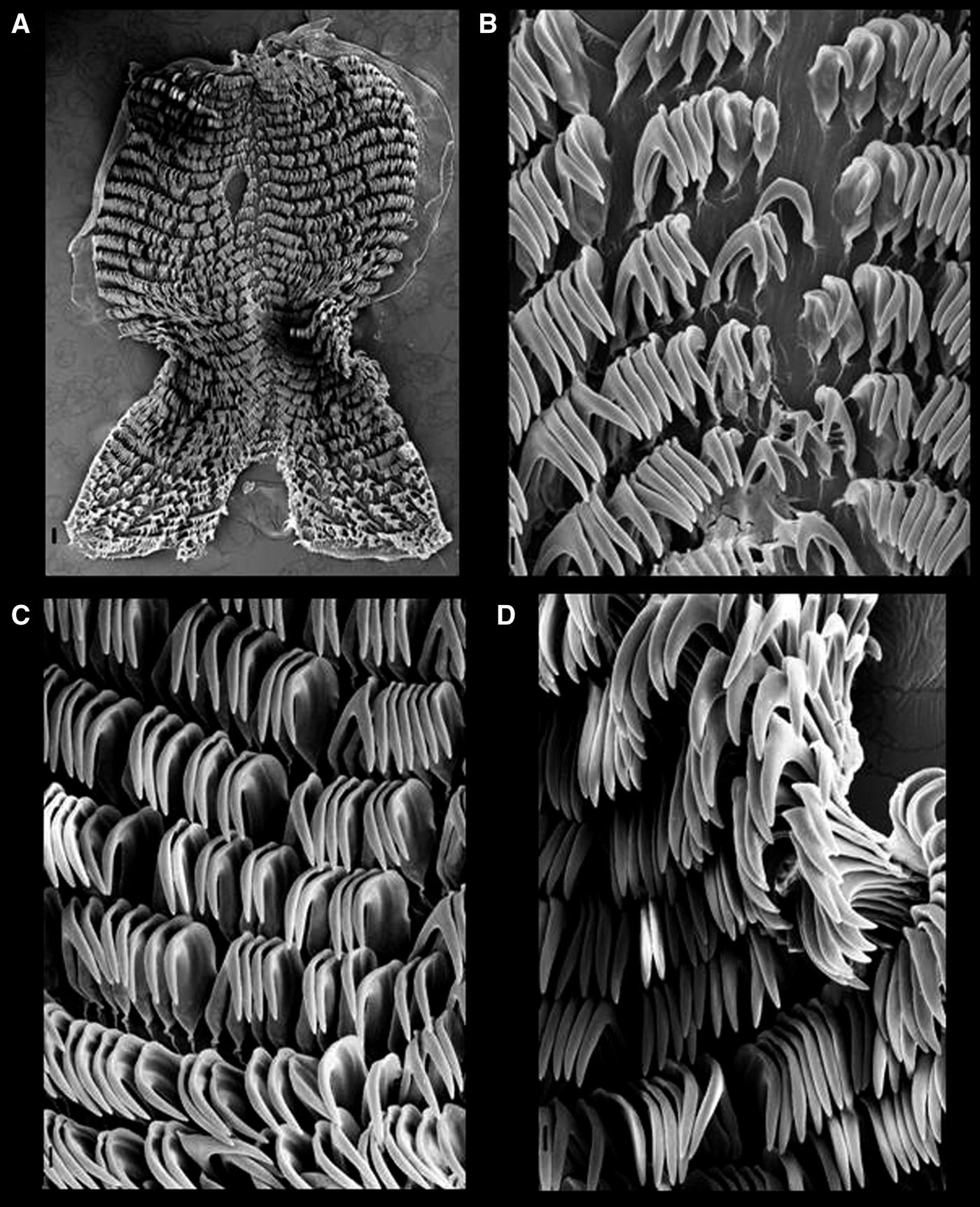
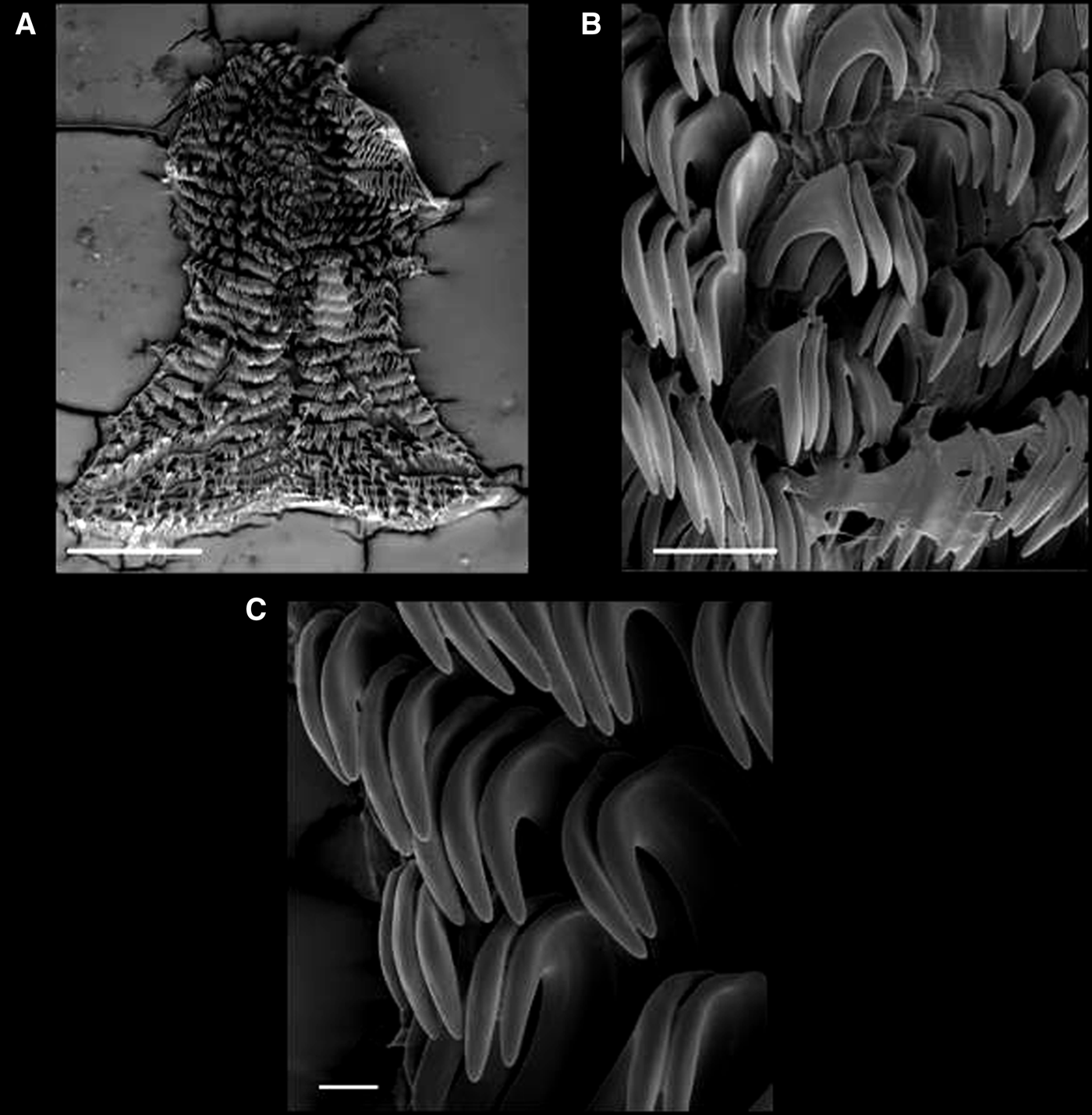
A–C: Doris verrucosa, radula in SEM (MZSP 99538). (A) Panoramic view. Scale: 500 μm. (B) Central view of innermost lateral teeth. Scale: 50 μm. (C) Outer lateral teeth. Scale bar: 20 μm.
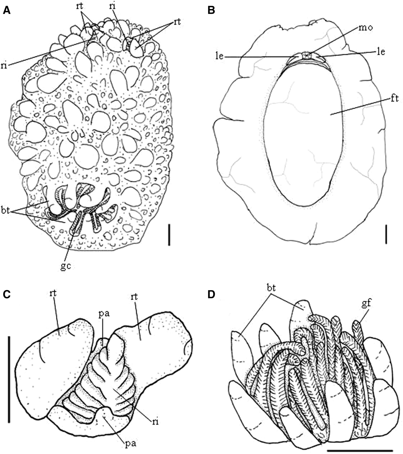
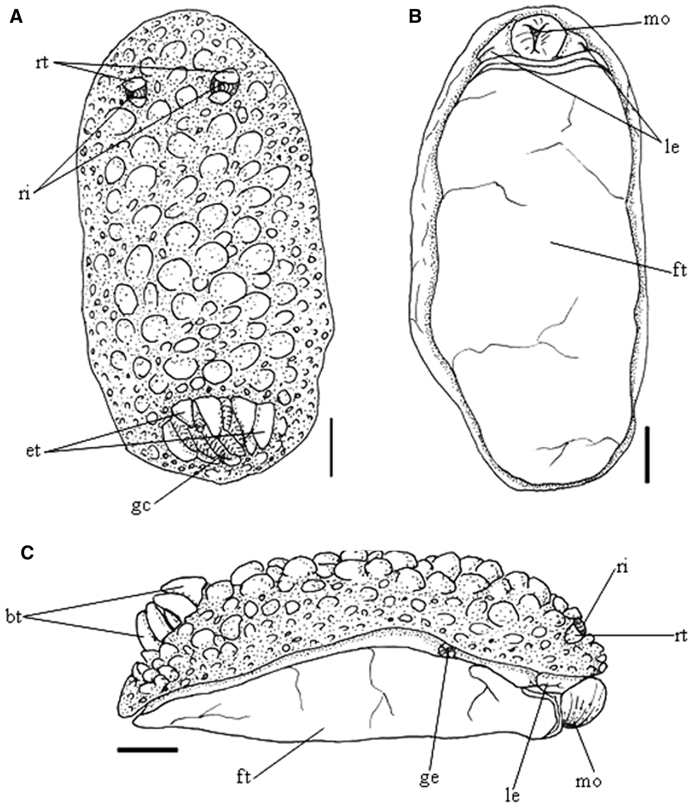
External morphology of Doris verrucosa. (A) Whole dorsal view. (B) Whole ventral view. Scale: 1 mm. (C) Rhinophoral cavity, dorsal view. (D) Gill circle, dorsal view. Scale bars: 0.5 mm.
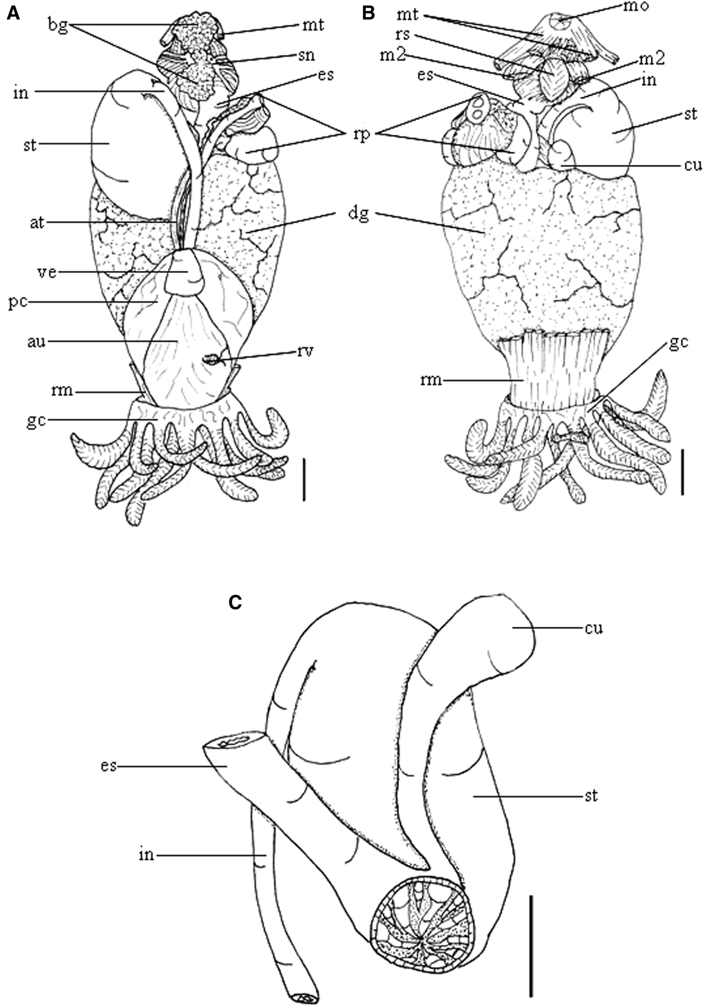
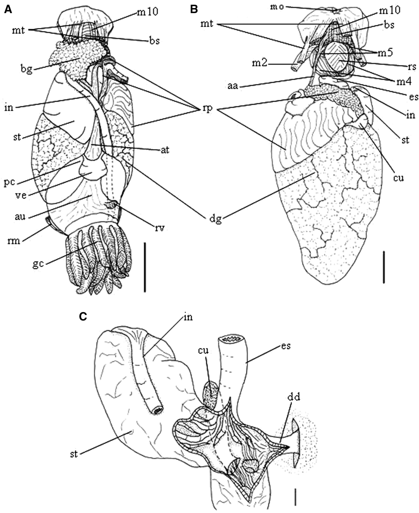
Visceral mass of Doris verrucosa, in situ. (A) Dorsal view. (B) Ventral view. (C) Medial digestive system, ventral view. Scale bars: 1 mm.
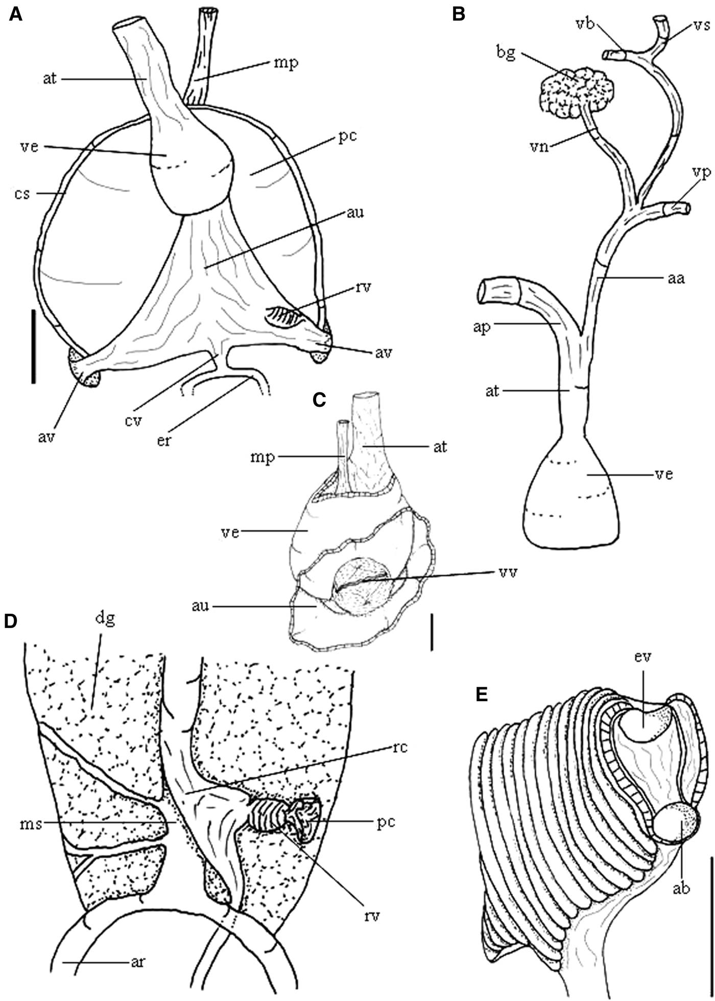
Doris verrucosa circulatory and excretory systems. (A) Organs involved by pericardium, dorsal view. (B) Main vessels in arterial circulation, dorsal view. (C) Detail of atrioventricular valve, posterior dorsal view. (D) Scheme of the organs of the excretory system, dorsal view. (E) Gill filament, transversally sectioned, apical side view, with highlighting of the afferent and efferent vessels. Scale bars: 1 mm.
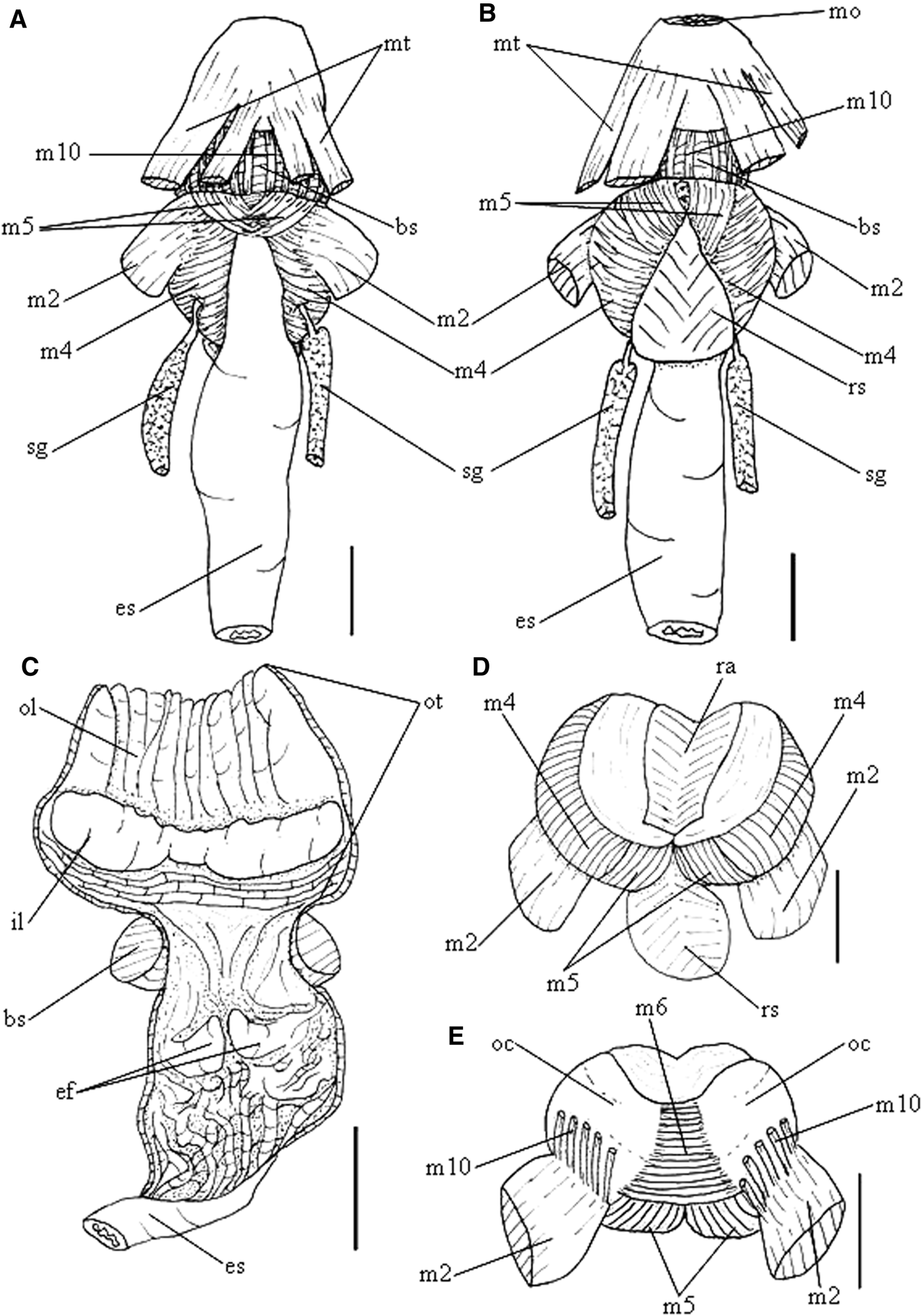
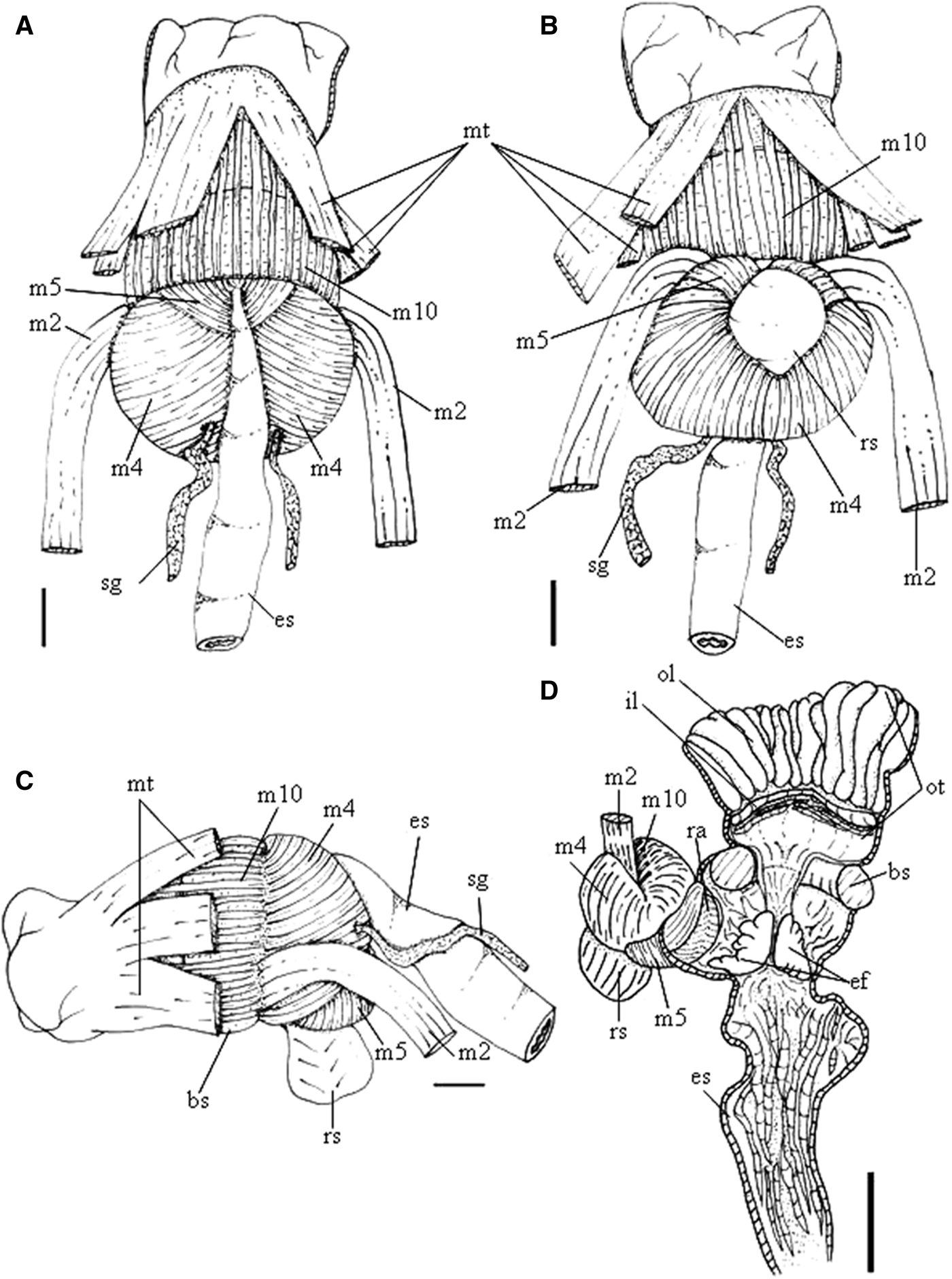
Doris verrucosa anterior digestive system. (A) Dorsal view. (B) Ventral view. (C) Longitudinally sectioned, ventral view odontophore removed. (D) Odontophore, ventral view, first layer of muscles and structures removed. (E) Same, dorsal view. Scale bars: 1 mm.
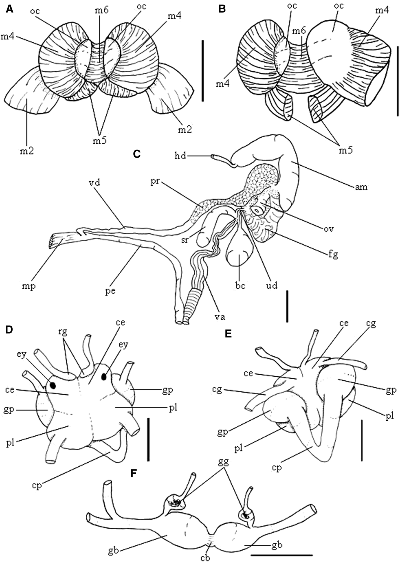
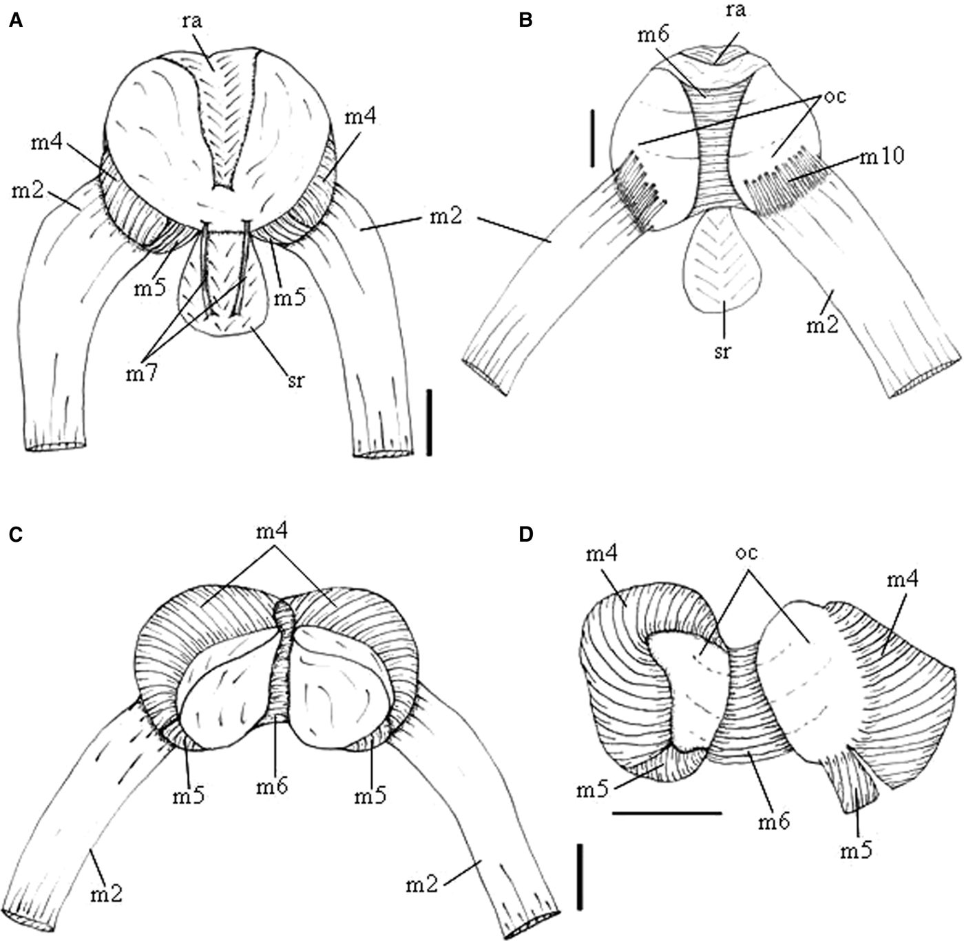
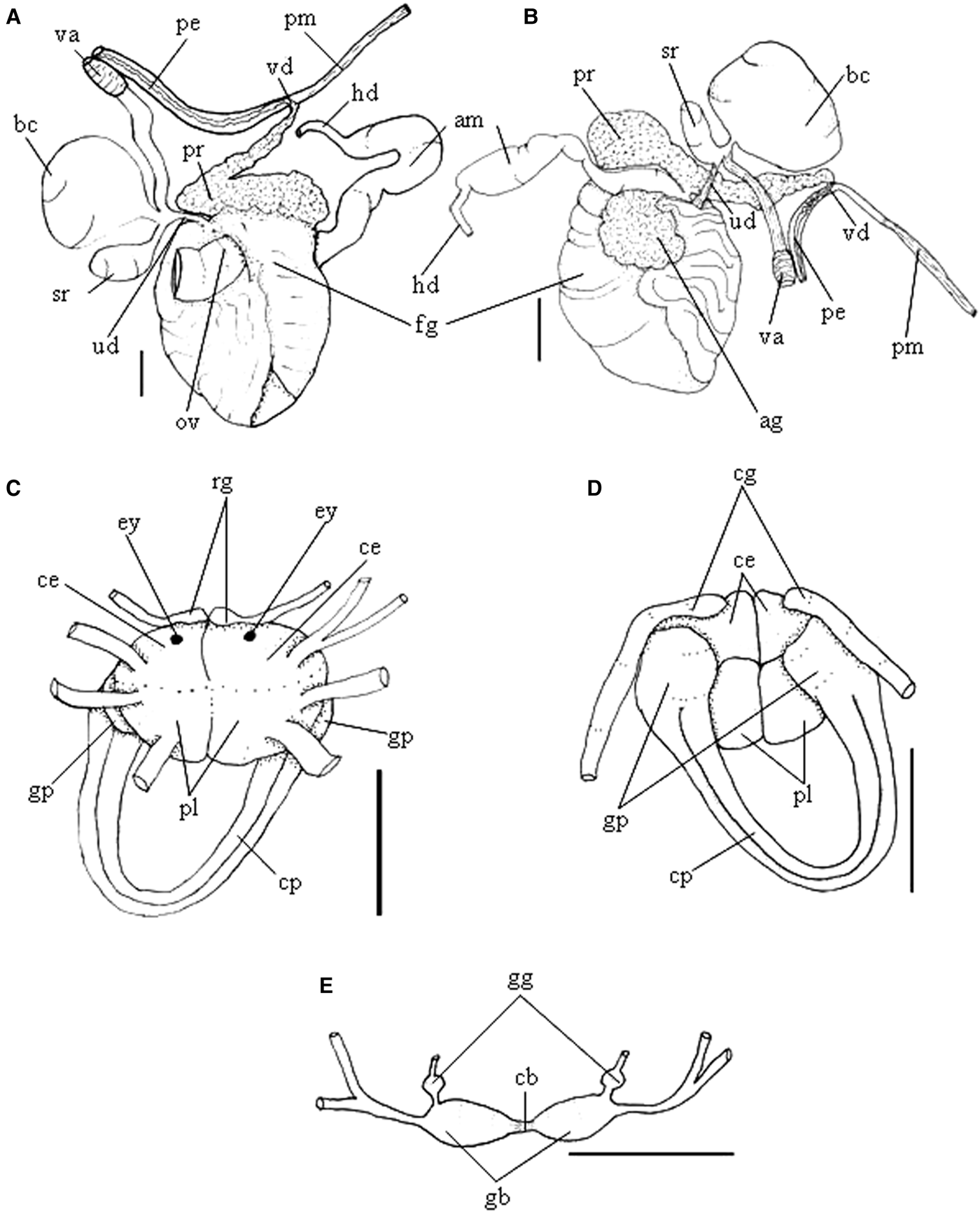
Doris verrucosa anatomy. (A) Odontophore, ventral view, radula removed, both cartilages deflected. (B) Same, ventral view, left m4 and m5 (right in figure) folded down to expose odontophore cartilage. (C) Reproductive system, dorsal view. (D) Nervous system, dorsal view. (E) Same, ventral view. (F) Detail of buccal and gastro-oesophageal ganglia, ventral view. Scale bars: 1 mm.
Doris verrucosa Linnaeus, Reference Linnaeus1758: 653; Reference Linnaeus1767: 1083; Pennant, Reference Pennant1777: 36; Cuvier, Reference Cuvier1804: 467 (pl. 1, figs 4–6); Rapp, Reference Rapp1827: 517; Delle Chiaje, Reference Delle Chiaje1828: 129 (pl. 38, figs 14, 23); Phillippi, Reference Phillippi1836: 104; d‘Orbigny, Reference Orbigny, Webb and Berthelot1839: 39; Fischer, Reference Fischer1867: 7; Eliot, Reference Eliot1910: 94; Pruvout-Fol, Reference Pruvot-Fol1934: 236; Marcus & Marcus, Reference Marcus and Marcus1967: 201 (fig. 1); Schmekel, Reference Schmekel1968: 169 (fig. 3); Franz, Reference Franz1970: 80; Marcus, Reference Marcus1972: 312; Marcus, Reference Marcus1977: 10; Schmekel & Portmann, Reference Schmekel and Portmann1982: 77; Bouchet & Valdés, Reference Bouchet and Valdés2000: 74; Valdés, Reference Valdés2002: 543 (figs 2–3); Valdés et al., Reference Valdés, Hamann, Behrens and Dupont2006: 168;
Doris derelicta Fischer, Reference Fischer1867: 7; Bouchet & Valdés, Reference Bouchet and Valdés2000: 75; Valdés, Reference Valdés2002: 546.
Doris biscayensis Fischer, Reference Fischer1872: 6; Valdés, Reference Valdés2002: 546.
Staurodoris verrucosa var. mollis Eliot, Reference Eliot1906: 338–339; Valdés, Reference Valdés2002: 546.
Staurodoris bobretzkii Gadzikiewicz, Reference Gadzikiewicz1907: 509; Valdés, Reference Valdés2002: 546.
Type material (D. verrucosa): neotype, MNHN 24625 (designated by Bouchet & Valdés, Reference Bouchet and Valdés2000).
Type locality (D. verrucosa): Castropol, Asturias, Atlantic Coast of Spain (Bouchet & Valdés, Reference Bouchet and Valdés2000).
Material examined: SPAIN, Western Andalusia, Sancti Petri, MZSP 99538, 2 specimens (L. Cervera coll., 29/vi/2010). ITALY, Naples, Gulf of Naples, MNRJ 2416, 3 specimens.
Redescription
External morphology (Figures 1, 2 & 4): Size 9-15-20 mm. Colour uniform yellowish or grey. Body oval, elongated, dorsum covered by tubercles of varying size and quantity; larger tubercles more elongated than rounded, located mainly in central dorsal region; tubercles decreasing in size toward mantle edge. Rhinophores with about 13 transverse lamellae, colour similar to body; rhinophoral sheaths with small papillae on anterior and posterior edges and two tubercles on left and right sides. Gill composed of 15–17 unipinnate branchial leaves, colour similar to body, arranged in circular fashion surrounding anus; branchial sheaths with eight tubercles, equidistant from each other, colour similar to body. Mouth opening in anterior ventral region, between anterior region of notum and foot. Digitiform tentacles absent. Lateral extensions of integument triangular, about half of size of mouth, each one with ventral furrow up to half of their extensions. Anterior border of foot grooved.
Haemocoel organs (Figure 5A, B): Pericardium and posterior half of visceral mass volume occupying ~1/3 of haemocoel. Buccal mass located anteriorly, occupying ~1/10 of haemocoel volume. Nervous system dorsal to buccal mass, covered by blood gland. Reproductive system on right side of animal; occupying ~1/10 of haemocoel volume. Stomach on left side, intestine with small curve at anterior portion, immersed in haemocoel, both occupying ~1/5 of haemocoel volume.
Circulatory and excretory systems (Figure 6A–E): Pericardial cavity dorsal and posterior to digestive gland, occupying ~1/2 of visceral mass volume, anterior to gill circle. Afferent and efferent branches located inside each gill filament, flowing from and to afferent and efferent branchial veins. Gill retractor muscle originating from base of gill circle, running longitudinally up to half of foot level, inserting into dorsal surface of foot. Auricle funnel-like (wider anteriorly) with thin walls. Ventricle slightly taller than wide, with thick muscular walls; auricle-ventricular valve clear, with two protruding tabs projected from auricle towards ventricle. There are three major vessels: (1) aortic trunk, located anteriorly to pericardium, connected to anterior ventricular region; (2) auricular vessels connecting lateral cavities of integument to auricle; (3) branchio-cardiac vessel located at posterior end of pericardium, connecting branchial efferent ring to auricle. Aortic trunk branched into anterior artery irrigating stomach, caecum and digestive gland; posterior artery irrigating reproductive system, buccal mass, odontophore and nervous system. Medial sinus connected to afferent branchial ring, irrigating entire digestive gland. Renal vesicle located on right dorsal side of pericardium, near base of auricle, connected to inner surface of pericardium; renal chamber elliptical, coloured light yellow, with longitudinal folds, ~1/4 the size of ventricle. Renal chamber extending from dorsal to medial sinus, previously connected to renal vesicle, extending posteriorly to centre of gill circle and opening in nephrostoma, next to base of anal papilla.
Digestive system (Figures 5C, 7A–E & 8A, B): Buccal mass occupying about 10% of visceral mass volume. Oral tube composed of outer lip, with pleats lengthwise; inner lip with transverse fold; mt, three pairs of retractor muscles of buccal mass, originating on oral tube, running dorsally and ventrally to oral tube, inserting on body side, about six times as wide and twice as long as m10. Odontophore oval, connected to oral tube by pair of ventral protractor muscles (m10); thin longitudinal, dorsal and ventrolateral protractors of oral sphincter, originating in anterior region of odontophore and inserted in posterior region of integument close to oral tube. Oral sphincter surrounding chitinous part of oral tube. Odontophore muscles: m2, pair of strong retractor muscles of buccal mass, occupying ~2/9 of odontophore, as wide as long, triangle-shaped, origin on anterior dorsal odontophore, running laterally to m4 and inserted ventrally on dorsal portion of foot; m4, pair of dorsal tensor muscles, strong and broad, occupying 4/9 of odontophore volume, 1/3 as long as wide, covering 2/3 of cartilage, inserted on ventral portion of subradular membrane; m5, pair of dorsal auxiliary tensor muscles, occupying 2/9 of odontophore volume, twice as long as wide, originating on most posterior region of odontophore cartilages, covering ~1/3 of posterior cavity of odontophore, as long as, but with ~1/2 of m4 width, inserting on ventral side of subradular membrane, around radular sac; m6, unpaired horizontal muscle, with transverse fibres connecting to median surface of left and right odontophore cartilages, occupying ~2/9 of length of odontophore, about same length and half as wide as m4, posterior portion ~twice as wide as anterior portion. Pair of odontophore cartilages elliptical, occupying ~2/9 of odontophore volume. Subradular membrane thin, strong, translucent. Radular sac ~1/3 as large as odontophore. Radular teeth (Figure 3A–C): rachidian teeth absent; formula 32 × 40.0.40 (in 9 mm long specimen). Each lateral tooth with broad base, tapering toward apex, hook-shaped, with single terminal cusp; outermost teeth thinner, inner base width about half of lateral teeth width, apex also hook-shaped, cusp simple and smooth. Pair of salivary glands long, tubular; duct inserting in anterior region of oesophagus, extending posteriorly to anterior region of digestive gland. Oesophagus simple, originating dorsally to odontophore, inserting directly in anterior region of stomach, internal longitudinal folds with same diameter along entire length. Stomach oval, ~30% of visceral mass volume, with folds at the centre of entire inner surface; longitudinal pleats thicker posteriorly, close to anterior region of intestine. Common opening for oesophagus, stomach and caecum located on digestive gland. Intestine with longitudinal folds along its entire length, diameter about half oesophagus diameter, but more uniform. Caecum as an elongated sac, located ventrally to stomach, opening on anterior portion of stomach, close to oesophageal insertion, ~1/2 length and ~1/3 of width of stomach; typically containing dark brown substance. Digestive gland dark beige, being largest organ of visceral mass and occupying ~40% of its volume, cone-shaped; anterior portion about twice as wide as posterior portion, inner face of gland sponge-like, bearing various ducts and a distinct main duct. Anus opening into anal papilla at centre of gill circle, ~1/6 of gill filament length.
Genital system (Figure 8C): Occupying ~10% of visceral mass volume, located between buccal mass and digestive gland, mainly on right side and dorsally. Genital opening on right side, anterior third of length of animal from head, located between foot and notum. Hermaphrodite duct thin, long. Ampulla located on female gland, elongated and tubular. Prostate tubular, glandular, length same as ampulla. Vas deferens about same length as penis, connected to penis's muscle. Penis's muscle connected to ventral portion of pericardium, near ventricle, roughly same length as penis. Penis muscular, cylindrical and elongated, about 2/3 of length of prostate. Gonad immersed in digestive gland. Female gland well-developed, rounded, occupying ~15% of reproductive system volume, divided into mucus gland (~2/3 of female gland, colour beige), and albumen gland (~1/3 of anteriormost region, dilated, irregularly shaped, colour dark brown). Oviduct occupying ~1/3 of female gland volume. Uterine duct thin, relatively short, length ~1/30 of penis length, located at base of vagina, inserted in female gland near oviduct. Seminal receptacle pyriform, as large as bursa copulatrix, length ~1/3 of vagina length, connected to vagina near uterine duct through short stalk. Bursa copulatrix rounded, length ~1/3 of vagina length, connected to vagina after seminal receptacle, also through short stalk. Vagina cylindrical, elongated, approximately as long as and twice as wide as penis, followed ventrally by prostate and located parallel to penis in genital opening.
Nervous system (Figure 8D–F): Located dorsally to odontophore, occupying ~1/20 of haemocoel volume, mostly covered by blood gland. Pair of cerebral and pleural ganglia fused with each other dorsally and ventrally. Pedal ganglia fused with cerebral and pleural ventrally, but not fused among themselves. Pedal commissure simple, surrounding oesophagus and salivary glands. Buccal ganglia short, located ventrally to odontophore, between radular sac and anterior portion of oesophagus, connected to cerebral ganglia through long and slender connective tissue, united to gastro-oesophageal ganglia by short connective tissue. Gastro-oesophageal ganglia length about 1/3 of buccal ganglia length, circular. Rhinophoral (olfactory) ganglia bulb-shaped, connected to anterior portion of cerebral ganglia. Dorsal eyes located on cerebral ganglia. Statocysts small and iridescent, located ventrally to pedal ganglia.
Distribution: Atlantic Coast of Europe and Mediterranean Sea (confirmed to Spain and Naples, Italy).
Habitat: Rocky subtidal region.
Doris januarii (Bergh,Reference Bergh1878) comb.
nov.
(Figures
9–15)
Doris januarii colour variation, living specimens. (A) Rio Arinquidá, Tamandaré/PE (MNRJ 12932). (B) Praia das Conchas, Cabo Frio/RJ (MZSP 96899). (C) Canal de Itajurú, Cabo Frio/RJ (MZSP 96900). (D) Araçá, São Sebastião/SP (MZSP 102797). Scale bars: 2 mm.
Doris januarii, radula in SEM. (MZSP 31513). (A) Panoramic view. Scale bar: 100 μm. (B) Central view of innermost lateral teeth. (C) Inside view of lateral teeth. (D) More external lateral teeth. Scale bars: 20 μm.
Doris januarii external morphology. (A) Dorsal view. (B) Ventral view. (C) Lateral right view. Scale bars: 2 mm.
Doris januarii haemocoel structure. (A) Whole dorsal view as in situ. (B) Same, ventral view. Scales: 2 mm. (C) Midgut, ventral view, with opening to digestive gland. Scales: 1 mm.
Doris januarii foregut. (A) Dorsal view. (B) Ventral view. (C) Lateral view (anterior region left). (D) Ventral view, longitudinal cut, odontophore deflected to right. Scale bars: 2 mm.
Doris januarii odontophore. (A) Ventral view, superficial layer of structures removed. (B) Same, dorsal view. Scales: 1 mm. (C) Same, ventral view, radula removed, cartilages deflected. (D) Same, ventral view, left m4 and m5 (right in figure) folded down to expose odontophore cartilage. Scale bars: 2 mm.
Doris januarii reproductive and central nervous systems. (A) Genital structures, dorsal view. (B) Same, ventral view. Scale bars: 2 mm. (C) Central nervous system, dorsal view. (D) Same, ventral view. (E). Detail of buccal and gastro-oesophageal ganglia, ventral view; Scale bars: 1 mm.
Staurodoris januarii Bergh, Reference Bergh1878: 583, pl. 63, fig. 24, pl. 64, figs 8–12.
Doris verrucosa: von Ihering, Reference von Ihering1886: 230; Reference von Ihering1915: 142; Marcus, Reference Marcus1955: 127 (figs 102–108); 1957: 420 (fig. 90); Rios, Reference Rios1985: 184 (pl. 64, fig. 925); Reference Rios1994: 212 (pl. 72, fig. 1036); Reference Rios2009: 426; García et al., 2008: 131; Agudo-Padrón et al., Reference Agudo-Padrón, Bleicker and Saalfeld2009: 10; Amaral et al., Reference Amaral, Migotto, Turra and Schaeffer-Novelli2010: 235 (non Linnaeus, Reference Linnaeus1758).
Doridigitata derelicta: Morretes, Reference Morretes1949: 116.
Type material: In ZMUC, probably lost (Valdés, Reference Valdés2002).
Type locality: Rio de Janeiro, Brazil.
Material examined: BRAZIL. Pernambuco: Tamandaré, Rio Arinquidá, MNRJ 12932, 2 specimens (L. Manzoni coll., 2/12/2007); Rio de Janeiro: Cabo Frio, Canal do Itajurú, MZSP 96000, 1 specimen (V. Padula coll., 18/5/2010), MZSP 96001, 2 specimens (V. Padula coll., 18/iv/2010), MZSP 96899, 1 specimen (V. Padula coll., 8/12/2007); Rio de Janeiro, Praia da Urca, MNRJ 10668, 2 specimens (R. Norelli coll., 10/5/1985); Angra dos Reis, Ilha do Cavaco, MNRJ 8197, 3 specimens (J.H. Leal coll., 1979); São Paulo: São Sebastião, MZSP 13296, 6 specimens; MZSP 31512, 3 specimens (1916), Praia do Varadouro, MZSP 31513, 6 specimens (25/3/1975), Araçá, MZSP 102797, 1 specimen (P. Lima coll., 12/12/2011), Praia do Segredo, MZSP 103203, 1 specimen (C.M. Cunha coll. 17/1/2012); Santos, Ilha de Urubuqueçaba, MZSP 39128, 1 specimen (C.M. Cunha coll. 3/9/2001); São Vicente, Ilha das Palmas, MZSP 75973, 14 specimens (Eurico coll., 26/7/1973)
Redescription
External morphology (Figures 9 & 11): Size 10-20-40 mm. Colouration uniform, but varying between yellowish, green, brown and grey. Body as in D. verrucosa. Rhinophores with about 9 lamellae, colour similar to body, rhinophoral sheaths as in D. verrucosa. Gill composed of 15–16 unipinnate branchial leaves, colour similar to body, arranged in circular fashion. Branchial sheaths rounded by eight tubercles, colour similar to body. Mouth opening in antero-ventral region, between anterior mantle edge and foot. Digitiform tentacle absent. Lateral extensions of integument triangle-shaped, width ~1/2 of mouth width, ventral furrow following up to half of each extension. Anterior border of foot grooved. Foot almost touching edge of mantle. Anus located in centre of gill circle.
Haemocoel organs (Figure 12A, B): General anatomy similar to D. verrucosa.
Circulatory and excretory systems (Figure 12A): Same arrangement as previous species, with pericardial cavity located behind digestive gland, before gill circle, occupying ~1/3 of haemocoel volume. Gill retractor muscles present. Auricle funnel-shaped, connection to gill circle twice as wide as portion connected to ventricle. Ventricle as wide as tall. Branching of aortic trunk with same arrangement as in D. verrucosa.
Digestive system (Figures 12C, 13 & 14): Buccal mass occupying ~10% of visceral mass volume. Buccal mass and odontophore similar to D. verrucosa, with following differences: m2, occupies ~3/5 of odontophore cartilages, approximately same width, three times as long and half as thick as described for D. verrucosa, originating in anterior portion of odontophore, running laterally to m4, inserted ventrally on dorsal part of foot; m4, occupies 3/5 of odontophore, two times as long as wide, rectangle-shaped, originated on side of odontophore cartilages, covering 4/5 of cartilage, inserted on ventral part of subradular membrane; m5, twice as long as wide, originated in posterior most region of odontophore cartilages, covering ~1/5 of posterior region of odontophore cartilages, with similar size and 1/4 as wide as m4, inserted on ventral side of subradular membrane, surrounding radular sac; m6, occupying ~1/2 of odontophore cartilages, posterior portion approximately three times wider than anterior portion, about half as thick as described for D. verrucosa; m7, pair of thin and short muscles, running parallel to ventral portion, originated near exposed portion of radula and inserting in posterior median region of radular sac. Radular sac ~3/4 of odontophore size. Radular teeth (Figure 10): rachidian tooth absent, formula 32 × 40.0.40 (in a 13 mm long specimen), to 47 × 57.0.57 formula (in a 30 mm long specimen). Lateral tooth with wide base, tapering at hook-shaped tip, with single cusp; outer teeth thinner, about half as wide as base, also with hook-shaped tip, cusp simple and smooth. Stomach oval, occupying ~15% of visceral mass volume with common opening to digestive gland located at connection to oesophagus and caecum. Intestine following same pattern described for D. verrucosa. Caecum elongated, located ventrally to stomach, without connection to intestine, size ~1/4 stomach size. Digestive gland with dark beige colouration, occupying 60% of visceral mass, cone-shaped, with anteriormost portion three times as wide as posterior, inner face sponge-like, with one main duct. Anus follows same pattern described for D. verrucosa, with papilla ~2/3 size of gill filaments.
Genital system (Figure 15A, B): occupying ~15% of visceral mass volume. Arrangement similar to D. verrucosa. Ampulla elongated, tubular, ~1/3 thinner than in D. verrucosa. Prostate tubular, glandular, approximately same length of ampulla. Vas deferent three times shorter than in D. verrucosa. Penis's muscle present. Penis muscular, cylindrical, elongated, ~1/2 size of prostate. Gonad immersed in digestive gland. Female gland well-developed, rounded, irregular, occupying ~70% of genital system, divided into mucous gland, occupying ~4/5 of female gland, with beige colour; albumen gland occupying ~1/5 of anteriormost region of female gland, dilated, irregular, with dark brown colour. Oviduct occupying ~1/5 of female gland. Uterine duct 1/3 longer than that of D. verrucosa. Seminal receptacle pyriform 1/3 of size of bursa copulatrix and half width, connected to vagina near uterine duct, through short stalk. Bursa copulatrix rounded, connected to vagina after seminal receptacle, also though short stem. Vagina cylindrical, elongated, 1/3 longer and same width of penis, running ventrally by the prostate, running parallel to penis up to genital opening.
Nervous system (Figure 15C–E): Located dorsally to odontophore, covered by blood gland, occupying about 1/20 of haemocoel volume. Pair of cerebral and pleural ganglia fused with each other dorsally and ventrally. Pedal ganglia fused to cerebral and pleural ventrally, but not fused among themselves. Pedal ganglia connected through double commissure, twice as long as in D. verrucosa, surrounding oesophagus and salivary glands. Buccal ganglia connected by short commissure, located ventrally to odontophore, between radular sac and anterior portion of oesophagus, connected to cerebral ganglia through long and slender connective tissue; gastro-oesophageal ganglia united by short connective tissue. Gastro-oesophageal ganglia size ~1/5 of buccal ganglia, spherical. Rhinophoral ganglia bulb-shaped, connected to anterior portion of cerebral ganglia. Dorsal eyes situated on cerebral ganglia closer to each other than in D. verrucosa. Statocysts small and iridescent, located inside pedal ganglia.
Distribution: From Pernambuco to São Paulo.
Habitat: Rocky subtidal region, near areas covered by the encrusting sponge Hymeniacidon heliphila Packer, 1910 during reproductive season, up to 12 m depth (Martins et al., Reference Martins, Pires, Lima, Cavalheri and Vieira2010).
DISCUSSION
External diagnostic features, such as the presence of two tubercles around the rhinophoral cavity and eight tubercles around the gill, have been reported in previous anatomical and phylogenetic papers for both Doris verrucosa (Schmekel, Reference Schmekel1968; Bouchet & Valdés, Reference Bouchet and Valdés2000; Valdés, Reference Valdés2002) and Doris januarii (Marcus, Reference Marcus1955). Both species also show lateral, triangle-shaped extensions with a ventral furrow around the mouth (Valdés, Reference Valdés2002) (Figures 4A & 11B).
Several differences between the European and Brazilian specimens, now considered respectively Doris verrucosa and D. januarii, are here considered diagnostic for the separation of both species, as follows: D. verrucosa has about 13 rhinophoral lamellae, while D. januarii shows about 9 lamellae. The tubercles covering the dorsum in D. verrucosa are more elongated and distant from each other (Figure 4A), while those of D. januarii are more rounded and closer to each other (Figure 11A). The foot of D. verrucosa is wide, but it does not reach the edge of the notum: it has the same width/length of the anterio-posterior proportion; on the other hand, D. januarii has a foot two or three times proportionally wider than that of D. verrucosa, with only a small area of the notum visible (Figures 4B & 11B).
Both species also have important anatomical differences in the anterior digestive system, particularly in the odontophore muscles: the m2 pair is very short and thick in D. verrucosa, while that of D. januarii is about four times more elongated and slender; the m5 pair in D. verrucosa covers the posterior half of the odontophore cartilage, while in D. januarii it covers only about 1/3 of the cartilage (Figures 8A, B & 14D); the m6 is thick and the anterior portion is three times smaller than the posterior, it is funnel-like in both D. verrucosa and D. januarii, but in the latter it is approximately half as wide (Figures 8B & 14D). Additionally, a pair m7 was only found in D. januarii (Figure 14A). In both species it was not possible to visualize the m3, m8 and m9 (Simone, Reference Simone2011), most probably because the nudibranch presents an odontophore musculature well reduced (Valdés, Reference Valdés2002). Still regarding the odontophore muscles, the terminology of Ponder et al. (Reference Ponder, Colgan, Healy, Nützel, Simone, Strong, Ponder and Lindberg2008) and Simone (Reference Simone2011) is applied, for better understanding the homology with other gastropod groups. However, this issue is not totally clarified. Nudibranch odontophores are poorly understood, and improving such knowledge is one of the goals of an ongoing project.
The lack of odontophore cartilages has been regarded as an apomorphic feature of heterobranchs (Haszprunar, Reference Haszprunar1988; Wägele et al., Reference Wägele, Klussmann-Kolb, Verbeek and Schrödl2013). However, clear odontophore cartilages have been commonly described for several higher heterobranchs (e.g. Simone, Reference Simone2010, for the pulmonate Olympus nimbus). Simone (Reference Simone2011) considered the presence of a single pair of cartilages a synapomorphy of Apogastropoda (Caenogastropoda + Heterobranchia), stressing that the presence of odontophore cartilages in basal heterobranchs is still a matter for debate. Odontophore cartilages are clearly present in Pulmonata and Nudibranchia, but appear to be absent, or reduced, in allogastropods and Aplysiomorpha (personal obs. in Aplysia depilans). This may indicate that the reduction of odontophore cartilages can be a synapomorphy of Heterobranchia, with several reversions in higher taxa. Anyway, and as referred above, this is a matter of an ongoing project.
The salivary glands are also different between the two species. Doris verrucosa has a pair of long and tubular glands and the salivary ducts tapers before inserting on the dorsal and anterior portion of the oesophagus (Figure 7A, B), while in D. januarii the glands have a similar shape as in D. verrucosa, but they do not taper toward the salivary duct (Figure 13A, B).
The anal papilla has different proportions in the two species in relation to the size of the gill filament. In D. verrucosa, the papilla is about 1/6 the length of the gill filaments, while in D. januarii the anal papilla is about 2/3 the length of the gill filaments.
The reproductive system in both species is triaulic, with three openings: vagina, penis and oviduct. It also has a semiserial arrangement in the connection between the uterine duct, seminal receptacle, bursa copulatrix and vagina, which is common in dorids (Odhner, Reference Odhner and Odhner1926; Schrödl, Reference Schrödl2003). The ampulla has the same shape, elongated and tubular in both species, but differs in length and width. Doris verrucosa has an ampulla with approximately the same length, but ~1/3 wider than that of D. januarii. The vas deferens in D. verrucosa is three times longer than that of D. januarii (Figures 8C & 15A). Interestingly, both species have the penis's muscle attached to the ventral portion of the pericardium. The penis in D. verrucosa is about twice as long as that of D. januarii, and both are spineless. The oviduct is 1/3 the size of the female gland in D. verrucosa, while that of D. januarii is 1/5 of the size of the female gland. The uterine duct also has differences in the proportions; in D. januarii (Figure 15B) it is almost twice as large as that of D. verrucosa (Figure 8C) and is easily distinguishable. In D. verrucosa the seminal receptacle and the bursa copulatrix have similar size; they differ in shape, pyriform in the receptacle and rounded in the bursa (Figure 8C); D. januarii has the same shape in these two structures as those of D. verrucosa, but the receptacle is about 1/3 of the size and half of the width of the bursa. In Figure 15A, the bursa copulatrix of D. januarii is about four times its volume, probably because it was filled with eggs.
Another important difference between D. verrucosa and D. januarii resides in the central nervous system, particularly in the pedal commissure. In D. verrucosa it is simple and short (Figure 8E; see also Hancock & Emblenton, Reference Hancock and Emblenton1852), while a double pedal commissure is clearly detected in D. januarii (Figure 15D). A small bifurcation of the left pedal commissure has been reported in the literature (Valdés, Reference Valdés2002, Figure 3E), a character that was not confirmed here. The presence of a double pedal commissure in D. januarii is still under additional investigation in order to determine if one of these commissures has any correlation with the parapedal or visceral commissures detected in some dorids (García et al., 1998; Valdés, Reference Valdés2002).
A character that is very difficult to identify is the presence of a fold at the upper lip in the anterior pedal border, because of the fixation. Marcus (Reference Marcus1955) described the anterior border as transversely grooved and with a longitudinal fold beginning in the middle of the upper lip and continuing on the sole where it ends in a shallow transverse fold (see figure 107). However, Marcus (Reference Marucs1957), analysed living specimens and concluded that the longitudinal fold, described in 1955, was a result of the fixation of the specimens.
Marcus & Marcus (Reference Marcus and Marcus1967) studied specimens from Sapelo Island (Georgia, USA) described the anterior pedal border as bilabiated and slightly notched, whereas it was not like that in any specimen examined in Marcus (Reference Marucs1957) and in this study. The lateral triangle-shaped extensions were described as an oral tentacle with barrel-shape, however, oral tentacle was defined with narrow basis and it is clearly distinct from the integument of the mouth lips (Dayrat, Reference Dayrat2010).
In the original description of Doris januarii (Bergh, Reference Bergh1878) only the radular teeth were additionally figured, but the external and internal features are the same as our analysed specimens.
The above-mentioned differences found among the analysed samples endorse the specific separation. Though D. januarii was described in the genus Staurodoris, the genus has been considered a synonym of Doris (Bouchet & Valdés, Reference Bouchet and Valdés2000), and no clear reason for changing this scenario was detected in this study.
Due to the absence of comparative anatomical studies of Doris from the Northern Atlantic, Caribbean and Africa, as well as the lack of material from those locations, we could not verify the presence of Doris verrucosa in those regions. However, if the differences among samples from Georgia, USA found by Marcus & Marcus (Reference Marcus and Marcus1967) were to be confirmed, a third species could be present in that region.
CONCLUSIONS
Differences regarding the odontophore, reproductive and nervous systems (i.e. pedal commissure), show that Doris specimens from the Brazilian coast are different from their European counterparts. In the face of that, Doris verrucosa Linnaeus, Reference Linnaeus1758 is here maintained as occurring on the Atlantic coast of Europe and Mediterranean Sea, while the former synonym, Doris januarii (Bergh, Reference Bergh1878), is now a new combination for specimens distributed on the Atlantic coast of Brazil. The analysis of additional specimens is still necessary to verify the supposed occurrences of D. verrucosa in the Northern Atlantic, Caribbean and Africa, as they could be distinct species.
ACKNOWLEDGEMENTS
We would like to thank Carlo Magenta Cunha for the help provided during the senior author's masters degree, to Alexandre Pimenta for the loaning of the MNRJ specimens, to Lucas Cervera (Cadiz, Spain) for the donated samples and Daniel C. Cavallari for the English tips.
FINANCIAL SUPPORT
This work was partially supported by Conselho Nacional de Desenvolvimento Científico e Tecnológico (CNPQ) proc. no. 134361/2010-5.