I. INTRODUCTION
Gold nanoparticles (AuNPs) have been the subject of extensive research due to their various applications in catalysis, biology, sensing, electronics, and photonics.Reference Rosi and Mirkin1–Reference Guo, Li, Li, Zhang, Jiang and Liu7 For example, it has been demonstrated that AuNPs could be used for delivery of genetic materials into cells through a biochemical strategy.Reference Ghosh, Kim, Han, Forbes and Rotello8–Reference Chou, Ming and Chan12 A gene delivery system based on AuNPs could be especially attractive because the nanoparticles can be readily conjugated with targeting biomolecules as well as genes at high packing densities, which therefore facilitates high delivery efficiency and low enzymatic degradation of modified DNA strands.Reference Guo, Huang, Jiang, Sun, Deng, Liang, Du, Xing, Zhao, Wang, Dong and Liang13–Reference Mirkin, Letsinger, Mucic and Storhoff15 Moreover, AuNPs exhibit neither high toxicity nor strong immunogenicity.Reference Phim, Kim and Nam14, Reference Connor, Mwamuka, Gole, Murphy and Wyatt16–Reference Qu, Wang, Hu, Cheang, Xing, Zhang and Xu18 However, the applications of AuNPs in biomedicine are limited because of their low stability and solubility in aqueous media. So far, various AuNPs composite nanostructures such as core–shell,Reference Botella, Corma and Navarro19–Reference Zhang and Guo22 yolk–shell,Reference Lu, Zhao, Yu, Dong, Shi, Hu, Xu, Wen and Yu23–Reference Wu and Xu25 and filmsReference Ung, Liz-Marzán and Mulvaney26 have been prepared. Among these materials, AuNPs/SiO2 composites attract much more attention due to the growing interest of silica nanoparticles for biological applications. The general synthetic method is to encapsulate presynthesized AuNPs with SiO2 or deposit AuNPs on functionalized SiO2 nanoparticles to obtain amino-functionalized silica.Reference Xu and Perry27, Reference Gutierrez, Hamoudi and Belkacemi28
Recent results show that silica nanoparticles, with ultralarge pore or surface functionalized by cationic amino groups, not only can bind and protect plasmid DNA from enzymatic digestion but also can transfect cultured cells and express encoded proteins.Reference Kim, Na, Kim, Ryoo, Cho, Lee, Jeon, Ryoo and Min29–Reference Kneuer, Sameti, Bakowsky, Schiestel, Schirra, Schmidt and Lehr32 Herein, we report a simple method for one-step synthesis of AuNPs/aminosilica composite nanospheres from HAuCl4 and N-(3-trimethoxysilylpropyl)-ethylenediamine (TMSen) without using other reducing agents and stabilizers. In this process, TMSen acts as the reducing agent and the source of aminosilica. It turns out that the as-prepared nanocomposites can complex with plasmid DNA and efficiently deliver the enhanced green fluorescence protein (EGFP) into cultured human umbilical vein endothelial cells (HUVECs).
II. EXPERIMENTAL
Chloroauric acid tetrahydrate and N,N-dimethylformamide (DMF) were of analytical grade and were commercially available from Shanghai Chemical Reagent Co. Ltd. (Shanghai, China). TMSen (90%) is from Alfa Aesar (Ward Hill, MA). All the chemicals were used as received without further purification.
In a typical procedure, 50 μL of 0.1 M aqueous solution of HAuCl4 was added to a 10 mL conical flask containing 5 mL of DMF, followed by adding 3.6 μL of TMSen. The conical flask was sealed after the mixture was well mixed by magnetic stirring and then maintained at 60 °C in an oven for 17–20 h. After being cooled to room temperature, the product was collected by centrifugation (15,000 rpm, 5 min) and washed several times with ethanol. The product was used for further characterization and gene transfection.
X-ray diffraction (XRD) patterns were recorded on a Philips X’Pert PRO SUPER x-ray diffractometer (Koninklijke Philips Electronics N.V., Amsterdam, Netherlands) equipped with graphite monochromatized Cu Kα radiation. Field-emission scanning electron microscopy measurements were performed on a field-emission scanning electron microanalyzer (JEOL-6700F, Tokyo, Japan). Transmission electron microscopy (TEM) and high-resolution transmission electron microscopy (HRTEM) images were obtained using a JEOL-2010 operated at an acceleration voltage of 200 kV. The energy-dispersive x-ray spectroscopy (EDS) analysis was also done using a JEOL-2010 TEM with an Oxford windowless Si (Li) detector equipped with a four-pulse processor. UV–Vis spectrum was recorded on a UV-2501PC/2550 spectrometer at room temperature (Shimadzu Corporation, Kyoto, Japan). ζ-potential measurements were carried out using a Beckman Delsa Nano C (Beckman Coulter Inc., Brea, CA) when the concentration of AuNPs/aminosilica composites is 1 g/L at 20 °C.
Complexation between AuNPs/aminosilica nanoparticles and DNA (gWiz β-gal plasmid, 6380 bp) was analyzed by 1% agarose gel electrophoresis. The DNA–AuNPs/aminosilica complexes were prepared according to the following process. Briefly, 1 μL of AuNPs/aminosilica solution (5 mg nanoparticles in 500 μL of water) was added into 1 μL of DNA aqueous solution (0.1 μg/μL) and 8 μL of double distilled water (pH = 7.4) in a 1.5 mL microcentrifuge tube. The AuNPs/aminosilica nanoparticles and DNA were mixed at the mass ratio of 100:1, 50:1, 30:1, 10:1, and 1:1 with a constant DNA concentration, respectively. Then, the resulting DNA–AuNPs/aminosilica complexes were allowed to stay at room temperature for 30 min to facilitate complexation. The samples were centrifuged at 5000 rpm for 5 min, and then, the suspensions were analyzed. The suspensions were loaded into 1% agarose gels prestained with ethidium bromide (EtBr, 0.1 μg/mL). The samples were electrophoresed at 100 V for 40 min in Tris–acetate (TAE) buffer [0.045 M TAE; 0.001 M ethylenediaminetetraacetic acid (EDTA)] and the bands were visualized on a UV transilluminator.
HUVECs were cultured in 75 cm2 flasks in Dulbecco’s modified Eagle medium (Gibco BRL, Life Technologies Corporation, Carlsbad, CA) supplemented with 10% fetal bovine serum, containing antibiotics (500 μg/mL penicillin and 500 μg/mL streptomycin) at 37 °C with 5% CO2 containing air. For transfection experiments, cells were trypsinized and seeded to three 6-well plates at a density of 2.5 × 10⁵ cells per well and then stayed overnight to achieve 60–80% confluence before the transfection procedure was performed. Cells were washed with phosphate-buffered saline (PBS) twice before the addition of 2 mL of Opti-MEM (Life Technologies Corporation, Carlsbad, CA). Twelve microliters of EGFP plasmid DNA [185 μg/mL, gWiz β-gal plasmid, 6380 bp, plasmid encoding EGFP (pEGFP) gene was obtained from Shanghai GeneChem Inc., Shanghai, China] was added to the mixture containing 12 μL double distilled water or Lipofectamine 2000 or AuNPs/aminosilica nanoparticles solution (5 mg of nanoparticles in 500 μL of aqueous Tris–HCl/EDTA buffer) and 18 μL of water (pH = 7.4) in three 1.5 mL centrifuge tubes. The contents were kept at room temperature for 30 min and then added into five wells with 7 μL per well. The cells were then incubated at 37 °C in 5% CO2 air. After 5 h, the cells were aspirated and replaced with culture medium. After 48 h, cells were assayed for EGFP expression by confocal laser scanning microscope (CLSM; Leica TCS NT; Leica, Wetzlar, Germany) and flow cytometry. Cells were collected by trypsinization, centrifuged (2000 × g, 3 min), then resuspended in PBS, and analyzed by flow cytometry (Beckman Coulter, Brea, CA). Another parallel experiment was performed, and the cells as-produced were mixed with pyridinium iodide (PI), through measuring the fluorescence of PI in cells; the cell viability was estimated.
III. RESULTS AND DISCUSSION
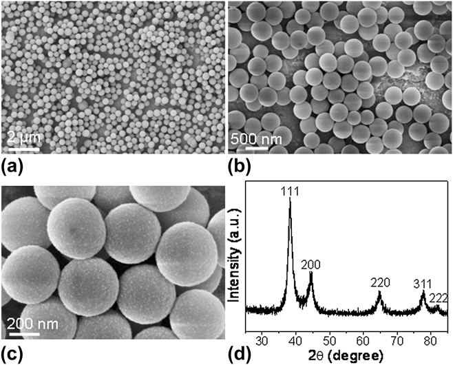
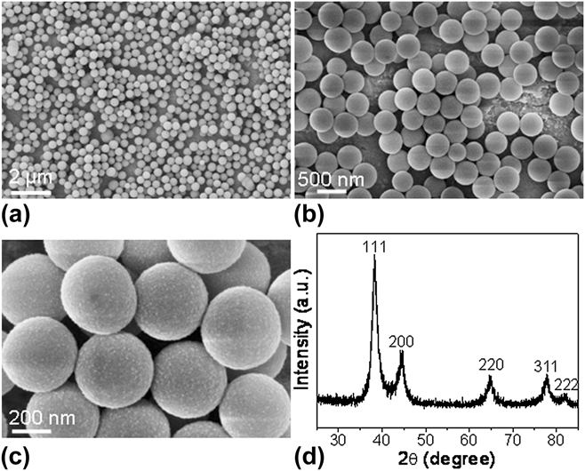
Figures 1(a)–1(c) show scanning electron microscopy (SEM) images of the typical AuNPs/aminosilica composite nanospheres obtained by aging the mixture of TMSen and aqueous solution of HAuCl4 in DMF at 60 °C for 18 h. It can be clearly seen that the nanocomposite spheres are highly uniform with an average diameter of about 300 nm. The surface of each sphere was decorated by Au nanoparticles, as clearly seen from Fig. 1(c). The XRD pattern of the obtained spheres is shown in Fig. 1(d). All peaks can be indexed to face-centered-cubic (fcc) gold with a calculated lattice constant of a = 4.09 Å, which is consistent with the standard literature value (JCPDS card no. 04-0784). In particular, all the diffraction peaks are broaden to some extent, indicating that these AuNPs have a small size. It is well documented that an amine-containing compound can serve as a kind of mild reducing agent for noble metal salts and the redox reaction rate can be accelerated by increasing the reaction temperature.Reference Sun, Dong and Wang33 The AuNPs produced in our composite nanospheres can also be ascribed to the direct redox reaction between TMSen and AuCl4−. Protonated free amine groups of TMSen can bind to gold complex ions through electrostatic interaction, resulting in the formation of AuNPs after the gold complex ions are reduced by amine groups. On the other hand, the produced Au nanoparticles can catalyze the hydrolysis of TMSen siloxane and form polymerized aminosilica, and finally, AuNPs/aminosilica composite nanospheres were observed.Reference Prasad, Stoeva, Sorensen, Zaikovski and Klabunde34
(a–c) SEM images of the obtained AuNPs/aminosilica nanocomposites at different magnifications and (d) XRD pattern of the sample.
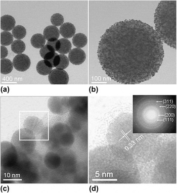
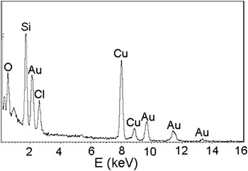
To further investigate the actual structure of the as-synthesized AuNPs/aminosilica composites, TEM and HRTEM measurements were carried out. As shown in Fig. 2(a), these particles are of spherical shape and their size distribution is relatively narrow, in agreement with the SEM observations. A large number of Au nanoparticles with the dark contrasts were embedded in each sphere [Fig. 2(b)]. Figures 2(c) and 2(d) show the HRTEM images of part of an individual sphere. Figure 2(d) gives the enlarged image of the square region in Fig. 2(c) to identify the lattice planes forming the Au nanoparticle. A d-spacing of 0.23 nm for adjacent lattice planes can be indexed to the (111) plane of gold. The TEM and HRTEM images also reveal that the AuNPs are well dispersed in the composite spheres. The average diameter of the AuNPs in the spheres is about 10 nm. In addition, EDS was performed to further confirm the existence of Au. As shown in Fig. 3, the as-synthesized spherical composites are composed of gold, silicon, carbon, and oxygen.
(a and b) TEM images of the typical sample, (c) HRTEM image of a typical sphere, and (d) enlarged image of the square region shown in (c). The insert in (d) is an electron diffraction pattern obtained on a typical composite sphere, in which all rings can be indexed to the fcc phase Au.

EDS spectrum of the obtained AuNP/aminosilica nanocomposite spheres. Cu came from the substrate.

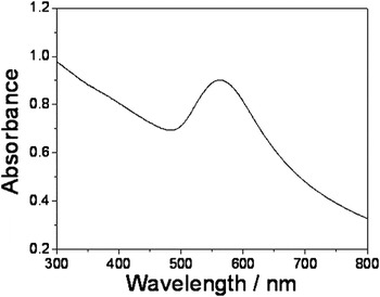
AuNPs show strong plasmon resonance absorption depending on their size and shape. Generally, the maximum plasmon band of spherical AuNPs falls between 520 and 530 nm.Reference Jana, Gearheart and Murphy35 We have measured the optical property of the ethanolic dispersion of AuNPs/aminosilica composite nanospheres by UV–Vis spectroscopy. Figure 4 shows the UV–Vis spectrum of the as-prepared nanospheres. Compared with AuNPs, the absorption band centered at 562 nm is redshift. The reason for the redshift is believed to be caused by the presence of organosilica between the Au nanoparticles because the refractive index of silica is higher than that of ethanol.Reference Lu, Yin, Li and Xia20
UV–Vis spectrum of the AuNPs/aminosilica nanocomposite spheres.

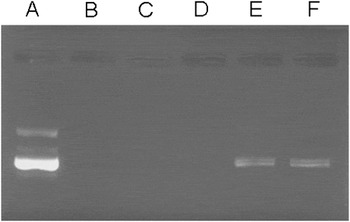
The ability of a gene delivery vector to produce a stable electrostatic complex with the negatively charged plasmid DNA is the basic requirement for efficient gene transfection. The surface charge and size of delivery nanoparticles are two important parameters in determining the efficiency of cellular uptake. ζ-potential measurements demonstrate that the obtained AuNPs/aminosilica composite nanospheres bear a positive surface potential with +66.5 mV, which can make them interact with the negatively charged DNA to form complexes. It was reported that amino cations are important in gene delivery.Reference Ghosh, Kim, Han, Forbes and Rotello8, Reference Roy, Ohulchanskyy, Bharali, Pudavar, Mistretta, Kaur and Prasad30–Reference Kneuer, Sameti, Bakowsky, Schiestel, Schirra, Schmidt and Lehr32, Reference Thomas and Klibanov36–Reference Sunshine, Green, Mahon, Yang, Eltoukhy, Nguyen, Langer and Anderson38 We think that the cationic amino groups can bind with the negatively charged DNA molecules. The nanoparticles are the composites of AuNPs and aminosilica. The existence of amino group enhances the interaction of these particles with DNA molecules. The comparable size of DNA and small nanocomposite particles facilitates their interaction. To confirm the complexation, a gel electrophoresis shift assay was performed. From the result of agarose gel electrophoresis (Fig. 5) by checking the intensities of the bands, it can be clearly seen that when AuNPs/aminosilica and DNA are mixed at a mass ratio of 30:1 or higher, all the DNA molecules have interacted with AuNPs/aminosilica nanocomposite spheres to form complexation.
Gel electrophoresis assay demonstrating AuNPs/aminosilica–DNA complexation. Lane A is DNA only. The next five lanes are AuNPs/aminosilica nanocomposite particles complexed with a constant amount of DNA at different mass ratios of composite to DNA with a constant DNA concentration, respectively (DNA: 10 mM, pH = 7.4): B, 100:1; C, 50:1; D, 30:1; E, 10:1; and F, 1:1. Binding is shown by the inhibition of DNA electrophoretic mobility.

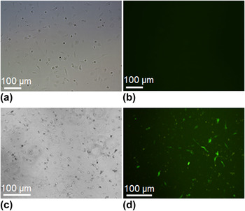
To confirm the delivered DNA is functional, the expression of pEGFP was checked by in vitro transfection of the cultured cells. The pEGFP composites were introduced into HUVECs for cell cultures and incubated in a humidified atmosphere with 5% CO2 at 37 °C for 5 h before aspirating. After the culture medium was renewed, the mixtures were incubated for a further 48 h allowing for EGFP gene expression, and the transfected cells were then imaged with a fluorescence microscope. Figure 6 shows CLSM images of untransfected HUVECs [Figs. 6(a) and 6(b)] and HUVECs transfected with pEGFP delivered with AuNPs/aminosilica composite nanospheres [Figs. 6(c) and 6(d)]. It can be observed that EGFP was successfully transfected by using AuNPs/aminosilica composite nanospheres as the delivery vector, and no obvious change was observed in the shape of the cell. No cellular fluorescence is observed from untransfected HUVECs. The confocal fluorescence image shown in Fig. 6(d) clearly shows that these AuNPs/aminosilica composite nanospheres can serve as effective gene delivery agents. The observed green cellular fluorescence is from the expressed pEGFP.
CLSM images of untransfected HUVECs and HUVECs transfected with EGFP delivered with AuNPs/aminosilica nanoparticles. (a) Bright-field microscopic image and (b) confocal fluorescence image of untransfected HUVECs; (c) bright-field microscopic image and (d) confocal fluorescence image of HUVECs transfected with EGFP.

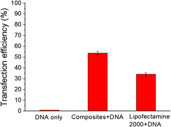
The transfection efficiency of the composite nanospheres with positively charged amino groups is demonstrated using HUVECs. Lipofectamine was selected as the positive control. Lipofectamine 2000 is a lipid-based transfection reagent capable of transfecting a wide range of cell types. The transfection efficiency was evaluated by calculating the percentage of the cells with green fluorescence using flow cytometry. Figure 7 shows the transfection efficiency in HUVECs by using the AuNPs/aminosilica composite nanosphere–DNA complexes, lipofectamine–DNA complexes (positive control), and pure DNA (negative control). The results of in vitro transfection experiments show that the obtained functional AuNPs/aminosilica composite nanospheres display higher transfection efficiency (54%), which is much higher than that of commercial Lipofectamine 2000 (34%).
Cell transfection efficiency (mean ± SEM, n = 6). The percentage of EGFP expression was determined by flow cytometry after transfection.

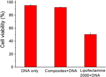
Tremendous attention has been paid on the biocompatibility of silica and results reveal that it is with a low level of cytotoxicity.Reference Kneuer, Sameti, Haltner, Schiestel, Schirra, Schmidt and Lehr39–Reference Kim, Kim, Lee, Kim, Kim, Yu, Song, Moon and Hyeon43 Cellular metabolic activity was measured by PI assays to evaluate possible toxicity that might arise from inorganic nanoparticles during transfection (see Sec. II). Cells that have lost membrane integrity cannot exclude PI. After binding to cellular DNA or double-stranded RNA, PI emits a red fluorescence. A high PI percentage indicates cell death. In our study, the AuNPs/aminosilica composite nanospheres exhibited much higher level of cell viability (93%) at a concentration of 100 μg/mL, close to the DNA control sample (Fig. 8). In contrast, for cells treated with Lipofectamine 2000, less than 55% were viable at a concentration of 25 μg/mL, showing a moderately toxic. Cationic lipids and liposomes are quite effective in gene delivery.Reference Gao and Huang44–Reference Torchilin46 However, highly negatively charged DNA can induce fusion of such liposomes to produce large particles, which could decrease the transfection efficiency and increase toxicity.Reference Matsui, Sando, Sera, Aoyama, Sasaki, Komatsu, Terashima and Kikuchi47 In general, these AuNPs/aminosilica composite nanospheres show no apparent toxicity and the cell growth is slightly affected, and cells remain nearly 100% viable relative to control cells. This could be attributed to the good biocompatibility of both AuNPs and aminosilica. It has been revealed that AuNPs can provide particularly attractive scaffolds for the manufacture of transfection agents.Reference Ghosh, Kim, Han, Forbes and Rotello8, Reference Rosi, Giljohann, Thaxton, Lytton-Jean, Han and Mirkin10, Reference Thomas and Klibanov36 Gold colloids are bioinert, nontoxic, and readily synthesized and functionalized.Reference Lu, Shi, Hu, Xu, Yu, Wen, Zhao, Xu and Yu2 They also provide a multifunctional platform for both therapeutic and diagnostic purposes.Reference Ghosh, Han, De, Kim and Rotello9 Moreover, through proper functionalization, these particles can be engineered to accumulate preferentially at tumor sites using targeting ligands, providing a powerful tool for cancer gene therapy.Reference Ghosh, Kim, Han, Forbes and Rotello8 Previous study found that polyethylenimine (PEI) conjugated to AuNPs exhibited six times higher transfection efficiency compared with PEI, indicating AuNPs play an important role in enhancing the transfection efficiency.Reference Thomas and Klibanov36
Cell viability assay results (mean ± SEM, n = 6). Cytotoxicity of Lipofectamine 2000 and nanocomposite particles was incubated with HUVECs. After 48 h incubation, cell viability was tested by PI assay at the end of transfection experiments using the optimum mass ratio. AuNPs/aminosilica nanocomposite spheres had negligible toxicity to HUVECs.

IV. CONCLUSIONS
In conclusion, uniform spherical AuNPs/aminosilica nanoparticles have been prepared by a facile one-step route. The gene delivery and expression process have been confirmed by confocal fluorescence microscopy. To the best of our knowledge, this is the first example of study on the preparation of stable AuNPs/aminosilica nanocomposite particles used for gene transfection. The transfection efficiency of AuNPs/aminosilica is higher than that of Lipofectamine 2000. Furthermore, the as-prepared nanospheres are quite safe confirmed by PI assays. This is important because many transfection reagents showed high toxicity like some cationic polymers and liposomes. Compared with cationic liposomes, inorganic AuNPs/organosilica composite nanoparticles have several main advantages: they can be produced on a large scale at low cost and can be easily functionalized. Moreover, they show high transfection efficiency, biocompatibility, and no apparent toxicity. Hence, the as-prepared AuNPs/aminosilica composite nanoparticles are promising for biomedical applications as a kind of safe biomaterials. Further developments such as the decrease of size and multifunctionalization of AuNPs/aminosilica nanocomposite particles will offer new possibilities in target-specific gene delivery and enhancing gene transfection efficiency.
ACKNOWLEDGMENTS
This work was supported by the National Basic Research Program of China (2011CB933700), the 100 Talents program of the Chinese Academy of Sciences, and the National Natural Science Foundation of China (20971118).