Introduction
Although bryophilous fungi are often host-specific (Hansruedi Reference Hansruedi1988), lichenized fungi seldom are (Sander Reference Sander1999; Peck et al. Reference Peck, Grabner, Ladd and Larsen2004; Cáceres et al. Reference Cáceres, Lücking and Rambold2007; Medina et al. Reference Medina, Lücking and Rojas2012; Rosabal et al. Reference Rosabal, Burgaz and Reyes2013; but see Conran Reference Conran1997 or Werth & Sork Reference Werth and Sork2010). During recent large-scale biodiversity inventories of the south-eastern United States and southernmost Chile, we discovered two crustose species with morphological characters that suggested placement in the genus Catinaria Vain. (see Hafellner Reference Hafellner1984; Ekman Reference Ekman1996; Smith et al. Reference Smith, Aptroot, Coppins, Fletcher, Gilbert, James and Wolseley2009). While both species were separated by a considerable geographical distance, they shared an unusual ecology in occurring only on the thalli of hepatics. Hepaticolous lichens have rarely been reported in the literature, but where they have they have been considered host specific (Döbbeler & Poelt Reference Döbbeler and Poelt1981; Döbbeler & Vězda Reference Döbbeler and Vězda1982; Stenroos et al. Reference Stenroos, Huhtinen, Lesonen, Palice and Printzen2009). Here we formally describe the two unusual species of Catinaria as new to science and document another example of the rare phenomenon of host specificity in hepaticolous lichens.
Material and Methods
All of the specimens used in this study are deposited in the herbarium of the New York Botanical Garden (NY). A list of comparative material of Catinaria species examined for this study is provided in Appendix 1. Specimens were examined dry using an Olympus SZ-STB dissecting microscope. Study of thallus anatomy and measurements of microscopic characters were carried out on sections prepared by hand with a razor and mounted in water or iodine (I), using an Olympus BX53 compound microscope equipped with a DP72 digital camera and CellSens imaging software. Chemistry was studied with standard spot tests (K, C, P, UV) following Brodo et al. (Reference Brodo, Sharnoff and Sharnoff2001). Thin-layer chromatography (TLC) was carried out on a limited number of specimens of C. brodoana using solvent C, following Culberson & Kristinsson (Reference Culberson and Kristinsson1970) as modified by Lendemer (Reference Lendemer2011). Specimens of C. radulae were not examined with TLC. In both cases detailed chemical study was precluded by the small size of specimens and overall limited material available for study.
Results and Discussion
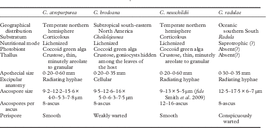
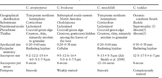
For this study we examined a total of 38 specimens of bryophilous fungi with dark brown-black biatorine apothecia, Catillaria-type asci, and 1-septate hyaline ascospores. All of the fungi occurred on thalli of liverworts and could be classified into two groups based on their host, geographical distribution, nutrition mode, and characters of the apothecia and ascospores (see Table 1). All of the specimens (n=35) from North America occurred on thalli of Cheilolejeunea and shared the characters of having inconspicuous thalli composed of goniocysts, apothecia with a distinctly cellular exciple, and ascospores with a weakly warted perispore. The remaining three specimens were all collected in southern South America on thalli of Radula and shared the characters of having seemingly immersed and non-lichenized thalli, with an exciple composed of radiating hyphae, and ascospores with a strongly warted perispore.
Comparison of Catinaria species currently recognized or described in this study
Based on the presence of Catillaria-type, dark brown-black biatorine apothecia, and relatively large, broadly ellipsoid, hyaline 2-celled ascospores, the bryophilous material examined for this study is likely most closely related to Catinaria as currently circumscribed (Hafellner Reference Hafellner1984; Ekman Reference Ekman1996; Smith et al. Reference Smith, Aptroot, Coppins, Fletcher, Gilbert, James and Wolseley2009). Historically, the genus Catinaria was circumscribed much more broadly than it is today because it originally included two very different species, one of which was much misunderstood (Ekman Reference Ekman1996; Ekman & Tønsberg Reference Ekman and Tønsberg1996; Fryday & Lendemer Reference Fryday and Lendemer2010). As such, the genus has a complex nomenclatural past that was resolved only when it was conserved with C. atropurpurea as the conserved type (Jørgensen & Santesson Reference Jørgensen and Santesson1993). The majority of species once placed in Catinaria are now treated as belonging to other genera such as Megalaria Hafellner (see Ekman Reference Ekman1996).
In its modern sense, Catinaria is restricted to two described species (C. atropurpurea (Scher.) Poelt & Vězda and C. neuschildii (Körb.) P. James) that differ primarily in the number of ascospores per ascus (Smith et al. Reference Smith, Aptroot, Coppins, Fletcher, Gilbert, James and Wolseley2009). The most common and widely distributed of the two taxa is also the type, C. atropurpurea, which has also been the subject of considerable taxonomic confusion (see Ekman Reference Ekman1996). In view of this confusion, rather than relying only on published literature, we compared the bryophilous material with more than 30 specimens of C. atropurpurea including an isotype, as well as three specimens of C. neuschildii.
In our study, which is the first to explicitly include type material, Catinaria atropurpurea is characterized by its crustose, minutely areolate to granular thallus, coccoid green-algal photobiont, absence of secondary compounds, biatorine apothecia with distinct persistent margins, a proper exciple composed of thin-walled, branched, radiating hyphae, hyaline 1-septate ascospores with a smooth rather than warted perispore, and Catillaria-type asci. Our interpretation of C. atropurpurea coincides with that of Smith et al. (Reference Smith, Aptroot, Coppins, Fletcher, Gilbert, James and Wolseley2009). Both of these interpretations differ from other published accounts (e.g. Hafellner Reference Hafellner1984), primarily in reporting smooth rather than warted ascospores. As has been noted by Smith et al. (Reference Smith, Aptroot, Coppins, Fletcher, Gilbert, James and Wolseley2009), there appear to be multiple taxa included within C. atropurpurea, including three potentially undescribed species with warted ascospores in Europe. Further study is needed to determine whether the material with warted ascospores illustrated by Hafellner (Reference Hafellner1984) belongs to one of these potentially undescribed species. While most of the specimens we examined were in agreement with the isotype of C. atropurpurea, there are three additional entities in North America that require investigation: one with small apothecia and ascospores on Populus from Michigan (e.g. Harris 712-B (NY!)), one with warted ascospores from the southern Appalachians (e.g. Tripp 5285 (NY!)), and one from Newfoundland on lignum with an inspersed hymenium (e.g. Lendemer 11098 (NY!)).
Taxonomic Section
Catinaria brodoana R. C. Harris & W. R. Buck sp. nov.
MycoBank No.: MB817132
Similar to Catinaria atropurpurea (Schaer.) Vězda & Poelt but differs by occurring on Cheilolejeunea sect. Leucolejeunea spp., by a thallus consisting of goniocysts, smaller, black apothecia, a cellular exciple and warted ascospores.
Type: USA, North Carolina, Washington Co., Bull Neck Swamp, Deep Creek Rd N of jct with Bear Lane, 35°56'56''N, 75°24'02''W, elev. 1 ft., swamp forest with Chamaecyparis, Taxodium and mixed hardwoods (Acer, Magnolia virginiana, Persea) with Lyonia-Ilex glabra understorey, on Cheilolejeunea clypeata, on Magnolia virginiana, 23 March 2013, Lendemer et al. 36377 (NY!—holotype).
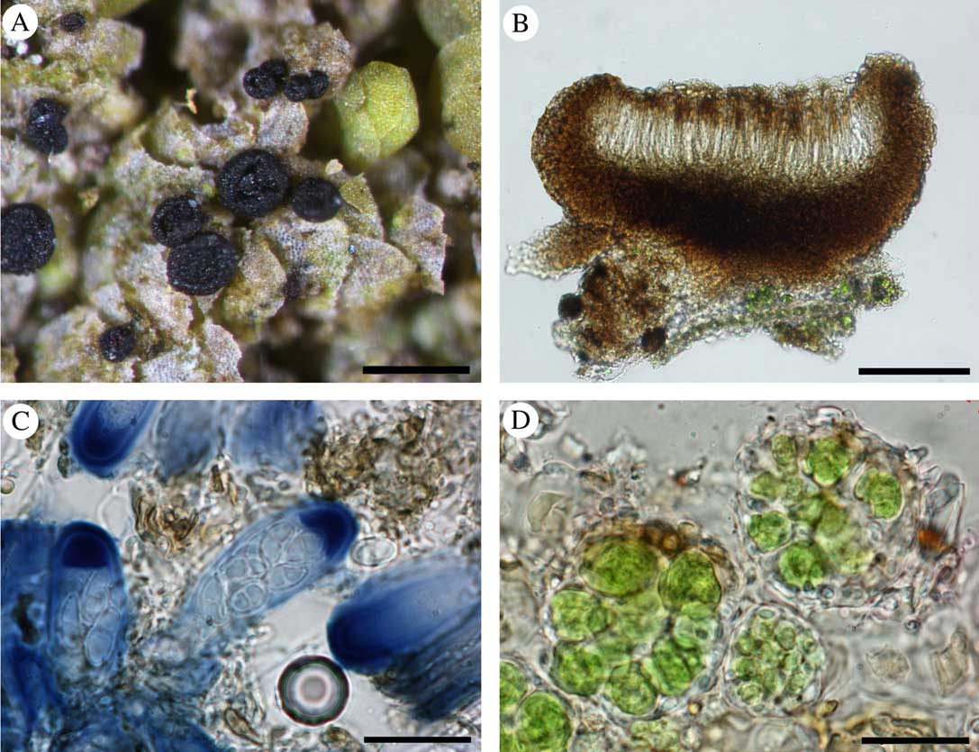
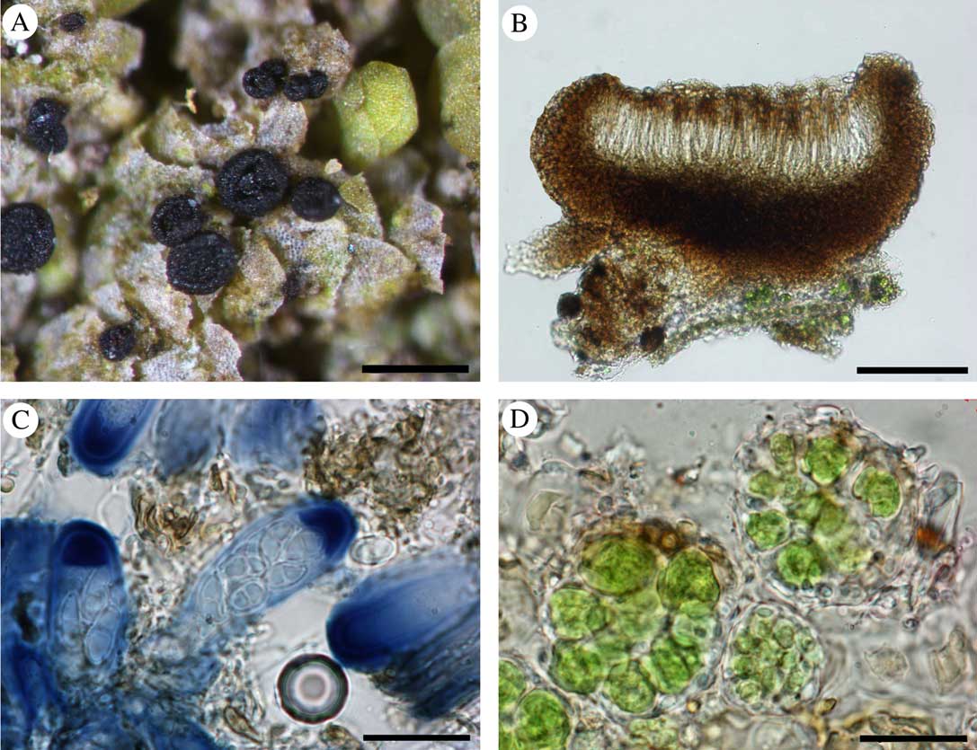
Catinaria brodoana, holotype. A, apothecia on host thallus with goniocysts barely visible as small green patches dispersed on the host; B, transverse section of apothecium mounted in water; C, asci mounted in KI; D, detail of goniocysts mounted in water. Scales: A=0·5 mm; B=100 μm; C & D=20 μm. In colour online.
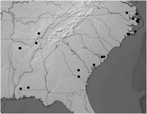
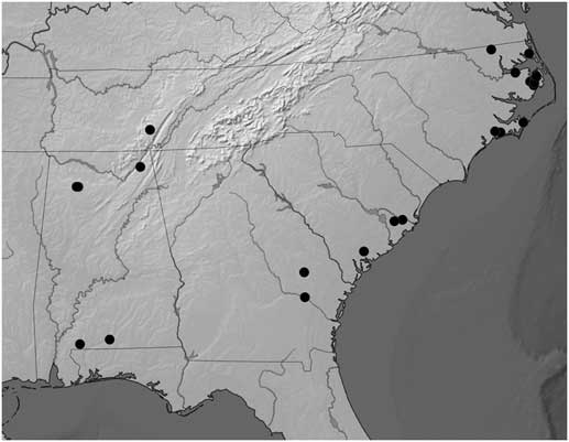
Known geographical distribution of Catinaria brodoana in Alabama, Georgia, North Carolina, South Carolina and Tennessee, as illustrated on a topographic map of south-eastern North America.
Thallus not readily visible and evident, consisting of globose goniocysts hidden among the leaves of the host liverwort; goniocysts globose, translucent to light greenish, 22–42 μm diam. Photobiont chlorococcoid, cells 6–9 μm diam.
Apothecia black, 0·20–0·35 mm diam., laminal on hepatic leaves,±turbinate when young, becoming disciform, strongly constricted at the base; margin raised, concolorous with disc, persistent; exciple brown but with blackish brown area below the hymenium,±cellular with dark brown pigment granules between the hyphae (pigment K−or K+darkening), 37–42 μm thick above, thickening to 70–80 μm below; epihymenium brown (pigment K−or K+darkening); hymenium hyaline, not inspersed with oil droplets, c. 70 μm tall; hypothecium greyish brown (pigment K−or K+darkening), thin, c. 25 μm in height; paraphyses unbranched, 2·0 μm wide, not expanded at the tips or becoming slightly expanded to 3·0 μm, often with brown pigment hoods surrounding the apical cell; asci Catillaria-type, narrowly clavate, c. 50–55×15–17 μm; ascospores 8 per ascus, hyaline, 1-septate, 9·5–12·6–16·0×5·0–6·3–7·5 μm, with weakly warted epispore.
Pycnidia not found.
Chemistry. No substances detected with standard spot tests or chromatography of selected specimens that were ample enough to allow minimal destructive sampling through removal of a chemical subsample. Spot tests: K−, C−, KC−, P−, UV−.
Etymology. We are pleased to name this new species in honour of Irwin (Ernie) Brodo (b. 1935), long-standing friend, colleague and specialist in crustose lichens.
Ecology and distribution. Based on the many collections cited here, Catinaria brodoana appears to be restricted to growing on species of Cheilolejeunea sect. Leucolejeunea (A. Evans) W. Ye et al. (Lejeuneaceae) in the south-eastern United States. This liverwort taxon has traditionally been recognized at the generic level (e.g. Hicks Reference Hicks1992), but recent molecular work (Ye & Zhu Reference Ye and Zhu2010; Ye et al. Reference Ye, Gradstein, Shaw, Shaw, Ho, Schäfer-Verwimp, Pócs, Heinrichs and Zhu2015) has placed it within a broadly circumscribed Cheilolejeunea. In the south-eastern United States, there are three species of Ch. sect. Leucolejeunea, Ch. clypeata (Schwein.) W. Ye & R. L. Zhu, Ch. conchifolia (A. Evans) W. Ye & R. L. Zhu and Ch. unciloba (Lindenb.) Malombe. We have found Catinaria brodoana on all three, but much more commonly on Ch. clypeata and Ch. conchifolia. These latter two species are endemic to the south-eastern United States (Hicks Reference Hicks1992), but Ch. unciloba is more widespread. We have no explanation as to why Ch. unciloba is not readily favoured as are the other two species, especially because it is said to intergrade with Ch. clypeata (Hicks Reference Hicks1992).
While Ch. clypeata in particular is widespread in the eastern United States, the hepaticolous lichen described here seems to have a much narrower distribution than its host. It appears to be widespread in the swamp forests of the south-eastern Coastal Plain, and the density of collections in the Mid-Atlantic Coastal Plain of North Carolina could either reflect a collection bias against this tiny species, or a greater abundance in the region as has been noted for other lichens and lichenicolous fungi (Lendemer & Allen Reference Lendemer and Allen2014; Lendemer et al. Reference Lendemer, Harris and Ruiz2016). Although we suspect that the populations of C. brodoana present in the low elevations of the southern Appalachian Mountains (Cumberland Plateau and Bankhead National Forest) are disjunctions from the Coastal Plain, as has been documented for other species (see Lendemer & Harris Reference Lendemer and Harris2015), it is also possible that the species occurs in the intervening Piedmont and has simply been overlooked.
Discussion. Catinaria brodoana differs significantly from C. atropurpurea (Table 1) in excipular type (±cellular vs. radiating hyphae) and thallus morphology (consisting of goniocysts vs. entirely immersed or with small areoles). Thus it is possible that C. brodoana should be placed in its own genus. However, given that Catinaria is clearly in need of additional study, and that C. brodoana has characters in common with C. atropurpurea (brown coloration of apothecial tissues, ascus type), we have decided that it is best placed in Catinaria. Given its unusual ecology, the only species that C. brodoana is likely to be confused with is C. radulae, which differs in occurring on a different group of host liverworts (Radula vs. Cheilolejeunea in C. brodoana), in lacking a lichenized thallus (vs. a lichenized thallus composed of goniocysts), excipular anatomy (radiating hyphae vs. cellular in C. brodoana), and geographical distribution (southern South America vs. south-eastern North America in C. brodoana).
Additional specimens examined. USA: Alabama: Covington Co., Conecuh National Forest, Solon Dixon Forestry Education Center, Cave Rd, on Cheilolejeunea unciloba on Ilex, 2007, Harris 53459 (NY); Escambia Co., Escambia Creek Wildlife Management Area, N of Conway Rd, 0·2 mi W of CR 27, on Ch. conchifolia on Persea, 2007, Harris 53356 (NY), 53372-A (NY); Jackson Co., Bryant Creek, Sand Mountain, SW of Pisgah, on Ch. clypeata on Liquidambar, 2000, Davison 5112 (NY); Winston Co., Bankhead National Forest, Sipsey Wilderness, Wolfpen Canyon, on Ch. clypeata on Fagus, 2002, Davison 6640 (NY); Bankhead National Forest, Sipsey Fork, on Ch. unciloba on Carpinus, 2016, Davison 9199 (NY), on Ch. clypeata on Fagus, Davison 9201D (NY), on Ch. clypeata on Fagus, Davison 9203 (NY), on Ch. clypeata on Fagus, Davison 9204B (NY). Georgia: Candler Co., Fifteenmile Creek Preserve, E side of Fifteenmile Creek just S of I-16, on Ch. clypeata on Gordonia, 2009, Lendemer 21727 (NY); Tattnall Co., Big Hammock Natural Area, blue and yellow trails, c.1 mi E of jct of CR 441 & GA 121/144/169, on Ch. conchifolia on Quercus, 2009, Lendemer 21381 (NY). North Carolina: Camden Co., North River Game Land, E end of Indian Island, N of Indian Island Rd/Sassafras Lane, 3 mi E of Sandy Hook Rd, on Ch. clypeata on Nyssa, 2012, Lendemer 31314 (NY); Carteret Co., Cedar Island National Wildlife Refuge, W of NC 12 along undeveloped trail, on Ch. clypeata on large Acer, 2013, Harris 59049-A (NY); Croatan National Forest, Juniper Branch of Southwest Prong of the Newport River, along FS 177/Millis Swamp Rd, 2·6 mi SW of jct w/ SR 1125/Nine Mile Rd, on Ch. clypeata on snag, 2013, Gibbons 202 (NY), 205 (NY), on Ch. clypeata on Acer, Lendemer 35505-A (NY); Dare Co., Alligator River National Wildlife Refuge, N of Possum Rd, 0·7 mi W of jct w/ Milltail Rd, on Ch. clypeata on Magnolia, 2012, Lendemer 3418 (NY); Alligator River National Wildlife Refuge, W of Milltail Rd, 0·4 mi N of bridge at Milltail Creek, 6 mi S of jct w/ US64, on Ch. conchifolia on dead Ilex, 2012, Lendemer 34297 (NY); Alligator River National Wildlife Refuge, W of H&B/Dry Ridge Rd, 0·5 mi S of jct w/ Alligator Rd, on Ch. clypeata on Magnolia, 2012, Lendemer 34318 (NY), on Ch. conchifolia on Acer, Lendemer 34340-A (NY); Alligator River National Wildlife Refuge, W of Whipping Creek Rd 0·5 mi N of jct w/ Chip Rd, on Ch. clypeata on Magnolia, 2013, Lendemer 36620 (NY); Alligator River National Wildlife Refuge, SE of jct of Butler Rd and Sandy Ridge Rd, 0·6 mi NW of jct w/ Milltail Rd, on Ch. clypeata on Liquidambar, 2014, Lendemer 42894 (NY); Gates Co., Chowan Swamp Game Land, Dowry/Tinkham Rd 0·2 mi W of Sandbanks Rd (CR 1200), 3 mi N of US 13 in Storys, on Ch. clypeata on large Fraxinus, 2012, Harris 57039-A (NY); Hyde Co., Alligator River National Wildlife Refuge, along Chip Rd 2 mi SW of jct w/ Whipping Creek Rd, on Ch. conchifolia, on Gordonia, 2014, Buck 63078 (NY), Lendemer 43151 (NY); Alligator River National Wildlife Refuge, Chip Rd, 3·2 mi S of jct w/ Whipping Creek Rd, W of S end of Swan Creek Lake, on Ch. clypeata on Acer, 2014, Lendemer 43260 (NY); Jones Co., Croatan National Forest, tributary to Hunters Creek, along SR 1101/Great Lake Rd, 0·1 mi E of jct w/ FS 134/Holston Hunter Rd, on Ch. clypeata on Ilex, 2013, Gibbons 292 (NY); Tyrrell Co., Emily and Richardson Preyer Buckridge Coastal Reserve, S of Grapevine Landing Rd/SR 1314, just W of Grapevine Landing, on Ch. clypeata on Nyssa, 2012, Lendemer 34721 (NY); Washington Co., Bull Neck Swamp, Deep Creek Rd, N of jct w/ Bear Land, on Ch. clypeata on Acer, 2013, Lendemer 36364-A (NY); Bull Neck Swamp, Deep Creek Rd 0·5 mi N of bridge over Deep Creek, on Ch. clypeata, on Nyssa, 2014, Lendemer 42958 (NY). South Carolina: Berkeley Co., Francis Marion National Forest, FS110/Walleye Rd, 1·0 mi NW jct w/ FS 118/Strawberry Rd, E of Walleye Bay and W of Big Ocean Bay, on Ch. conchifolia on Quercus, 2013, Lendemer 41203 (NY); Francis Marion National Forest, vicinity of Pitch Landing at terminus of FS 192, W of Echaw Creek, on Ch. clypeata on Quercus, 2013, Lendemer 41556 (NY); Colleton Co., Donnelley Wildlife Management Area, 0·1 mi NE of Pineland Rd, 0·6 mi N of jct w/ Blocker Run Rd, on Ch. conchifolia on Acer, 2013, Lendemer 41826-A (NY). Tennessee: Grundy Co., Savage Gulf State Natural Area, Collins West Access, above Collins River, below trail to Suter Falls, on Ch. clypeata on dead Acer, 2001, Davison & Nordman 5383 (NY).
Catinaria radulae R. C. Harris & W. R. Buck sp. nov.
MycoBank No.: MB817133
Similar to Catinaria atropurpurea but differs by occurring on Radula flavifolia, by smaller, black apothecia and by warted ascospores.
Type: Chile, Prov. Antártica Chilena, Comuna Cabo de Hornos, Parque Nacional Alberto de Agostini, Isla Grande de Tierra del Fuego, E of arm of Seno Mama, 54·5833°S, 71·5695°W, elev. 15 m, Magellanic tundra, on Radula flavifolia, on underside of branches and trunk of Drimys winteri, 4 February 2011, B. Shaw 14149A (NY!—holotype).
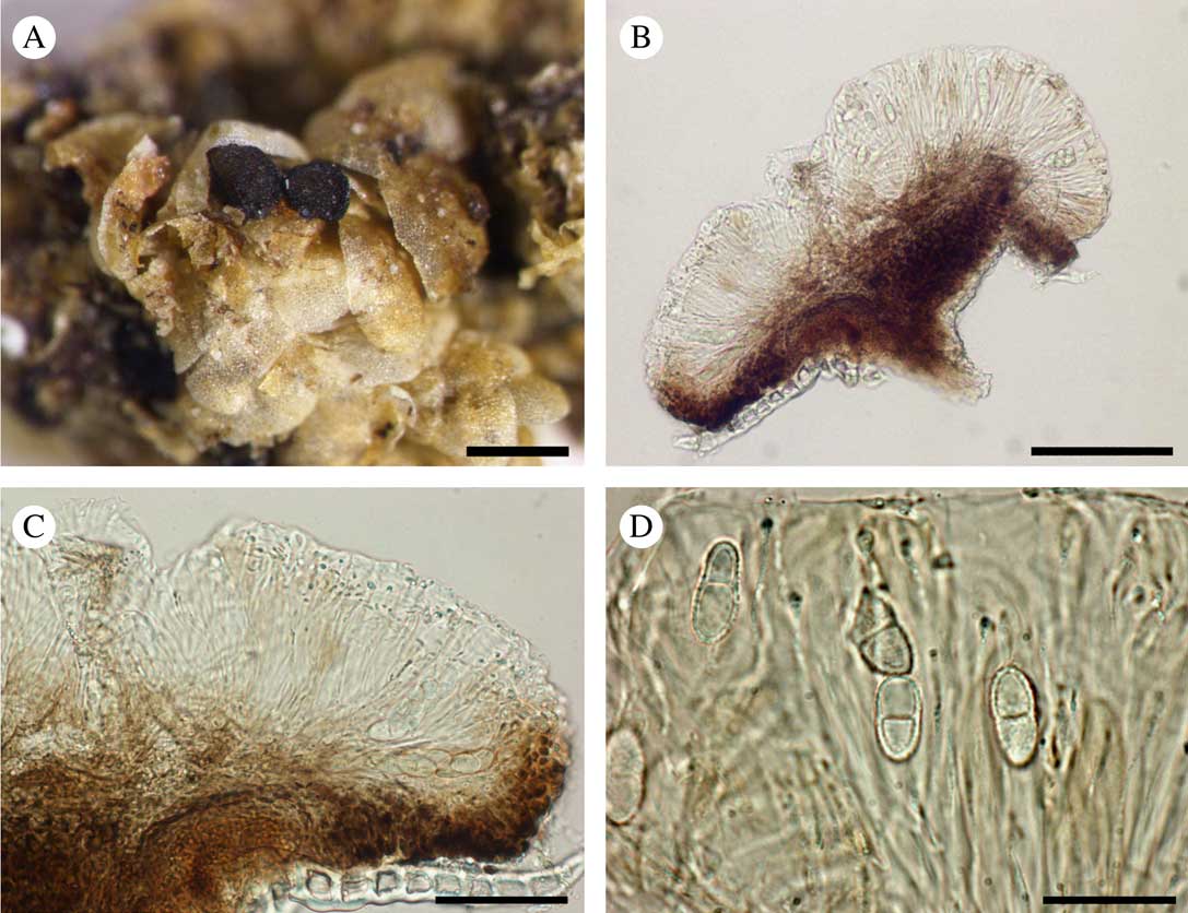
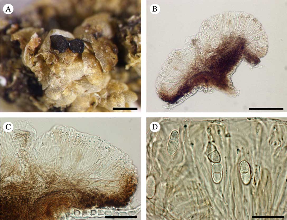
Catinaria radulae, holotype. A, apothecia on host thallus; B, transverse section of apothecium illustrating pigmentation of the tissues; C, detail of transverse section of apothecium mounted in water; D, ascospores mounted in water. Scales: A=0·5 mm; B=100 μm; C=50 μm; D=20 μm. In colour online.
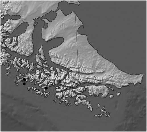
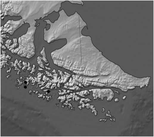
Known geographical distribution of Catinaria radulae as illustrated on a topographic map of the Cape Horn Archipelago and surrounding region.
Lichenized thallus not observed. Photobiont absent.
Apothecia black, disciform, c. 0·30–0·35 mm diam.; margin slightly raised, concolorous with disc; exciple brown (pigment K− or K+ darkening), of weakly radiating, brown-capped hyphae, c. 50 μm thick; epihymenium hyaline; hymenium hyaline, not inspersed with oil droplets, c. 75 μm tall; hypothecium brownish (pigment K− or K+ darkening), thin, c. 25 μm tall; paraphyses unbranched, 2·0 μm wide, not expanded at tips or slightly expanded to 3·0 μm, occasionally with brown pigment hoods surrounding the apical cell; ascus Catillaria-type, narrowly clavate, c. 35×12 μm; ascospores 8 per ascus, hyaline, 1-septate, 12·5–17·5×6–7 μm, with conspicuously warted epispore.
Pycnidia not found.
Chemistry. No substances detected with standard spot tests: K−, C−, KC−, P−, UV−. Available material too limited to permit comprehensive study with chromatography.
Etymology. The new species is named in honour of its seemingly obligate host, the liverwort Radula flavifolia (Hook.f. & Taylor) Gottsche et al.
Ecology and distribution. Catinaria radulae is so far known only from the Cape Horn Archipelago in southernmost Chile where it was collected at three different sites on three different islands. Although a number of Radula species were found to be infected with bryophilous fungi during the Cape Horn inventory, the new taxon was found only on R. flavifolia. We have also examined Radula material from further afield, including from the Brunswick Peninsula (Engel Reference Engel1978), but these efforts did not locate more of the Catinaria.
Discussion. Catinaria radulae is a very perplexing species as there is no obvious lichenized thallus in the material examined. Thus it is possible that it represents the first non-lichenized member of the genus, although confirmation of this would require the discovery of additional and more abundant material to permit further study. Morphologically, C. radulae is very similar to C. brodoana in overall aspect, but it differs in its excipular type (±radiating vs. cellular in C. brodoana) and more conspicuously warted ascospores. The two species also occur on different hosts, are entirely allopatric, and appear to have different lifestyles (non-lichenzed vs. lichenized in C. brodoana). Despite the anomalous absence of a photobiont in C. radulae, the placement in Catinaria is supported by similarities to the apothecial anatomy of C. atropurpurea, including the exciple composed of radiating hyphae.
Additional specimens examined. Chile: Prov. Antártica Chilena: Comuna Cabo de Hornos, Parque Nacional Alberto de Agostini, NE peninsula of Isla Londonderry, Puerto Fortuna, on Radula flavifolia on Drimys winteri, 2012, Shaw 15651A (NY); NE shore of Isla Basket, on N shore of peninsula terminating at Punta Berta, on Radula flavifolia, 2005, Engel 26078A (NY).
This study is a result of National Science Foundation Awards #1145511 (to JCL & RCH) and #9488380 (to WRB & J. Engel). WRB also thanks Matt von Konrat, Juan Larraín and Laura Briscoe at the Field Museum (Chicago) for bringing him there but nevertheless allowing him to scan Cape Horn Radula during some of the time he should have been identifying Bolivian mosses. We thank Peter Döbbeler for his comments on infected Chilean Radula and Paul Davison for donating some of his collections to NY.
Appendix 1. Selected specimens of Catinaria examined for comparison with the two new species described herein.
Catinaria atropurpurea. Canada: Nova Scotia: Queens Co., Thomas H. Raddall Provincial Park, on Quercus, 1999, Harris 42907 (NY).—Switzerland: “sylvae bernenses”, Schaerer s.n.=Lichenes Helvetici Exs. IX: 206 (NY, isotype).—USA: Delaware: Sussex Co., Cape Henlopen State Park, on Acer, 2012, Hodkinson 18863A (NY). Kentucky: Letcher Co., Bad Branch Nature Preserve, on Liriodendron, 1991, Harris 27000 (NY). Massachusetts: Barnstable Co., Bass River off Cranberry Lake, on Quercus, 23 viii 1974, Buck s. n. (NY). Michigan: Baraga Co., Sturgeon River Campground 5 mi NE of Sidnaw, on Populus, 1972, Harris 7917 (NY). Minnesota: Mahnomen Co., Little Elbow Lake State Park, on Populus, 1974, Wetmore 23002C (NY). New Jersey: Ocean Co., Manahawkin Wildlife Management Area, on Quercus, 2009, Lendemer 20078 (NY). North Carolina: Haywood Co., Great Smoky Mountains National Park, Big Fork Ridge, on dead Tsuga, 2015, Lendemer 44573A (NY). South Carolina: Charleston Co., Santee Coastal Reserve Wildlife Management Area, on Taxodium, 2013, Buck 61895 (NY). Tennessee: Sevier Co., Great Smoky Mountains National Park, Chimney Tops Trail, on Rhododendron, 2010, Buck 56233A (NY). Virginia: Patrick Co., Blue Ridge Parkway, Rock Castle Gorge Loop Trail, on Liriodendron, 1995, Harris 36643 (NY). West Virginia: Pendleton Co., Monongahela National Forest, Fanny Bennett Hemlock Grove, on Fagus base, 2001, Buck 38934 (NY).
Catinaria neuschildii. Canada: Ontario: Lake Timagami, Bear Island, on Acer, 1946, R. F. Cain 26731 (NY).—Sweden: Smäland: Almesåkra, 1875, P. G. Thoerin s. n. (NY).—USA: Missouri: Greene Co., Wilson’s Creek National Battlefield, along Wire Rd, on Ulmus, 2000, Harris 44134 (NY).