While enjoying a walk in the park with your son, you suddenly notice a young woman with a sad expression on her face who is sitting on a bench reading a letter. A wave of melancholy consumes you, and your son and you both express a wish to console the woman. This natural tendency to share and understand the emotions and feelings of others in relation to oneself, whether one actually witnesses another person's expression, perceived it from a photograph, read about it in a fictive novel, or imagined it, refers to the phenomenological experience of empathy.
Various domains of psychology suggest that one function of empathy is to promote social interaction. For example, social psychologists regard empathy as a proximate factor motivating prosocial behavior (Batson, Reference Batson1991; Davis, Reference Davis1994). Similarly, a large tradition in developmental science has been to study the onset and development of empathy, as some theorists suggest that empathy plays a crucial role in moral development, motivating prosocial behavior and inhibiting aggression toward others (e.g., Hoffman, Reference Hoffman, Stipek and Bohart2001; Miller & Eisenberg, Reference Miller and Eisenberg1988). Indeed, empathy develops from infancy, and by 2 years of age most children manifest prosocial helping responses' to others' distress (Zahn-Waxler & Radke-Yarrow, Reference Zahn-Waxler and Radke-Yarrow1990). In contrast, certain developmental disorders, such as autism spectrum disorder (ASD) and conduct disorder (CD) are marked by empathy deficits that likely influence their antisocial responses to other's distress, albeit with aloof apathy or active aggression, respectively.
The link between empathy and social interaction likely derives from the relationship between empathy and intersubjectivity. It has been postulated that empathy is a primary source of intersubjectivity, as the sense of shared experience is a prerequisite for understanding what drives other people's intentions, emotions, and motivations (Gallagher, Reference Gallagher2001; Meltzoff & Decety, Reference Meltzoff and Decety2003; Trevarthen & Aitken, Reference Trevarthen and Aitken2001). That is, intersubjectivity, the ability to share the subjective states of others and resonate with their perspective, strongly relies on the ability to read (in the sense of reacting and understanding) others' emotions to determine their psychological state. Indeed, the absence of intersubjectivity deprives individuals of the opportunity to develop prosocial behaviors, empathy, and moral judgments that are important byproducts of developing social cognition (Rochat & Striano, Reference Rochat, Striano and Rochat1999).
In addition to intersubjectivity, empathy is also phenomenologically tied to psychological constructs that may be partly innate in humans, offering further evidence of the evolutionary basis of forming social bonds, and the role of empathy in this process. Indeed, some of the basic building blocks of empathy, such as emotion sharing and an ecological sense of self, seem to be present in the first days of life, suggesting a neurobiologically based predisposition for humans to be connected to others (Rochat, Reference Rochat2002). These processes prepare the individual for later empathic connections through affective interaction with others. Humans are indeed social animals, and virtually all of their actions are directed toward or are produced in response to others (Batson, Reference Batson1990). Humans rely on others for survival and are endowed with a motivation to form and maintain strong interpersonal relationships, what Baumeister and Leary (Reference Baumeister and Leary1995) have termed “the need to belong.”
Recent data from cognitive neuroscience also offer new insights regarding the neural mechanisms and brain areas that underpin empathy (Decety & Jackson, Reference Decety and Jackson2004, Reference Decety and Jackson2006; Decety & Lamm, Reference Decety and Lamm2006; Leiberg & Anders, Reference Leiberg and Anders2006). The goal of this paper is to address the underlying cognitive–neural architecture that instantiates empathy and to highlight the dysfunction of these processes in developmental disorders marked by social–cognitive impairments. Based on empirical findings from cognitive neuroscience and developmental science, we argue that a number of components contribute to the experience of empathy: (a) affective sharing, a bottom-up process grounded in perception–action coupling and potentially underpinned by mirror neuron systems; (b) the ability to differentiate oneself from a perceived target, which relies on a sense of agency, self-, and other awareness, and likely involves frontoparietal and prefrontal circuits; and (c) executive functions instantiated in the prefrontal cortex (PFC), which operate as a top-down mediator, helping to regulate emotions and yield mental flexibility (Figure 1). Taken together, drawing from these multiple sources of data help paint a more complete picture of the phenomenological experience of empathy, as well as the respective mechanisms driving the phenomenon.
Schematic representation of bottom-up (i.e., direct matching between perception and action) and top-down (i.e., regulation and control) information processes involved in empathy. These two levels of processing are interrelated. The low level, which is automatically activated (unless inhibited) by perceptual input, accounts for emotion sharing. Executive functions, implemented in the prefrontal cortex, serve to regulate both cognition and emotion, notably through selective attention and self-regulation. This metalevel is continuously updated by bottom-up information, and in return controls the lower level by providing top-down input. Thus, the top-down regulation, through executive functions modulates low levels and adds flexibility, making the individual less dependent on external cues. The metacognitive feedback plays a crucial role in taking into account one's own mental competence in order to react (or not) to the affective states of others.

A Clarification of Terms
Empathy is a loaded term, with various definitions roaming the literature. Broadly construed, empathy has been defined as an affective response stemming from the understanding of another's emotional state or condition similar to what the other person is feeling or would be expected to feel in the given situation (Eisenberg, Shea, Carlo, & Knight, Reference Eisenberg, Shea, Carlo, Knight, Kurtines and Gewirtz1991). In line with this conception, empathy can concede an interaction between two individuals, with one experiencing and sharing the feeling of the other (Feshbach, Reference Feshbach, Bohart and Greenberg1997). Other theorists more narrowly define empathy as one specific set of congruent emotions, those feelings that are more other focused than self-focused (Batson, Fultz, & Schoenrade, Reference Batson, Fultz and Schoenrade1987). Similarly, according to Hoffman (Reference Hoffman2000), empathy refers to the psychological processes that allow a person to experience feelings more congruent with another's situation than with his own situation.
Many developmental theories highlight the role of empathy in moral development, suggesting that when humans experience others' emotions of distress they are motivated to respond with prosocial help (Eisenberg, Spinrad, & Sadovsky, Reference Eisenberg, Spinrad, Sadovsky, Killen and Smetana2006). However, whether experiencing others' emotional states entails prosocial responding is unclear. During perspective-taking tasks (using cognitive means to adopt another person's point of view), social psychological research demonstrates that when individuals imagine how the other person would feel in a given situation versus how they would feel in the same situation, different emotions arise: the individuals are prone to feel sympathy for the other in the former, whereas the latter can lead to personal distress, that is, a self-oriented aversive emotional response such as anxiety or discomfort (Batson, Early, & Salvarani, Reference Batson, Early and Salvarani1997). Personal distress may lead an observer to relieve her own stress, and not necessarily help the other. Thus, it seems that the cognitive means used to assess an automatic shared affective state with another's distress influences the likeliness to respond prosocially. In the following sections, we review the affective and cognitive components that give way to empathy, reviewing first the automatic proclivity to share emotions with others, and the cognitive process of perspective taking and executive control, which allow individuals to be aware of their intentions and feelings and keep separate self and other perspectives.
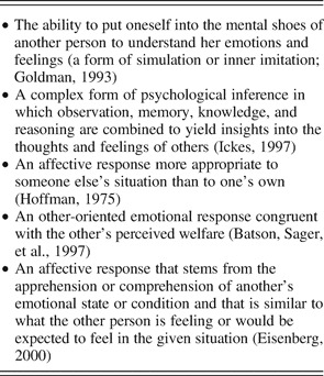
Here we will consider empathy as a kind of induction process by which emotions, both positive and negative, are automatically shared. Empathy can be the source of an emotional response emanating from the self and directed to the other, a conceptualization congruent with many scholars (see Table 1). It is important that such a definition stresses the distinction between empathic concern and personal distress, both of which spring from empathy but have different goals and consequences. Furthermore, we will consider the construct of empathy within an overarching conceptual framework. This framework suggests that empathy involves parallel and distributed processing in a number of dissociable neurocomputational mechanisms (Figure 1). Shared neural circuits, self-awareness, mental flexibility, and emotion regulation constitute the basic macrocomponents of empathy, which are mediated by specific and interacting neural circuits, including aspects of the PFC, insula, limbic system, and frontoparietal networks. Consequently, this model assumes and predicts that dysfunction in each of these macrocomponents may lead to an alteration of the experience of empathy, and correspond with selective social cognitive disorders depending on which aspect is disrupted (Decety & Moriguchi, Reference Decety and Moriguchi2007).
Definitions of empathy

Note: These definitions point to an emotional experience that is more congruent with another's situation than with one's own. Another important aspect of the construct of empathy is that it must involve some sort of self–other differentiation, which makes it distinct from related reactions such as emotional contagion.
In the following sections, we marshal support for this model by discussing first the behavioral evidence and then the putative neural mechanisms that underpin them. We also suggest that inadequacies in these mechanisms may help account for various social–cognitive disorders. These sections are organized according to each major aspect that contributes to empathy: emotion sharing, self- or other awareness, and executive control and emotion regulation. We conclude by speculating on the consequences of the experience of empathy in moral reasoning. We believe that combining developmental science with cognitive neuroscience can provide a more comprehensive understanding of empathy and related emotions, which also has the potential for generating new hypotheses regarding social–cognitive disorders and thus contributes to better treatment and intervention in developmental psychopathology.
Emotion Sharing
The automaticity of emotion sharing
Bodily expressions help humans and other animals communicate various types of information to members of their species. Specifically, emotional expression and perception play pivotal roles in human social interaction (Schulkin, Reference Schulkin2005). Emotions are short-lived psychological–physiological phenomena that represent efficient modes of adaptation to changing environmental demands. It has long been suggested that emotion expression is an evolutionary adaptation that facilitates survival (Darwin, Reference Darwin1872). Such a claim is supported by the observation that rules govern emotional expressions, which can be elicited by simple stimuli, as in the example of disgust in the presence of bitter taste, as well as the speculation that detection of emotional expression offers clear adaptive advantages, particularly in the formation and maintenance of social relationships.
Emotional expression not only informs an individual of another's subjective (and physiological) experience but also serves as a sort of social glue maintaining emotional reciprocity among dyads and groups. Emotional contagion, defined as the tendency to automatically mimic and synchronize facial expressions, vocalizations, postures, and movements with those of another person and, consequently, converge emotionally with the other (Hatfield, Cacioppo, & Rapson, Reference Hatfield, Cacioppo and Rapson1994) is a social phenomenon of shared emotional expression that given its automaticity occurs at a basic level outside of conscious awareness.
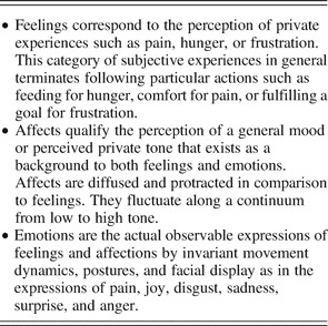
From infancy, complex facial motor patterns permit infants to match facial emotion expressions with others (e.g., Field, Woodson, Greenberg, & Cohen, Reference Field, Woodson, Greenberg and Cohen1982; Haviland & Lelwica, Reference Haviland and Lelwica1987). Very young infants are able to send emotional signals and to receive and detect the emotional signals sent by others. Shortly after birth, healthy infants convey facial expressions of interest, sadness, and disgust (Field, Reference Field and Eisenberg1989). Likewise, discrete facial expressions of emotion have been identified in newborns, including joy, interest, disgust, and distress (Izard, Reference Izard1982). These findings suggest that subcomponents of full emotional expressions are present at birth (Table 2), supporting the possibility that these processes are hard wired in the brain. It has been suggested that infant arousal in response to feelings, affects, and emotions signaled by others serves as an instrument for social learning, reinforcing the significance of the social exchange, which then become associated with the infant's own emotional experience (Nielsen, Reference Nielsen2002). Consequently, infants would come to experience emotions as shared states and learn to differentiate their own states, in part, by witnessing the resonant responses they elicit in others. This automatic emotional resonance between other and self provides the basic mechanism on which social cognition in general and empathy in particular later develops.
Classification of three basic categories of subjective experiences

Note: Rochat and Striano (1999) introduced a useful classification of three basic categories of subjective experiences which are often confused in the literature, and which include feelings, affects and emotions.
Infant affective resonance manifests in infant cry reactions to peer crying. One-day-old infants selectively cry in response to the vocal characteristics of another infant's cry, a finding that led to the speculation that from birth infants are endowed with an innate precursor of empathic distress (Hoffman, Reference Hoffman1975). Moreover, infants exposed to newborn cries cry significantly more often than those exposed to silence and those exposed to a synthetic newborn cry of the same intensity (Sagi & Hoffman, Reference Sagi and Hoffman1976). The finding demonstrates that infants' auditory perception of another's aversive affective state elicits the same distressful emotional state in the self. Importantly, this reaction exists before infants develop a sense of others as physical entities distinct from the self (Hastings, Zahn-Waxler, & McShane, Reference Hastings, Zahn-Waxler, McShane, Killen and Smetana2006). This convergence between the self and other's aversive affective experience reflects the instantiation of the initial building block that precedes the experience of empathy: a behavior matching response to other's emotional states.
Infants experience emotion contagion through interaction with their caretakers. Such a behavior scaffolds what Bowlby (Reference Bowlby1958) termed attachment, that is, the inclination to seek proximity to another person and feel secure in the presence of that person. Attachment theory fits neatly with evolutionary theory, which contends that kin-related altruism and reciprocal altruism (a potential result of empathy) may reflect activation of brain processes that mediate social attachments (Panksepp, Reference Panksepp1986). By the first months of infanthood, infants recognize and mimic the distinct emotions of their mothers, a behavior that ultimately facilitates attachment (Haviland & Lelivica, 1987). This resonance or echoing of affect, feelings, and emotions that takes place in the reciprocal interaction between infants and their caretakers is a necessary element for the development of empathy and advanced social cognition (Rochat & Striano, 1999).
The primary source of intersubjectivity impacts the quality of the infant's affective state. For example, 3-month-old infants of depressed mothers are less inclined to match emotional states with their mother than 3-month-old infants of nondepressed mothers. For instance, depressed mothers and their infants synchronize negative expression states more frequently than nondepressed mothers and their infants (Field, Healy, Goldstein, & Guthertz, Reference Field, Healy, Goldstein and Guthertz1990). This seems to indicate that under normal conditions, mothers stimulate and arouse their infant while modulating their infant's behavior. Through this process, mother and infant's emotional expression synchronize. Emotional unavailability and affective unresponsiveness may lead to the lack of such emotional modulation among the depressed mother–infant dyad. The greater matching of negative emotion among depressed mother–infant dyad likely reflects experienced emotion contagion of negative affect. This interpretation is in line with findings related to atypical autonomic arousal in anxiously attached children, a likely aftermath attachment style of infants raised by depressed mothers. Autonomic arousal is associated with degree of facial mimicry, with anxiously depressed children showing increased arousal in response to negative facial expressions (Rogeness, Cepeda, Macedo, Fischer, & Harris, Reference Rogeness, Cepeda, Macedo, Fischer and Harris1990).
Infants of healthy, nondepressed mothers show similar behaviors to the infants of depressed mothers in experiments using perturbation tests in which researchers disrupted the flow of contingent expression modulation between healthy mothers and infants. In the still or blank face test (Tronick & Weinberg, Reference Tronick, Weinberg, Murray and Cooper1997) a mother who previously established a protoconversational flow with her infant arrests her expression, and looks to the infant without a response to the infant's behavior. Infants show several petitions for communication via smiling, vocalizing, and gesturing. When the mother continues to adopt a “still face,” the infants show eye contact avoidance and distress, much like the infant's reactions to depressed mother's despondent affective state.
Results from the still face perturbation test have been replicated, however, using a double-video DTV link so that infants who are a few weeks old and mothers could communicate live (Weinberg & Tronick, Reference Weinberg and Tronick1996). After rhythmic communication stabilized, a 1-min recording of the mother's behavior was rewound and replayed. In this case, the mother's behavior was no longer contingent with the infant's movements. The infants intermittently tried to interact with the taped behavior, and showed confusion when the mother failed to respond with similar timing or appropriate expression, and eventually showed prolonged distress and avoidance. It is of interest that replay of the infant's behavior to the mother caused the mother to feel uncomfortable, and verbal reports demonstrate that she worried that the infant was unable “to connect.” It should be noted that many scholars remain skeptical of such findings related to early infant intersubjectivity as measured by video dialogue between infant and mother. Attempt to replicate these results show mixed success, and furthermore, researchers of the reproduced studies interpret the infants' interactive behaviors as nothing more than social contingency outside of intersubjectivity (for a review, see Rochat, Reference Rochat1999).
In sum, the developmental data suggest that the mechanism subserving emotion (as the observable expressions of feelings and affects) sharing between infant and caretaker is immediately present from birth. Newborns are innately and highly attuned to other people and motivated to socially interact with others. From the earliest months of their lives, infants engage with other people and with the actions and feelings expressed through other people's bodies (Hobson, Reference Hobson2002; Rochat & Striano, Reference Rochat and Striano2002). Such a mechanism is grounded in the automatic perception–action coupling of sensorimotor information, which seems present at birth in some form. This mimicry between self and other is critical for many facets of social functioning. For instance, it facilitates attachment and provides information about the other's emotional state. Mimicry also constitutes a primary source of interpersonal engagement with others, what has been termed primary intersubjectivity (Gallagher & Meltzoff, Reference Gallagher and Meltzoff1996; Gallagher, Reference Gallagher2004). This mechanism provides the foundation for understanding that others are “like me,” and underlie the development of theory of mind and empathy for others (Meltzoff & Decety, Reference Meltzoff and Decety2003). In the following section, we discuss in detail the mechanism that drives emotional sharing and mimicry, the direct link between perception and action.
Perception–action coupling mechanism and the mirror neuron system
The automatic mapping between self and other is supported by considerable empirical literature in the domain of perception and action, which has been marshaled under the prominent common-coding theory. This theory claims that somewhere in the chain of operation that leads from perception to action, the system generates certain derivatives of stimulation and certain antecedents of action that are commensurate in the sense that they share the same system of representational dimensions (Prinz, Reference Prinz1997). The core assumption of the common coding theory is that actions are coded in terms of the perceivable effects (i.e., the distal perceptual events) they should generate. Performing a movement leaves behind a bidirectional association between the motor pattern it was generated by and the sensory effects that it produces. Such an association can then be used backward to retrieve a movement by anticipating its effects (Hommel, Musseler, Aschersleben, & Prinz, Reference Hommel, Musseler, Aschersleben and Prinz2001). These perception–action codes are also accessible during action observation, and perception activates action representations to the degree that the perceived and the represented actions are similar. Such a mechanism has also been proposed to account for emotion sharing and its contribution to the experience of empathy (Decety, Reference Decety2002; Decety & Jackson, Reference Decety and Jackson2004; Preston & de Waal, Reference Preston and de Waal2002). In the context of emotion processing, it is posited that perception of emotion activates in the observer the neural mechanisms that are responsible for the generation of similar emotion. It should be noted that a similar mechanism was previously proposed to account for emotion contagion. Indeed, Hatfield et al. (Reference Hatfield, Cacioppo and Rapson1994) argued that people catch the emotions of others as a result of afferent feedback generated by elementary motor mimicry of others' expressive behavior, which produces a simultaneous matching emotional experience.
Neurophysiological evidence for this perception–action coupling comes from electrophysiological recordings in monkeys in which a unique class of visuomotor neurons have been found in the ventral premotor and posterior parietal cortices. These neurons, called mirror neurons, are active during a specific motor action and the perception of the same action made by another individual (Rizzolatti, Fogassi, & Gallese, Reference Rizzolatti, Fogassi and Gallese2001). Evidence for the existence of mirror neurons in humans is more indirect, and principally relies on functional neuroimaging studies that indicate that the neural circuits involved in action execution overlap with those activated when actions are observed (Blakemore & Decety, Reference Blakemore and Decety2001; Decety & Grèzes, Reference Decety and Grèzes2006), as well as transcranial magnetic stimulation (TMS) and motor-evoked potentials (MEP) studies that show changes in the excitability of the observer's brain regions that encode the execution of observed actions (Fadiga & Craighero, Reference Fadiga and Craighero2004). This shared neural network for action production and observation includes the premotor cortex, the inferior frontal gyrus, the parietal lobule, the supplementary motor area, and the cerebellum. Recent neuroimaging experiments demonstrate that the mirror–neuron system is flexible, and that experience and motivation modulate its functioning. For instance, regions that belong to the mirror–neuron system showed greater hemodynamic response when hungry participants were presented with videos of people grasping food. In contrast, decreased activity was detected in these regions when participants were in a satiated state (Cheng, Meltzoff, & Decety, Reference Cheng, Meltzoff and Decety2007). In addition, a number of neuroimaging studies have shown that similar brain areas, pertaining to the same network are reliably activated during imagining one's own action, imagining another's action, and imitating actions performed by a model (Decety & Chaminade, Reference Decety and Chaminade2003; Decety & Grèzes, Reference Decety and Grèzes2006). For instance, a similar neural network is engaged when individuals observe or imitate emotional facial expressions (Carr, Iacoboni, Dubeau, Mazziotta, & Lenzi, Reference Carr, Iacoboni, Dubeau, Mazziotta and Lenzi2003). Within this network, there is greater activity during imitation, compared with observation of emotions, in premotor areas including the inferior frontal cortex, as well as in the superior temporal cortex, insula, and amygdala. Such shared neural circuits reflect an automatic transformation of other people's behavior (actions or emotions) into the neural representation of one's own behavior, and provides a functional bridge between first and third person perspectives, culminating in empathic experience (Decety & Sommerville, Reference Decety and Sommerville2003; Sommerville & Decety, Reference Sommerville and Decety2006).
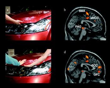
The perception of other people in pain has revealed to be of particular importance for the investigation of the neural mechanisms underlying empathy. Pain is a window through which one can obtain a detailed view of the cognitive and neurophysiological mechanism underlying the experiences of empathy and sympathy. The perception of pain in others thus constitutes an ecologically valid way to investigate the mechanisms underpinning the experience of empathy for two main reasons: first, most humans understand what is “pain”; it is a common and universal experience; and understands what are its physical and psychological manifestations; second, we have good knowledge about the neurophysiological pathways that are involved in processing nociceptive information that include the somatosensory cortex, the supplementary motor area (SMA), the anterior midcingulate cortex (aMCC), the insula, the periaqueductal gray (PAG), and thalamus. Numerous functional magnetic resonance imaging (fMRI) studies have shown that when we perceive other people in pain, the neural circuits underpinning the processing of first-hand experience of pain are activated in the observer (for a meta-analysis, see Jackson, Rainville, & Decety, Reference Jackson, Rainville and Decety2006). In one recent study, typically developing middle school-aged children were scanned while observing dynamic visual stimuli depicting other people in pain (Decety, Michalska, & Akitsuki, Reference Decety, Michalska and Akitsuki2008). Results show that neural circuits subserving the processing of nociceptive information are recruited by the sight of other people in pain (Figure 2). Such a pattern of activation in children should not be surprising given the behavioral and physiological data that document that affective sharing and vicarious emotional arousal, especially in response to others distress, is hard wired and functional very early in life (e.g., Eisenberg & Eggum, 2008; Hoffman, Reference Hoffman2000). This rudimentary capacity for resonating with the pain of others may trigger empathic distress in the observer, and provides the affective and motivational base for moral development (Hoffman, Reference Hoffman and Eisenberg1982).
In this fMRI study, 17 typically developing children (9 ± 1 years) were scanned while presented with short dynamic (2.2-s duration) visual stimuli depicting painful and nonpainful situations (Decety et al., Reference Decety, Lamm, Decety and Ickes2008). These situations involved either (a) a person whose pain was caused by accident or (c) a person whose pain was intentionally inflicted by another individual, as well as situations without any pain with one or two agents for baseline. Consistent with previous fMRI studies of pain empathy with adults, the perception of other people in pain in children was associated with increased neurohemodynamic activity in the neural circuits involved in the processing of first-hand experience of pain, including the insula, somatosensory cortex (not shown), the aMCC, PAG, and SMA. (b) It was important that when children observed an individual harming another, regions of the prefrontal cortex that are consistently involved in representing social interaction and moral behavior such as the temporoparietal junction (not shown), (d) the paracingulate cortex (PCC), and orbitofrontal cortex (OFC) were also recruited. Of interest, children indicated in the postscan debriefing that they thought that the situations in which pain was caused by another person were unfair, and they asked about the reason that could explain this behavior.

It can be speculated that the perception–action coupling physiological mechanism is already present at birth and develops gradually through experience and exposure to actions performed by self and others (Lepage & Theoret, Reference Lepage and Theoret2007). This will account for neonate imitation as demonstrated by the work of Meltzoff and Moore (Reference Meltzoff and Moore1997). Recent empirical findings offer evidence for a mirror neuron system encoding perceived and executed human action in the child's developing brain. For instance, one study recorded electroencephalographic signals via intracranial electrodes from a 36-month-old child with epilepsy while the infant observed an experimenter either drew with his right hand or kept his right hand still (Fecteau et al., Reference Fecteau, Carmant, Tremblay, Robert, Bouthillier and Theoret2004). Cortical areas responding to the observation of biological movements partially overlapped with those that were active during the execution of the same movements. An electrophysiological study demonstrated that the neural response to the processing of biological motion was in place by 8 months (Hirai & Hiraki, Reference Hirai and Hiraki2005).
Mirror neuron dysfunction in ASD
Burgeoning research efforts suggest that an aberrant mirror neuron system may contribute to motor and social problems experienced in ASD. Research with humans using TMS demonstrates selective changes in the amplitude of the MEPs (M1) during action observation (e.g., Fadiga, Fogassi, Pavesi, & Rizzolati, Reference Fadiga, Fogassi, Pavesi and Rizzolatti1995). To build on this finding, TMS was applied over the motor cortex of adults with ASD and matched healthy controls. Compared to the controls, individuals with ASD showed significantly less M1 amplitude change during the observation of transitive, meaningless finger movements (Theoret et al., Reference Theoret, Halligan, Kobayashi, Fregni, Tager-Flusberg and Pascual-Leone2005). In contrast, observation of the finger movements in control subjects selectively modulated the excitability of the motor cortex in areas delivering signals to the muscles concerned with the observed action. The weaker M1 modulation in individuals with ASD suggests that the less mirror neuron activation in the motor cortex may be partly responsible for a cascade of deficits in social cognition.
The fMRI experiments are in line with these TMS findings, and indicate abnormal activation of mirror neuron systems during imitation in adults with Asperger syndrome (Nishitani, Avikainen, & Hari, 2003) and reduced functional connectivity in mirror neuron system areas (Villalobos, Mizuno, Dahl, Kemmostsu, & Muller, Reference Villalobos, Mizuno, Dahl, Kemmotsu and Muller2004). In attempt to examine a potential link between mirror neuron dysfunction and developmental delay of social cognitive skills, one fMRI study found a lack of activation in the inferior frontal gyrus (a key mirror neuron area) in children with ASD compared to controls during the observation and imitation of basic facial emotion expression (Dapretto et al., Reference Dapretto, Davies, Pfeifer, Scott, Sigman and Bookheimer2006). However, this finding was recently challenged by Bastiaanen, Thioux, and Keysers (Reference Bastiaanen, Thioux and Keysers2008, April), who scanned a group of 17 adults with ASD during the observation of dynamic facial expressions, including disgust. The authors found that ASD participants activate their mirror system not less, but more strongly than controls when observing dynamic facial expressions.
Structural neuroanatomical evidence also implicates aberrations in mirror neuron systems in ASD. One morphometric study reported locally diminished gray matter in adults with high-functioning ASD in areas incorporated in the mirror neuron system compared to controls matched for gender, age, intelligence quotient, and handedness (Hadjikhani, Joseph, Snyder, & Gager-Flushber, Reference Hadjikhani, Joseph, Snyder and Gager-Flushberg2005). Cortical thinning of the mirror system was correlated with the severity of ASD symptoms (as measured scores on the Autism Diagnostic Interview—Revised), and cortical thinning was also seen in areas engaged in emotion recognition and social cognition. Thus, irregular thinning of cortical areas that implement mirror neurons and the broader network of cortical areas subserving social cognition may contribute to the emotional deficits characteristic of autism, such as problems engaging in intersubjective transactions and displays of empathic responding. It should be noted, however, that in the tables included in the paper, not only mirror neuron cortical areas, but all areas of the brain, show a significant reduction of gray matter. Thus, one needs to be cautious with these new findings, as cortical thinning may not be specific to areas where mirror neurons are located. In fact, an earlier study using magnetoecephalography failed to find any difference in motor cortex activation between individuals with ASD and healthy controls while observing action (Avikainen, Kulomaki, & Hari, Reference Avikainen, Kulomaki and Hari1999), a result that is in contradiction with the study by Theoret and colleagues (2005).
Perhaps more convincing evidence of anomalies in the mirror neuron system in ASD derives from EEG studies that examine mu rhythm in sensorimotor areas. Robust evidence suggests that the magnitude of mu rhythm in sensorimotor areas is strongly suppressed during the execution and observation of an action in adults (e.g., Muthukumaraswamy, Johnson, & McNair, Reference Muthukumaraswamy, Johnson and McNair2003) and typically developing children (Lepage & Theoret, Reference Lepage and Theoret2006). In children and adults with high-functioning ASD, however, mu rhythm suppression occurs when individuals observe their own action, however it fails to suppress during the observation of other persons' action (Oberman et al., Reference Oberman, Hubbard, McCleery, Altschuler, Ramachandran and Pineda2005), suggesting dysfunction in mirror neuron systems. Moreover, this result did not significantly correlate with age, implicating that this deficit manifests early and shows little improvement with age. Bernier, Dawson, Webb, and Murias (Reference Bernier, Dawson, Webb and Murias2007) extended these results to show that in individuals with high-functioning ASD, the degree of mu wave suppression during action observation is associated with behavioral assessments of imitation ability. That is, less mu suppression correlates with poorer imitation abilities, and is most robust for facial imitation skills.
Together, these recent findings, which need to be replicated, seem to suggest that dysfunctions of the mirror neuron system may hamper the normal development of self–other connectedness, creating a cascade of deficient processes that lead to social deficits, including empathy. However, it is worth noting that this account is still debated, and that the results from a recent fMRI study do not support a global failure of the mirror neuron system in children with autism (Hamilton, Brindley, & Frith, Reference Hamilton, Brindley and Frith2007). In the following section we consider the implications of perception–action coupling deficits in a behavior that is considered a manifestation of emotion contagion, which is facial mimicry.
Facial mimicry of emotions in children with ASD and developmental coordination disorder (DCD)
An effective means to measure the role of perception–action coupling in emotion sharing is via electromyography (EMG) recording of the activation of specific facial muscles in response to viewing other people's facial expressions. Facial mimicry has been defined narrowly as the congruent facial reactions to the emotional facial displays of others, and is thus an expressive component (Hess & Blairy, Reference Hess and Blairy2001). More broadly construed, emotion contagion is an affective state that matches the other's emotional display. Thus, facial mimicry can be conceived of as a physical manifestation of emotion contagion, and it occurs at an automatic level in response to viewing others' emotions (Bush, Barr, McHugo, & Lanzetta, Reference Bush, Barr, McHugo and Lanzetta1989).
Individuals with ASD are often reported to lack automatic and spontaneous mimicry of facial expressions. A recent study measured adolescents and adults with ASD and controls' automatic and voluntary mimicry of emotional facial expressions via EMG recordings of the cheek and brow muscle regions while participants viewed still photographs of happy, angry, and neutral facial expressions (McIntosh, Reichmann-Decker, Winkelman, & Wilbarger, Reference McIntosh, Reichmann-Decker, Winkelman and Wilbarger2006). The cheek and brow muscles of individuals with ASD failed to activate in response to the videos, indicating that they did not automatically mimic the facial expressions, whereas the muscles of the normally developing controls showed activation. It is important that both groups showed evidence of successful voluntary mimicry. Difficulties in mimicking other people's emotional expression may thus prevent individuals with ASD from the afferent feedback that informs them of what others are feeling (Rogers, Reference Rogers, Nadel and Butterworth1999). Indeed, in real-life situations, individuals with ASD are likely drawing on distinct cognitive processing when gauging others' emotional states (Baron-Cohen, Reference Baron-Cohen2002).
Other developmental disorders known for their motor deficits are less obviously tied to problems with emotion sharing and empathy. For example, DCD is characterized by delayed motor development and weak motor skills, as well as poor social skills, including deficient empathy (Gillberg, Reference Gillberg1992). It is plausible that poor motor skills are the primary problem, because a child with motor deficits may be shunned from social inclusion. Alternatively, the child may purposefully engage him/herself in activities beyond the realm of physical activity, for example, indulge in mathematics. Thus, weak social skills may develop by default as much of social interaction relies on motor skills, especially in childhood. However, children with DCD show information processing deficits, specifically in visual–spatial processing (Wilson & McKenzi, Reference Wilson and McKenzie1998). Such a weakness would influence perception–action coupling of emotional expression. Therefore, the mechanism underlying their difficulty in coordinating movements may also contribute to their inability empathize.
One study tried to tease out the nature of the relationship between motor skills and social skills in children with DCD (Cummins, Piek, & Dyck, Reference Cummins, Piek and Murray2005). In a sample of 39 children with DCD and 39 normally developing children, children with motor problems performed worse on scales that measured the capacity to recognize static and dynamic facial expressions of emotion. What is more, when visuospatial processing was controlled, this difference remained; thus, the child's motor ability was a significant predictor of social behavior. Children with DCD's motor impairments may negatively influence their ability to calibrate sensorimotor information about their own body, and thus hinder activation of shared motor responses between self and other during interactions of emotion expression.
The shared neural representations account suggests that problems with one's own motor or body schematic system may undermine capacities for understanding others. Consequently, it is possible that developmental problems involving sensory–motor processes may have an effect on the capabilities that make up “primary intersubjectivity,” or the ability to react contingently to others' emotional expressions (Trevarthen & Aitken, Reference Trevarthen and Aitken2001), and therefore the child's ability to resonate emotionally with others. It thus seems plausible that the defects in social and sensory–motor problems in ASD and DCD may, in part, reflect a disturbed motor representation matching system at the neuronal level. This speculation not only helps explain problems in primary intersubjectivity, but also the other sensory–motor symptoms of autism: oversensitivity to stimuli, repetitious and odd movements, and possibly, echolalia (Gallagher, Reference Gallagher2004).
Self- and Other Awareness
Although emotion contagion provides the observer with direct information of the other's emotional state, this process only accounts for what has been termed “motor empathy,” or “empathic mimicry.” However, that the observation of an emotion elicits the activation of analogous motor representation in healthy observers, begs the question why there is not complete overlap between internally generated and externally engendered motor representations.
In a complete empathic experience, observers must be able to separate themselves from others and have some minimal mentalizing ability. This aspect is a landmark of mature empathic experience (Eisenberg et al., Reference Eisenberg, Spinrad, Sadovsky, Killen and Smetana2006; Zahn-Waxler & Radke-Yarrow, Reference Zahn-Waxler and Radke-Yarrow1990). Affective sharing must be modulated and monitored by the sense of whose feelings belong to whom (Decety & Jackson, Reference Decety and Jackson2004). Thus, self-awareness generally and agency in particular are crucial aspects in promoting a prosocial regard for the other rather than a desire to escape aversive arousal. Phenomenologically speaking, self-awareness pertains to the embodied, and contextually embedded first-person point of view in subjective experience. In a similar vein, research in the neurosciences and developmental science use the term agency to describe the ability to recognize oneself as the agent of an action, thought, or desire, which is crucial for attributing a behavior to its proper agent.
Developmental work demonstrates that infants come into the world with an ecological sense of self, that is, the self as perceived in relation to the physical environment (Neisser, Reference Neisser1991). The ecologic sense of self is analogous to what phenomenologists term, prereflective self-awareness, or the subjective, qualitative “feel” of entertaining experiences (Gallagher, Reference Gallagher2000). An implicit, ecologic sense of self develops from birth, prior to an explicit (conceptual) manifestation of self-knowledge by the second year, and this sense of self is discriminated from the sense of others (Rochat & Striano, Reference Rochat and Striano2000). By 2 months, infants become incrementally systematic and deliberate in the exploration of their own body and the perceptual consequences of self-produced action. For example, infants delineate between perceptual events that are self-generated or not self-generated. In one study, Rochat and Hespos (Reference Rochat and Hespos1997a) tested whether newborn infants within 24 hr of birth discriminate between double touch stimulation specifying themselves and external (one way) tactile stimulation indicating nonself objects via the robust rooting response manifest by healthy infants from birth. Recording the frequency of the rooting in response to external or self tactile stimulation indicated that newborns are inclined to manifest rooting responses almost three times more often in response to external compared to self-stimulation. The finding suggests that from birth, infants discriminate between intermodal invariants that specify self-compared to external stimulation. Thus, infants develop an understanding of their own body as a differentiated entity, situate, and agent in the environment.
The study by Martin and Clark (Reference Martin and Clark1982) is also of special interest; they tested 1-day-old babies reactions to audiotapes of neonatal crying, the crying of an 11-month-old, and the newborn's own crying. They not only replicated Simmer's results that infants cry in response to other infant cries but also showed the more interesting trait that newborns did not respond to the sound of their own cries. Another investigation, conducted by Dondi, Simion, and Caltran (Reference Dondi, Simion and Caltran1999), also demonstrated that newborns are able to discriminate their own and other infants' cries. These results suggest that there is some self-other distinction already functioning from birth.
By 3 months of age infants become aware of their own body as a dynamic and organized entity with specific featural characteristics. In another series of studies, infants faced two on-line video images presented on a split screen. Infants viewed a split videotape screen showing contingent movements of the body from the waist down (Morgan & Rochat, Reference Morgan and Rochat1997). One view showed infant's their own legs as they would be specified via direct visual proprioceptive feedback, whereas the other showed experimentally modified on-line view of their own legs. From 3 months of age, infants look significantly longer at the unfamiliar view of the legs that violates visual proprioceptive feedback. Thus, by this age infants experience an intermodal calibration of the body, developing an intermodal body schema that serves as a perceptual based “protorepresentation” of the body.
Although the above data highlights that infant representations of self and other actions are distinct, research also suggests that infants form shared representations of their own and others' actions. Neonates imitate the actions of others in a flexible and goal-directed way, suggesting that infants represent the other as “like me” (e.g., similar to the self in some respect; Meltzoff & Brooks, Reference Meltzoff, Brooks, Malle, Moses and Baldwin2001). Further evidence suggests that infants may productively use information from their own action capacities to understand the actions of others (Woodward, Sommerville, & Guajardo, Reference Woodward, Sommerville, Guajardo, Malle, Moses and Baldwin2001). Affective sharing among infants (reviewed above) also highlights the automatic overlap between other and self in infancy, which provides the basis for the development of intersubjectivity and social cognition.
A sense of agency can also be traced to infancy. From birth, infants learn to be effective in relation to objects and events. Within hours following birth, neonates can learn to suck in certain ways and apply specific pressures on a dummy pacifier to hear their mother's voice or see their mother's face (Decasper & Fifer, Reference DeCasper and Fifer1980; Walton, Bower, & Bower, Reference Walton, Bower and Bower1992). The finding suggests that infants manifest a sense of themselves as agentive in the environment. Furthermore, by 2 months of age, infants also show positive affect, such as smiling and pleasure expression, when they accomplish causing an auditory and visual event (by activating a music box by pulling a cord attached to a limb). When the cord is then furtively disconnected from the box, hindering infants' effectiveness, they switch expressions from pleasure to anger (Rochat & Striano, Reference Rochat and Striano2000).
In sum, the studies reviewed indicate that in addition to the early roots of perception–action coupling leading to emotional expression, a sense of self, agency, and other distinction emerge early in infancy. An ecologic sense of self develops immediately via proprioceptive calibration of sensory–motor experiences. Both this resonance mechanism and an ecological sense of self situate the individual in the social environment and account for the duality of human beings who are strongly motivated to be connected to others as well as to retain independence and autonomy. In the following section, we will highlight that primacy of the self-experience permeates throughout development, and can be seen in findings from cognitive neuroscience studies showing immediate activation of self-produced actions prior to other-produced actions. In addition, we provide neurophysiological evidence for a cerebral mechanism specifically devoted to self–other distinction.
Cognitive neuroscience of self–other awareness and agency
One role that cognitive neuroscience can contribute to the study of the self and other is to ground in physiological mechanisms the distinct dimensions, aspects, and characteristics of the self and other to help address the potential separability or relatedness of each component part of self-processing. It has been proposed that nonoverlapping parts of the neural circuit mediating shared representations (i.e., the areas that are activated for self-processing and not for other processing) generates a specific signal for each form of representation (Jeannerod, Reference Jeannerod1999). This set of signals involved in the comparison between self-generated actions and actions observed from others ultimately allow the attribution of agency (comparison between efferent motor signals and afferent sensory signals). It has also been suggested that the dynamics of neural activation with the shared cortical network is an important aspect to distinguish one's own actions from the actions of others, and that the latency difference between the changes in activity elicited by the perception of self versus others' actions reflects the calibration process of shared representations (Decety & Jackson, Reference Decety and Jackson2004; Jackson & Decety, Reference Jackson and Decety2004). Furthermore, the fact that the onset of the hemodynamic signal is earlier for the self than for the others (Jackson, Brunet, Meltzoff, & Decety, Reference Jackson, Brunet, Meltzoff and Decety2006; Grèzes, Frith, & Passingham, Reference Grèzes, Frith and Passingham2004) may be considered as a neural signature of the privileged and readily accessible self-perspective.
Accumulating evidence from neuroimaging studies in both healthy people and psychiatric populations, as well as lesion studies in neurological patients, indicates that the right inferior parietal cortex, at the temporoparietal junction (TPJ) with the posterior temporal cortex, plays a critical role in the distinction between self-produced actions and actions generated by others (Blakemore & Frith, Reference Blakemore and Frith2003; Jackson & Decety, Reference Jackson and Decety2004). In addition, some recent data suggest that this region is specifically involved in theory of mind (Apperly, Samson, Chiavarino, & Humphreys, Reference Apperly, Samson, Chiavarino and Humphreys2004; Saxe & Wexler, Reference Saxe and Wexler2005). The TPJ is a heteromodal association cortex, which integrates input from the lateral and posterior thalamus, as well as visual, auditory, somesthetic, and limbic areas. It has reciprocal connections to the PFC and to the temporal lobes. Because of these anatomical characteristics, this region is a key neural locus for self-processing that is involved in multisensory body-related information processing, as well as in the processing of phenomenological and cognitive aspects of the self (Blanke & Arzy, Reference Blanke and Arzy2005). Its lesion can produce a variety of disorders associated with body knowledge and self-awareness such as anosognosia, asomatognosia, or somatoparaphrenia (Berluchi & Aglioti, Reference Berluchi and Aglioti1997). For instance, Blanke, Ortigue, Landis, and Seeck (Reference Blanke, Ortigue, Landis and Seeck2002) demonstrated that out-of-body experiences (i.e., the experience of dissociation of self from body) can be induced by electrical stimulation of the right TPJ. Of interest, one study found aberrant white matter adjacent to the TPJ, as well as in the ventromedial prefrontal cortices and anterior cingulate gyri (Barnea-Goraly et al., 2003) in children with ASD. Thus, deficits in self-other processing in individuals with HFA may be due to, in part, structural differences in pertinent brain areas or in abnormal connectivity between these areas.
In addition, a number of functional imaging studies point out the involvement of the right TPJ in the process of agency (i.e., the awareness of oneself as an agent who is the initiator of actions, desires, thoughts, and feelings). In one fMRI study, participants were instructed to open and close their hand slowly and continuously (0.5 Hz), whereas this movement was filmed and projected to them online onto a screen (Leube et al., Reference Leube, Knoblich, Erb, Grodd, Bartels and Kircher2003). The authors reported a positive correlation between the extent of temporal delay between hand movement and its visual feedback and the hemodynamic increase in the right TPJ. In another fMRI study, Farrer and Frith (Reference Farrer and Frith2002) instructed participants to use a joystick to drive a circle along a T-shaped path. They were told that the circle would be driven either by themselves or by the experimenter. In the former case, subjects were requested to drive the circle, to be aware that they drove the circle, and thus to mentally attribute the action seen on the screen to themselves. In the latter case, they were also requested to perform the task, but they were aware that the experimenter drove action seen on the screen. The results showed that being aware of causing an action was associated with activation in the anterior insula, whereas being aware of not causing the action and attributing it to another person was associated with activation in the right TPJ. It is interesting that individuals experiencing incorrect agency judgments, as it can be the case in schizophrenia, feel that some outside force is creating their own actions. One neuroimaging study found hyperactivity in the right TPJ when patients with schizophrenia experienced alien control during a movement selection task compared with healthy controls (Spence et al., Reference Spence, Brooks, Hirsch, Liddle, Meehan and Grasby1997). Such delusions of control may arise due to a disconnection between frontal brain regions, where actions are initiated, and parietal regions where the current and predicted states of limbs are represented.
Another study used a device that allowed modifying the participant's degree of control of the movements of a virtual hand presented on a screen (Farrer, Franck, Georgieff, Frith, Decety, & Jeannerod, Reference Farrer, Franck, Georgieff, Frith, Decety and Jeannerod2003). Experimental conditions varied to the degree of distortion of the visual feedback provided to the participants about their own movements. Results demonstrated a graded hemodynamic activity of the right TPJ that parallels the degree of mismatch between the executed movements and the visual reafference. Strikingly, such a pattern of neural response was not detected in schizophrenic patients who were scanned under the same procedure (Farrer et al., Reference Farrer, Franck, Frith, Decety, Damato and Jeannerod2004). Instead, an aberrant relationship between the subject's degree of control of the movements and the hemodymamic activity was found in the right TPJ and no modulation in the insular cortex. Additional evidence for the contribution of the right TPJ in self-awareness and the sense of agency derives from studies on imitation that document the selective involvement of this region during reciprocal imitation, in which it may be difficult to keep track of agency, that is, who is imitating whom (Chaminade & Decety, Reference Chaminade and Decety2002; Decety, Chaminade, Grèzes, & Meltzoff, Reference Decety, Chaminade, Grèzes and Meltzoff2002). Results from these studies provide strong support for the implication of the right TPJ in the process of self-agency by demonstrating a clear dissociation between the left and the right TPJ. When participants imitated the other, the left TPJ was strongly engaged, whereas greater activation was detected in the right TPJ when they were being imitated. Only this later condition involved discrepancies between predicted outcomes of the action performed by the participants and those perceived.
The right TPJ is also selectively activated when participants are asked to mentally simulate actions from someone else's perspective but not from their own (Ruby & Decety, Reference Ruby and Decety2001). Similarly, this region was specifically involved when participants imagined how another person would feel in everyday life situations that elicit social emotions (Ruby & Decety, Reference Ruby and Decety2004) or painful experiences (Jackson et al., Reference Jackson, Brunet, Meltzoff and Decety2006; Lamm, Batson, & Decety, Reference Lamm, Batson and Decety2007) but not when they imagined these situations for themselves. Such findings point to the similarity of the neural mechanisms that account for the correct attribution of actions, emotions, pain, and thoughts to their respective agents when one mentally simulates actions for oneself or for another individual. Further, they support a crucial role for the right TPJ, not only in mental state processing, but also in lower level processing, including reorienting attention to salient stimuli (Decety & Lamm, Reference Decety and Lamm2007).
Other areas implicated in self-processing, such as the medial PFC, posterior cingulate cortex, and precuneus, have been shown to be active when individuals are at rest and deactivate during cognitively demanding tasks (Raichle et al., 2001). It has been hypothesized that this “resting state” network contribtes to self-reflective thought, social perceptions, and theory of mind (Gusnard, Akbudak, Shulman, & Raichle, Reference Gusnard, Akbudak, Shulman and Raichle2001). It is of interest that in line with the social deficits observed in ASD, individuals with ASD demonstrate atypical resting state activation in these networks (Kennedy, Redcay, & Courchesne, Reference Kennedy, Redcay and Courchesne2006). However, further research is needed to understand the nature of the relationship between self-experience, the resting state networks, and social behavior in typically developing children, and those with ASD.
Self, other, agency, and intersubjective exchange in ASD
A mature sense of self-, agency, and other awareness are crucial for full-blown empathy, as they allow the observer to move beyond shared representations and accurately gauge the other's emotional state in relation to oneself, in other words engage in intersubjective exchanges. According to clinical descriptions, autistic children show problems in thinking, relating, and communicating to the world as well as moving through alternative perspectives that others entertain (Hobson & Meyer, Reference Hobson, Meyer, Rogers and Williams2006), all of which we suggest draw from more than basic level motor resonance, as they include an agentive stance over one's actions and an ability to interpret others as agentive actors as well.
Empirical evidence supports the suggestion that children with ASD fail to effectively discriminate the actions of the self and other. A recent study examined self–other orientation (identification) among 16 older children with autism, and 16 comparison, developmentally delayed children matched for age and IQ (Meyer & Hobson, Reference Meyer and Hobson2004). Participants performed four object-oriented tasks such as rolling a wheel and stacking objects. Lines on the floor segregated the child's and experimenter's “personal space,” and tasks were either directed toward the child's “personal space” or toward the adult's “personal space.” After the model, children were encouraged to imitate actions with a toy. In comparison to control participants, children with autism performed significantly fewer responses that modeled the self–other orientation to the object. Instead, a significant portion of the children with ASD showed “geometric repetition.” That is, they accurately imitated the experimenter's actions (i.e., rolling the wheel); however, they did so without either a “self”-orientation or an “other” norientation. For example, children with ASD would roll a wheel horizontally in the center of the testing space, disregarding the experimenters' wheel rolling either toward themselves or the child. These findings suggest that it is not motor mimicry that is problematic in autism per se, but instead, specific impairments in intersubjective imitation. Children in the control group did not show this pattern of responding, which was interesting. Instead, they fell into one of two categories of responding: either consistently adopting appropriate self- or other orientation, or showing a mix a strategies, including not imitating at all. The authors speculated that the findings reflect the failure of identification in autism, highlighting correspondence between performance on this task and the capacity to comprehend others' perspectives and to mentally shift from one perspective to another. The finding is in line with other data suggesting an autism-specific difficulty in accurately imitating the orientation of an action in relation to the model's body (e.g., Ohta, Reference Ohta1987; Smith & Bryson, Reference Smith and Bryson1998). However, future research should replicate these findings with an additional control group of typically developing children, as it is unclear whether observed differences reflect unique responding in children with ASD or unique responding in children with developmental, cognitive delay.
Further evidence of weak self–other differentiation among children with ASD derives from a study utilizing a visual perspective taking task (Lee, Hobson, & Chiat, Reference Lee, Hobson and Chiat1994). During such a task autistic children were significantly less prone to use the pronoun “me” to answer whether they were the subject in a photograph. Moreover, autistic subjects were less likely than controls to employ the pronoun “you” to refer to the experimenter. The experimenters suggest that these results do not reflect vocabulary or semantic problems, as the children with ASD showed high verbal ability, which was measured by the British Picture Vocabulary Scale. Instead, it was suggested that failures to use pronouns reflect deficits in perspective taking. Person pronoun use is among the few trademarks signifying self-consciousness, others including self-recognition in mirrors and demonstration of self-conscious emotions such as shame (Zelazo, Reference Zelazo2004). By age 2, executive function allows individuals to control aspects of conscious awareness, including conscious control of emotion, thought, and action. Thus, executive functions may allow individuals to regulate their egocentric bias, and engage in accurate perspective taking. In the case of ASD, problems seem to reflect not a bias in one's own perspective per se, but a weakness in adopting a conscious perspective albeit personal or other oriented. Thus, it is likely that empathy deficits in autism reflect problems at each or several of the components integral to empathy. In the following section, we discuss the crucial role of executive function in mature empathy.
Mental Flexibility and Self-Regulation
Given the sharedness of the representations of one's own emotional states and others, as well as similarities in brain circuits involved during first- and third-person perspective taking, it would seem difficult not to experience emotional distress while viewing another's distressed state and personal distress does not contribute to the empathic concern and prosocial behavior (Batson et al., Reference Batson, Lishner, Carpenter, Dulin, Harjusola-Webb and Stocks2003; Decety & Lamm, 2008). Indeed, distress in the self can hinder one's inclination to soothe the other's distress. However, it would not be adaptive if this automatic sharing mechanism between self and other was not modulated by cognitive control and metacognition. It is necessary that executive functions work in a top-down fashion to regulate our proclivity to be biased in our self-perspective while gauging another persons' emotional state, and promoting a sympathetic regard for the other rather than a desire to escape aversive arousal (Decety, Reference Decety, Malle and Hodges2005). Ventral and dorsal regions of the PFC have been associated with response inhibition and self-control, which are key components of emotion regulation (i.e., adjustments in type, magnitude, and duration of emotional responses that are made to meet personal, situational and interpersonal demands; Ochsner & Gross, Reference Ochsner and Gross2005). Support for this hypothesis in the domain of pain empathy comes from a recent fMRI study in which physicians who practice acupuncture were compared to naïve participants while observing animated visual stimuli depicting needles being inserted into different body parts including the mouth region, hands, and feet. Results indicate that the anterior insula, periaqueducal gray, and anterior cingulate cortex (ACC; i.e., neural regions that belong to the pain matrix) were significantly activated in the control group, but not in the physician group, who instead showed activation of the dorsal and ventral medial regions of the PFC, as well as the right TPJ, involved in emotion regulation and metacognition (Cheng et al., Reference Cheng, Lin, Liu, Hsu, Lim and Hung2007).
It is of interest that the development of self and other mental state understanding is functionally linked to that of executive functions, that is, the processes that serve to monitor and control thought and actions, including self-regulation, planning, cognitive flexibility, response inhibition, and resistance to interference (Russell, Reference Russell1996). There is increasingly clear evidence of a specific developmental link between the development of mentalizing (i.e., the process of making sense of mental states in oneself and other persons) and improved self-control at around the age of four (e.g., Carlson & Moses, Reference Carlson and Moses2001). Improvement in inhibitory control corresponds with increasing metacognitive abilities (Zelazo, Craik, & Booth, Reference Zelazo, Craik and Booth2004), as well as with maturation of brain regions that underlie working memory and inhibitory control (Tamm, Menon, & Reiss, Reference Tamm, Menon and Reiss2002). A series of studies by Posner and Rothbart (Reference Posner and Rothbart2000) strongly suggest that executive regulation undergoes dramatic change during the third year of life.
The PFC develops slowly compared to other regions during ontogeny, and reaches its maturation only late in adolescence (Bunge, Dudukovic, Thomason, Validya, & Gabrieli, Reference Bunge, Dudukovic, Thomason, Vaidya and Gabrieli2002). Evidence for this delayed maturation is provided by measures of myelination, gray matter reduction, synaptogenesis, and resting metabolism (Huttenlocher & Dabholkar, Reference Huttenlocher and Dabholkar1997). Imaging studies indicate that prefrontal areas do not attain full maturations prior to adolescence (Paus et al., Reference Paus, Zijdenbos, Worsley, Collins, Blumenthal and Giedd1999; Sowell, Thompson, Holmes, Jernigan, & Toga, Reference Sowell, Thompson, Holmes, Jernigan and Toga1999). Childhood cognitive development relates to maturation of the middorsolateral frontal cortex as well as the ACC, which are critical for the development of executive functions, especially the dramatic increase in children' ability to suppress external influences (Paus et al., Reference Paus, Zijdenbos, Worsley, Collins, Blumenthal and Giedd1999). Specifically, changes in cerebral blood flow, which reflects synaptic activity, in the PFC, almost doubles from age 0 to 2 years (Chiron et al., Reference Chiron, Raynaud, Maziere, Zilbovicius, Laflamme and Masure1992). Further, the frontal association cortex is the last area to increase blood flow from infancy to childhood, and reaches adult values only by adolescence (Takahashi, Shirane, Sato, & Yoshimoto, Reference Takahashi, Shirane, Sato and Yoshimoto1999). Direct support for age-related changes in brain activity associated with metacognition is provided by a neuroimaging investigation of theory of mind in participants whose age ranged between 9 and 16 years (Moriguchi, Ohnishi, Mori, Matsuda, & Komaki, Reference Moriguchi, Ohnishi, Mori, Matsuda and Komaki2007). Both children and adolescents demonstrated significant activation in the neural circuits associated with mentalizing tasks, including the TPJ, the temporal poles, and the medial PFC. Furthermore, the authors found a positive correlation between age and the degree of activation in the dorsal part of the medial PFC. Impairment of the medial/cingulate PFC is commonly associated with deficits in social interaction and self-conscious emotions (Sturm, Rosen, Allison, Miller, & Levenson, Reference Sturm, Rosen, Allison, Miller and Levenson2006). Such patients may become apathetic, disinterested in the environment, and unable to concentrate their attention on behavioral and cognitive tasks. It has also been suggested that frontal damage hinders perspective taking ability (Price, Daffner, Stowe, & Mesulam, Reference Price, Daffner, Stowe and Mesulam1990). The current understanding of the role of the PFC in executive functioning fits well with developmental research, which indicates that empathic concern is strongly related to effortful control and self-regulation, with children high in effortful control expressing greater sympathy and less personal distress (Rothbart, Ahadi, & Hershey, Reference Rothbart, Ahadi and Hershey1994). Effortful control may support empathy and prosocial behavior by allowing the child to attend to the thoughts and feelings of another without becoming overwhelmed by their own distress (Posner & Rothbart, Reference Posner and Rothbart2000).
Adopting another's perspective is integral to human empathy and is linked to the development of moral reasoning (Kohlberg, Reference Kohlberg and Lickona1976), altruism (Batson, Reference Batson1991) and a decreased likelihood of interpersonal aggression (Eisenberg et al., Reference Eisenberg, Spinrad, Sadovsky, Killen and Smetana2006). Of special interest are findings from social psychology that document the distinction between imagine the other and imagine oneself (Batson, Sager, et al., Reference Batson, Sager, Garst, Kang, Rubchinsky and Dawson1997). These studies show that the former may evoke empathic concern (defined as an other-oriented response congruent with the perceived distress of the person in need), whereas the latter induces both empathic concern and personal distress. This observation may help explain why empathy, or sharing someone else's emotion, need not yield prosocial behavior. If perceiving another person in an emotionally or physically painful circumstance elicits personal distress, then the observer may tend not to fully attend to the other's experience and as a result lack sympathetic behaviors.
The effect of perspective taking to generate empathic concern was documented in a study conducted by Stotland (Reference Stotland and Berkowitz1969). In his experiment, participants viewed an individual whose hand was strapped in a machine that participants were told generated painful heat. One group of subjects were instructed to watch the target person carefully, another group of participants were instructed to imagine the way the target felt, and the third group was instructed to image themselves in the target's situation. Physiological (palm sweating and vasoconstriction) and verbal assessments of empathy demonstrated that the deliberate acts of imagination yielded a greater response than passive viewing. Empathy specifically seems to be sensitive to perspective taking, as demonstrated by a series of studies demonstrate the effectiveness of perspective-taking instructions in inducing empathy (Batson, Sager, et al., 1997) and that empathy-inducing conditions do not compromise the distinction between the self and other (Batson, Sager, et al., 1997, but see Cialdini, Brown, Lewis, Luce, & Neuberg, Reference Cialdini, Brown, Lewis, Luce and Neuberg1997, for a different account of empathy and self-other merging).
A recent study by Lamm et al. (Reference Lamm, Batson and Decety2007) investigated the distinction between empathic concern and personal distress combining a number of behavioral measures and event-related fMRI. Participants were asked to watch a series of videoclips featuring patients (their face only) undergoing painful medical treatment either with the instruction to put themselves explicitly in the shoes of the patient (“imagine self”), or, in another condition, to focus their attention on the feelings and reactions of the patient (“imagine other”). Behavioral measures confirmed previous social psychology findings that projecting oneself into an aversive situation leads to higher personal distress and lower empathic concern while focusing on the emotional and behavioral reactions of another's plight is accompanied by higher empathic concern and lower personal distress (e.g., Batson et al., Reference Batson, Lishner, Carpenter, Dulin, Harjusola-Webb and Stocks2003). Neuroimaging data were consistent with such findings. Both the self and other' perspectives were associated with hemodynamic signal increase in the neural regions that belong to the pain matrix including the insula and ACC. However, the self-perspective evoked stronger hemodynamic responses in brain regions involved in coding the motivational–affective dimensions of pain, including bilateral insular cortices and aMCC. In addition, the self-perspective led to stronger activation in the amygdala, a limbic structure that plays a critical role in fear-related behaviors, such as the evaluation of actual or potential threats. It is of interest that the amygdala receives nociceptive information from the spinoparabrachial pain system and the insula, and its activity appears closely tied to the context and level of aversiveness of the perceived stimuli (Zald, Reference Zald2003). Imagining oneself to be in a painful and potentially dangerous situation thus triggers a stronger fearful and/or aversive response than imagining someone else to be in the same situation. Alternatively and less specifically, the stronger involvement of the amygdala might also reflect a general increase of arousal evoked by imagining oneself to be in a painful situation. Regarding the insular activation, it is worth noting that it was located in the middorsal section of this area. This part of the insula plays a role in coding the sensory–motor aspects of painful stimulation, and it has strong connections with the basal ganglia, in which activity was also higher when adopting the self-perspective. Taken together, activity in this aspect of the insula possibly reflects the simulation of the sensory aspects of the painful experience. Such a simulation might both lead to the mobilization of motor areas (including the SMA) to prepare defensive or withdrawal behaviors, and to interoceptive monitoring associated with autonomic changes evoked by this simulation process.
Various domains of research suggest that mental flexibility to adopt another person's point of view is an effortful and controlled process. Moreover, the capacity to take the conceptual perspective of the other is thought to be a necessary component in the fully developed, mature theory of mind. Developmental research indicates that perspective taking develops progressively. In the affective domain, children demonstrate emerging awareness of the subjectivity of other people's emotions around 18 months. By this age, infants understand that they should provide an experimenter with a piece of food that the experimenter reacts to with apparent happiness (e.g., broccoli) rather than one that the experimenter previously reacted to with disgust (fish crackers), even if the infant prefers the latter food (Repacholi & Gopnik, Reference Repacholi and Gopnik1997). In contrast, 14-month-olds fail to demonstrate this understanding. This is the first empirical evidence that infants of this age have at least a limited ability to reason nonegocentrically about people's desires (Flavell, Reference Flavell1999).
Executive functions not only facilitate perspective taking, but also control attention and metacognitive capacities, both of which facilitate prosocial responding in reaction to another's distress. Attention to others and the environment occurs when individuals are able to attend to external stimuli and disregard to some extent their self-experience. Metacognitive capacities allow for recursive thinking about the self's actions, and are thus linked to emotions such as shame or guilt which emerge as a result of causing another's distress. Children first demonstrate responses to the distress of others with other-focused behaviors like concern, attention to the distress of the other, cognitive exploration of the event, and prosocial interventions around the second year of life. At this age children manifest a self-concept and self-conscious emotions, and children's reparative behaviors after they cause distress in the other emerge (Zahn-Waxler & Radke-Yarrow, Reference Zahn-Waxler and Radke-Yarrow1990). A longitudinal study of young children's development of concern for others' distress showed that prosocial behaviors, such as hugs and pats, emerge around the beginning of the second year of life, increasing in intensity throughout this year and sometimes provide self-comfort. However, by the end of the second year, prosocial behaviors appear to be more appropriate to the victims needs, are not necessarily self-serving, and children's emotions appear to be better regulated (Radke-Yarrow & Zahn-Waxler, Reference Radke-Yarrow, Zahn-Waxler, Staub, Bartal, Karylowski and Raykowski1984).
The ability to regulate emotions may be subject to individual differences, and may interact with the degree to which individuals experience emotions. Eisenberg and her colleagues (Reference Eisenberg, Fabes, Murphy, Karbon, Maszk and Smith1994) proposed a model suggesting an interaction between the intensity at which emotions are experienced and the extent to which individuals can regulate their emotions. In line with her model, multimethod regression analysis of empathy-related responses combining self-report measures and facial muscle activity in response to empathy-inducing videos (of impoverished children), suggest that increased emotional intensity and decreased regulation on standard self-report measures predict personal distress in response to viewing the video vignettes. These interactions are first seen in infancy, as findings from infant development demonstrate that 4-month-olds low in self-regulation are prone to personal distress at 12 months of age (Ungerer et al., Reference Ungerer, Dolby, Waters, Barnett, Kelk and Lewin1990). In childhood, individuals with increased levels of emotional intensity (based on self-report, teacher–parent report, and autonomic measurements) and weak regulation are prone to personal distress in response to another's predicament, as they become overwhelmed due to their vicariously induced negative emotions (Miller & Eisenberg, Reference Miller and Eisenberg1988).
Mental flexibility deficits in developmental disorders of empathy
Children with empathy deficits likewise show deficits in executive function and children with ASD specifically show deficits in mental flexibility (Bennetto, Pennington, & Rogers, Reference Bennetto, Pennington and Rogers1996; Minshew, Meyer, & Goldstein, Reference Minshew, Meyer and Goldstein2002; Ozonoff & McEvoy, Reference Ozonoff and McEvoy1994; Ozonoff, Pennington, & Rogers, Reference Ozonoff, Pennington and Rogers1991). A series of studies found that when an experimenter feigns distress in a room where children were playing, children with ASD looked to the experimenter much less than healthy and mentally retarded children (Corona, Dissanayake, Arbelle, Wellington, & Sigman, Reference Corona, Dissanayake, Arbelle, Wellington and Sigman1998; Dissanayake & Sigma, & Kasari, Reference Dissanayake, Sigman and Kasari1996; Sigman, Kasari, Kwon, & Yirmiya, Reference Sigman, Kasri, Kwon and Yirmiya1992). However, when Blair (Reference Blair1999) replicated such studies, but controlled for executive function demands of attention, children with ASD performed similar to healthy children. That is, when experimenters' feigned distress was unambiguous and took place under conditions of low distractibility, children with ASD showed autonomic responses similar to controls. In studies measuring facial mimicry, when given ample time, individuals with ASD do show affective compensatory tactics to accomplish emotion reading; and in emotion recognition tasks, they show activation in brain areas related to intentional attentional provision and categorization (Hall, Szechtman, & Nahmias, Reference Hall, Scezchtman and Nahmias2003). These data indicate that alongside bottom-up information processing deficits (e.g., affective mimicry), top-down executive control is also impaired in individuals with ASD.
Empathy deficits in Asperger syndrome also seem to reflect poor executive function. A case study of two adolescents with Asperger syndrome with severe inabilities for emotional and cognitive aspects of empathy suggests that these weaknesses do not reflect significantly worse emotion recognition or cognitive perspective taking per se, but instead, reveal poor integration of the cognitive and affective components of empathy (Shamay-Tsoory, Tomer, Yaniv, & Aharon-Peretz, Reference Shamay-Tsoory, Tomer, Yaniv and Aharon-Peretz2002).
Indeed, these executive function deficits in ASD have been documented by fMRI experiments using cognitive tasks. Aberrant activity in prefrontal, frontal, as well as atypical frontoparietal interactions, have all been documented in fMRI experiments probing executive function in ASD (Kana, Keller, Minshew, & Just, Reference Kana, Keller, Minshew and Just2007; Silk et al., Reference Silk, Rinehart, Bradshaw, Tonge and Egan2006). It is likely that deficits in mental flexibility are grounded in atypical structure and function in prefrontal, frontal, and parietal lobes and contribute to perspective-taking difficulties observed in individuals with ASD.
It is noteworthy that violent offenders, and children with aggressive behavior problems, experience deficits in empathy and empathic concern, although the result of the lack of empathy manifests in behavior differently than that seen in ASD or DCD. The former responds aggressively to others' distress (Arsenio & Lemerise, Reference Arsenio and Lemerise2001), whereas the later simply lack prosocial behavior. The distinction can be understood as the difference between apathy and hostility, both of which are categorized as unempathetic in the traditional sense, although one is “passive,” and the other is “active.” Individuals with ASD seem to lack either an interest or capacity to resonate emotionally with others, or engage in intersubjective transactions (Gallagher, Reference Gallagher2001). In contrast, children with developmental aggression disorders react aggressively to the observation of others' distress.
CD is a mental disorder of childhood and adolescence that is characterized by a longstanding pattern of violations of rules and laws. Symptoms of CD include aggression, frequent lying, running away from home overnight, and destruction of property. CD is important partly because it is the major childhood precursor to antisocial personality disorder in adulthood (Lahey, Loeber, Burke, & Applegate, Reference Lahey, Loeber, Burke and Applegate2005). Children with aggressive behavior problems show deficits in regulating emotions, which may result in harmful patterns of interpersonal behavior. Lewis, Granic, and Lamm (Reference Lewis, Granic and Lamm2006) reviewed several of their recent studies investigating individual and developmental differences in cortical mechanisms of emotion regulation, corresponding with different patterns of interpersonal behavior. Their methods include event-related potentials and cortical source modeling, using dense-array EEG, as well as videotaped observations of parent–child interactions, with both normal and aggressive children. By relating patterns of brain activation to observed behavioral differences, the authors found (a) a steady decrease in cortical activation subserving self-regulation across childhood and adolescence, (b) different cortical activation patterns as well as behavioral constellations distinguishing subtypes of aggressive children, and (c) robust correlations between the activation of cortical mediators of emotion regulation and flexibility in parent–child emotional communication in children referred for aggressive behavior problems.
Emotion is normally regulated in the human brain by a complex circuit that includes several regions of the PFC (dorsal and ventral), the amygdala, hypothalamus, ACC, insula, and ventral striatum. Descending pathways from orbitofrontal and medial prefrontal cortices, which are also linked with the amygdala, provide the means for speedy influence of the PFC on the autonomic system, in processes underlying appreciation and expression of emotions (Barbas, Saha, Rempel-Clover, & Ghashghael, Reference Barbas, Saha, Rempel-Clover and Ghashghaei2003). It is of interest that key areas found to be functionally or structurally impaired in antisocial populations include dorsal and ventral regions of the PFC, amygdala, hippocampus, TPJ, and ACC (Raine & Yang, Reference Raine, Yang and Patrick2006). Raine has hypothesized that the rule-breaking behavior common to antisocial, violent, and psychopathic individuals may in part be attributable to impairments in some of the structures (dorsal and ventral PFC, amygdala, and TPJ) or dysfunction in amygdala–orbitofrontal cortex (OFC) structural connectivity, normally subserving moral cognition and emotion.
In contrast, impulsive aggression may be the product of failed emotion regulation. Impulsive aggression is associated with a low threshold for activating negative affect and with failure to respond appropriately to the anticipated negative consequences of behaving aggressively. Davidson, Jackson, and Kalin (Reference Davidson, Jackson and Kalin2000) have proposed that the mechanism underlying suppression of negative emotion is via an inhibitory connection from regions of the PFC to the amygdala.
In addition to functional brain abnormalities corresponding with emotion dysregulation and empathy deficits in CD, impulsive aggression in CD is associated with decreased noradrenergic (NA) function, which also correlates with poor empathic ability in this population (Raine, Reference Raine1996). In fact, a low resting heart rate, a partly heritable trait reflecting fearlessness and stimulation seeking, at 3 years of age predicted aggressive behavior at 11 years of age (Raine, Venables, Mednick, & Sarnoff, Reference Raine, Venables, Mednick and Sarnoff1997). Children with clinical levels of behavior problems, often a precursor to the development of CD, show increased disregard for others, for example, anger, avoidance, and/or amusement by another's distress, a negatively toned response pattern that differs significantly from normal children's responses. It is likely that decreased NA function, which is associated with aggressive behavior, contributes to these antisocial reactions.
One way to determine whether CD is associated with low arousal or poor regulation is to investigate patterns and changes in autonomic nervous system. Sympathetic activation or parasympathetic inhibition leads to changes in cardiac functioning, which can be interpreted as bodily cues of discomfort or distress and a need for action. Porges (Reference Porges1996) has found that parasympathetic nervous system functioning, as reflected in heart rate variability (heart rate [HR] variability measures the variability in HR associated with breathing and indexes an individual's competency to physiologically and behaviorally reacts to external stimuli) influenced by the vagal system, is related to the control of attention, emotion and behavior. Porges suggests that the tonus of the vagus nerve provides a theoretical basis for the child's ability to focus attentional processes, inhibit irrelevant activity, regulate emotion, and appropriately engage with the environment (Porges, Reference Porges1995).
In the domain of empathy, the work of Eisenberg and Fabes (1994) suggests that deceleration of HR may be associated with attention to others that characterizes empathic concern, and HR deceleration is also associated with an increase in desire to help and comforting. A number of studies indicate that antisocial behavior, and CD are associated with, and predictable from, low resting HR in children, adolescents, and adults (e.g., Lahey, Hart, Pliszka, Applegate, & McBurrett, Reference Lahey, Hart, Pliszka, Applegate and McBurnett1993; Raine, Venables, & Sarnoff, 1997). Because low resting HR is associated with greater aggression, and aggression and concern for others are inversely related, it has been predicted that high HR should predict greater concern for others. One study found that HR was positively correlated with concerned responses toward adults who were simulating injuries (Zahn-Waxler, Cole, Welsh, & Fox, Reference Zahn-Waxler, Cole, Welsh and Fox1995). However, Calkins and Dedmond (Reference Calkins and Dedmon2000) reported that aggressive children displayed no physiological indicators of underarousal, as indexed by resting HR. The authors did find, however, that these children displayed poor behavioral and physiological regulation, as indexed by a lack of HRV during challenging situations. This latter finding supports the idea of a failure of self-regulation in relation to empathy and aggression. Cardiac vagal regulation was found to differentiate among children at risk for behavior problems (Calkins, Graziano & Keane, Reference Calkins, Graziano and Keane2007). In that study (a sample of 335 children), there was a trend for the children at risk for externalizing problems to display less vagal withdrawal than the control group. Future research should ultimately clarify the relationship between structural/functional differences in patterns of brain activation with NA function in individuals with CD.
The maturation of executive functions, including emotion regulation (subserved by the dorsal and ventral PFC and their connections with the amygdala) by 2 years of age contributes to the development of prosocial behaviors. Conversely, if executive functioning is not intact, self and other perspectives may not be regulated and individuals may over- or underidentify with an observed target. In the case of childhood aggression and CD, it is likely that poor executive control and dysfunction of emotion regulation contributes to empathy deficits, although other factors (NA function) also contribute to reactionary behaviors.
Conclusions
We have argued that empathy depends upon both bottom-up processes, which are driven by emotion expressions, and top-down processes, including self-regulation and executive control. These different aspects are underpinned by distinct neural systems that develop at different stages. Notably, emotion sharing relies on the perception–action coupling mechanism, which seems functional very early in development, and allows the newborn to implicitly share subjective bodily experiences with others. Controlled processes, subserved by the PFC, develop later and play a major role in metacognition, including taking into account a cognitive representation of one's own mind and other's mind, aspects that are necessary for social emotions which require self-monitoring (see Figure 1). We have also shown that developmental disorders related to empathy reflect dysfunction of these different aspects. It is noteworthy that the current knowledge in social cognitive neuroscience is at a preliminary stage, and many findings need to be reproduced. In relation to the goal of this paper, ties drawn between developmental and neuroscientific evidence in emotion sharing and empathy can only be loosely strung together, as many neuroscience findings pertain to adult participants, not children. Future studies should aim to incorporate children in the investigation of the neural basis of empathy, especially because contradictions surround the few neuroscience studies that examine emotion sharing in children.
Finally, the model reviewed here offers interesting insights for debates surrounding the naturalization of normative ethics. Empathy has been associated with the propensity to respond to another's predicament in a prosocial, or “moral” way. Yet, an understanding of what motivates us to feel empathy in the sense of caring for the other (Batson et al., Reference Batson, Lishner, Carpenter, Dulin, Harjusola-Webb and Stocks2003) and then help them is unclear. In fact, humans fail to consistently respond to others' negative situations prosocially across contexts. Further research in child development and cognitive neuroscience may help elucidate these interrelationships and hopefully offer a better understanding of emotional resonance, empathy, and their relationship with prosocial, moral reasoning (and the lack thereof). The results of a recent fMRI study on empathy and theory of mind with children seem to indicate that there is little overlap in the brain circuits associated with empathy for pain and moral reasoning, although this does not mean that these circuits do not communicate (Decety et al., 2008). In this study, children watched situations involving either a person whose pain was caused accidentally or a person whose pain was intentionally inflicted by another individual, as well as situations without any pain with one or two agents. It is important that when children observed an individual intentionally harming another, regions of the PFC that are consistently involved in representing social interaction and moral behavior such as the temporoparietal junction, the paracingulate cortex (PCC), and OFC were recruited (see Figure 2). It is of interest that children indicated in the postscan debriefing that they thought that the situations in which pain was caused by another person were unfair, and were asking about the reason that could explain this behavior. Evidence from moral neuroscience suggests a critical role of a corticolimbic network subserving moral judgment. This network includes the medial OFC, the TPJ, the amygdala, and anterior PCC (Moll, de Oliveria-Souze, & Eslinger, Reference Moll, de Oliviera-Souza and Eslinger2003). Furthermore, the monitoring of outcomes that relate to punishments and rewards is linked to activity in the OFC (Kringelbach & Rolls, Reference Kringelbach and Rolls2004). It is worth emphasizing that the regions selectively associated with the perception of an agent harming the other belong to the neural systems underlying moral thinking.
We have argued that genuine empathy goes beyond emotion sharing and a simple resonance of affect between the self and other. It depends crucially on self–other awareness and on the ability to regulate one's own emotional state, allowing proper identification of the other's condition and freeing up resources for coping with another's distress in prosocial ways. Successful emotion regulation in infancy is essential for the development of the ability to control one's own arousal and, with the sense of agency, be able to tag the aroused state as indicative of the state of the other. Children as well as adults who become overaroused by another's distress, due to lack of emotion regulation and/or self–other distinction, may use up too many cognitive resources dealing with their own emotion and fail to act in a prosocial fashion (Nielsen, Reference Nielsen2002).
Limitations of the scope
It is important to note several limitations of our review. The first limitation pertains to the extent to which conclusions can be drawn from the relationship between the development of empathy in healthy children and the manifestation of empathy deficits that occur in developmental disorders. We covered findings from infant development to address the innateness of the interacting components of empathy; however, most of the findings reviewed from developmental disorders pertain to observations from early childhood. This limitation in part reflects the difficulty to obtain infant data across developmental disorders as they are often not diagnosed until childhood. Some analysis of infant behavior of children diagnosed with developmental disorders comes from home video analysis (see Baranek, Reference Baranek1999), although such studies are sparse, and findings are ambiguous due to weak scientific control.
Second, the relationship between neural evidence and behavioral data is indirect, and thus speculations may be far reaching. Neuroimaging data help to answer fundamental questions about the mechanisms subserving imitation. However, their interpretation in relating structure to function should be done with caution. It is difficult to derive the computational function of an area without taking into account its extrinsic and intrinsic connectivity, the distribution of receptor types, and the information processing of the intrinsic neurons. Such information is generally lacking. In addition, a set of cortical areas may be active in a wide range of functions from action perception to empathy and theory of mind, but across those functions the networks in which they participate may be quite different (see Cacioppo et al., Reference Cacioppo, Berntson, Lorig, Norris, Rickett and Nusbaum2003). More empirical data from both developmental science and cognitive neuroscience in the future will contribute to a fuller understanding of the mechanisms involved in empathy and their developmental time course. Furthermore, brain imaging investigations of emotional processes often draw from limited methodologies that may overlook flaws in the operational definitions of the emotional phenomena studied. For example, many fMRI experiments require participants to identify the emotion in static faces or short animations, implicitly considering that emotion recognition can be equated with emotional experience. Clinical investigations with neurological patients, however, indicate that these processes (emotion recognition and experience) involve quite different neural substrates (Levenson, Reference Levenson, Coan and Allen2007). Future neuroimaging studies of emotion in general, and empathy in particular, should base stimuli on the discrete emotional experience examined. Meeting this goal would be facilitated by the further collaboration between cognitive neuroscientists and developmental psychologists.
Third, and finally, empathy is a complex construct and the above model does not account for all that empathy entails. The phenomenological experience of empathy and its role in initiating prosocial, empathic reactions likely draws on several interacting factors (and complicated distributed brain networks) not mentioned in our review. For example, motivation likely influences empathic accuracy: people who are motivated to produce empathically accurate responses to another's predicament are less susceptible to social inference biases such as the fundamental attribution error (Fletcher, Reeder, & Bull, Reference Fletcher, Reeder and Bull1990; Tetlock, Reference Tetlock1985). In addition, the type of rapport between observer and target influences the intensity of emotion contagion between members of a dyad, with patients and therapists, mothers and infants, and spouses, and even individuals who perceive others as similar to themselves ranking high in autonomic synchrony (Levenson & Ruef, 1997).
Another important area of interest regarding the contribution of empathy to the development of intersubjectivity is its relation to the so-called attachment system described by Bowlby (Reference Bowlby1958), that is, an innate psychobiological system that motivates infants to seek proximity to people who will protect them. One can argue that the mechanisms that underpin the development of empathy, especially through interaction with caregivers who are responsive, partly overlaps, and are functionally linked with promoting optimal functioning of the attachment system. The consequences of this link between empathy and attachment are paramount. Indeed, Mikulineer and Shaver (Reference Mikulineer and Shaver2005) have documented the idea that people who have the benefits of secure social attachments find it easier to perceive and respond to other people suffering, compared to those who have insecure attachments.
Personality traits, temperaments, and cultural norms of emotional display also contribute to the degree to which empathy may be experienced in the observer (Eisenberg & Fabes, 1994; Posner & Rothbart, Reference Posner and Rothbart2000). Likewise, children from cultures that promote reciprocal relations and cooperation tend to be better at perspective-taking tasks than children living in individualistic cultures (Eisenberg, Bridget, & Shepard, Reference Eisenberg, Bridget, Shepard and Ickes1997).
Social psychologists emphasize the role of situational context as opposed to personality in the experience of empathy (or the absence of it), although many recognize the combination of situation and personality as the best predictor of social behavior (Fiske, Reference Fiske2004). Although situational context is important, creating ecologically valid situations in a laboratory setting or in an MRI scanner poses a challenge. Theoretically, assessing personality traits and correlating them with task performance and biological markers should pose no great challenge. These constructs, however, are rarely stable and they are dependent on many variables. Thus, designing ecologically valid experiments remains a challenging process, especially with children.
A current aim in cognitive neuroscience is to study the interaction between affect and cognition. Empathy, a valuable social phenomenon, exemplifies this complex relationship because it draws on aspects such as emotion sharing and self-regulation. In addition, affective and social cognitive developmental neuroscience offers promising insights for both our understanding of typical and psychopathological social behavior.