Introduction
Cholesteatoma may be classified as either congenital or acquired. To be considered congenital, a cholesteatoma must be demonstrably unconnected with chronic suppurative otitis media. Derlaki and ClemisReference Derlaki and Clemis1 proposed three criteria for the diagnosis of congenital cholesteatoma: (1) development behind an intact tympanic membrane; (2) no previous history of aural infection; and (3) origin from embryonal inclusion of squamous epithelium or undifferentiated tissue, which changes into squamous epithelium within the developing temporal bone.
The common sites for congenital cholesteatoma are the petrous apex and the middle-ear space. Expansion of the lesion may lead to secondary infection, and this, together with the commonness of a past history of otitis media, can make it difficult to be certain that the lesion is a genuine example of congenital cholesteatoma. Cholesteatoma may also develop in association with congenital malformations of the ear.Reference Mills and Graham2 In some cases, these lesions may be true congenital cholesteatomas derived from embryonic cell rests.
This pathological approach to classification is not very useful in clinical practice. The otoscopic classification proposed by TosReference Tos and Pfaltz3 has the great advantage of being more readily applicable in everyday clinical situations. Mills and PadghamReference Mills and Padgham4 have proposed a modification of Tos's classification to include cases with an intact tympanic membrane (Table I). This type of cholesteatoma is relatively uncommon and may be congenital or acquired. Congenital cases usually present in childhood and 80 per cent are located in the anterior portion of the middle-ear space, if they are confined to one quadrant of the middle ear.Reference Postic, Korman, Samadi and Wetmore5 However, two reports from Japan indicate that, in that part of the world, many patients have disease in the posterior portion of the middle ear.Reference Kashiwamura, Fukuda, Chida and Matsumura6, Reference Kojima, Tanaka, Shiwa, Sakurai and Moriyama7 The clinical features, extent and management of congenital cholesteatoma has been well documented.Reference Postic, Korman, Samadi and Wetmore5–Reference Benhammou, Nguyen, El Makhloifi, Charachon, Reyt and Schmerber13
Modified Tos otoscopic classification of cholesteatoma

Published with permission.Reference Mills and Padgham4 TM = tympanic membrane
Much less has been written about acquired, intact tympanic membrane cholesteatoma. Some cases are iatrogenic, but not all follow previous surgical intervention. This paper presents the author's experience of intact tympanic membrane cholesteatoma over a 22-year period, with particular reference to the location, clinical features and treatment of this type of disease.
Materials and methods
Data had been recorded prospectively on all cholesteatoma cases treated by the author since 1986, in Dundee (1986–1998) and Edinburgh (1998–2008), Scotland, using a computer database (Lotus Approach). The cases had been classified using the modified Tos otoscopic classification proposed by Mills and PadghamReference Mills and Padgham4 (Table I). Cases with an intact tympanic membrane were selected for particular attention. Patients under the age of 16 were excluded because, although the author's Dundee practice had included children, his Edinburgh practice included only adults. The extent of the disease was described using the staging system proposed by Saleh and Mills (Table II).Reference Saleh and Mills14 The following items were investigated: presenting complaints; any previous otological surgery; site and extent of the disease; condition of the ossicular chain; and the surgical technique employed. The occurrence of residual disease following treatment was also noted. Where appropriate, comparisons were made between patients with disease behind an intact drum and the rest of the group.
Stage ossicular chain complications staging system for cholesteatoma

* Complications: lateral semicircular canal fistula, total sensorineural hearing loss, lateral sinus thrombophlebitis, intracranial abscess, meningitis. Published with permission.14 N/A = not applicable
Results
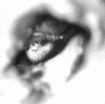
Two hundred and sixty-five new cases of cholesteatoma presenting in adult life (i.e. aged >16 years) were seen during the study period. Of these, 17 (6 per cent) had disease behind an intact tympanic membrane, with no evidence of an attic or pars tensa retraction pocket. This group did not include residual cholesteatomas following intact canal wall or open cavity mastoidectomy. Figure 1 shows the appearance of a typical case at tympanotomy. The lesions were ‘pearls’ of varying size, surrounded by a well defined sac. The ages of the intact tympanic membrane patients varied between 31 and 75 years (mean = 47 years), compared with an age range of 16 to 86 years for the group as a whole (mean = 41 years). There were five men and 12 women in the intact tympanic membrane group (ratio 1:2.4). This compares with a male to female ratio of 1.5:1 for all other cholesteatoma cases.
Cholesteatoma in the posterosuperior quadrant of the left middle ear. The lesion has a well defined sac. The chorda tympani can be seen running over the surface of the sac.

Amongst patients with cholesteatoma behind an intact tympanic membrane, the most common presenting complaint was deafness alone (n = 12, 71 per cent). Two patients complained of deafness and discharge and one of deafness and dizziness. In four cases, there was a white mass visible behind the tympanic membrane. Nine patients had undergone a previous tympanoplasty operation for non-cholesteatomatous, chronic, suppurative otitis media, while one had had a previous grommet insertion in the affected ear. Therefore, 10 cases (59 per cent) were almost certainly iatrogenic.
The disease was situated in the posterior portion of the middle ear in all intact tympanic membrane cases. In 11 cases (65 per cent), disease was confined to the middle ear (S1), in two there was also attic involvement (S2) and in three there was extension to the mastoid process (2 S3, 1 S4). The incus was eroded in 13 cases, while six had loss of the stapes arch and three erosion of the malleus. In all cases with ossicular erosion, the disease was closely related to the involved ossicles. There was a lateral canal fistula in one case.
Simple excision of the disease via a tympanotomy was undertaken in 10 intact tympanic membrane cases (59 per cent). This was combined with an atticotomy in three cases, while two underwent an intact canal wall mastoidectomy and two an modified radical mastoidectomy. In five patients, an ossiculoplasty was carried out at the same time as the cholesteatoma excision, while in a sixth a reconstruction was performed at a planned ‘second look’ operation.
Follow-up data were available at one year in seven cases, two years in three, three years in two and five years in one. All patients with at least one year's follow up had dry ears at one year. At one year, a mean air–bone gap of less than 10 dB was achieved in 58 per cent of ears, but only 33 per cent had mean hearing losses of less than 30 dB in the operated ear. At the time of writing, residual disease had been identified in two cases: in the mastoid process of one patient with extensive disease treated by intact canal wall mastoidectomy, and in the middle ear of the other patient. Residual disease rates in the intact tympanic membrane cholesteatoma group and in the remaining cholesteatoma cases are presented in Table III.
Residual disease in cholesteatoma patients* with intact tympanic membrane vs other types

* Patients aged 16 years and older. †n = 118; ‡n = 147; **n = 265. TM = tympanic membrane
Discussion
Given that there is no infallible means of determining whether or not a cholesteatoma in the middle-ear space is congenital or acquired, it may be better to describe all these lesions as ‘intratympanic cavity cholesteatoma’ or ‘intact tympanic membrane cholesteatoma’. They should not however be confused with intratympanic membrane cholesteatoma (‘keratin pearls’), which also occur.Reference Jaisinghani, Paparella and Schachern15, Reference Rappaport, Browning, Davis and Intratympanic16 However, the fact that ‘congenital’ cholesteatomas are found in the anterior middle ear (when confined to one site), while the lesions described in this paper were exclusively in the posterior middle ear, implies a different pathogenesis. The Japanese reports cited earlier,Reference Kashiwamura, Fukuda, Chida and Matsumura6, Reference Kojima, Tanaka, Shiwa, Sakurai and Moriyama7 which found the site of origin to be the posterior middle ear, may indicate a racial difference, or signify that the lesions described were not congenital in origin.
There are various mechanisms by which intact tympanic membrane cholesteatoma might develop. In post-surgical cases, it is likely that the surgeon inadvertently introduced squamous epithelium into the middle-ear space. In cases with no such past history, a narrow-necked retraction pocket might have become detached from the tympanic membrane, which then healed leaving a keratin cyst deep to it. A third possibility is ingress of squamous epithelium from the edge of a perforation, again with subsequent healing of the tympanic membrane defect. Chole and TinlingReference Chole and Tinling17 studied the development of intratympanic membrane and intratympanic cavity cholesteatoma in Mongolian gerbils. They reported that breaks in the basal lamina allowed pseudopods of epithelial cells to extend into the lamina propria. The basal lamina later reconstituted itself.
Perhaps the most clinically relevant finding of the present study is that intact tympanic membrane cholesteatoma can present with conductive hearing loss as the only complaint. In a minority of cases, there was otoscopic evidence of the disease process, but in most the diagnosis was only made at tympanotomy. This should be considered when deciding whether or not to carry out a tympanotomy in patients with conductive deafness, especially in those who have had previous ear surgery. A computed tomography scan would be a reasonable alternative to surgery if there was concern about intact tympanic membrane cholesteatoma in the absence of another clear indication for operative intervention. Because this type of disease has a predilection for the posterior portion of the middle-ear space, ossicular erosion is likely to occur resulting in conductive deafness. The hearing results in this series were indifferent, but the primary aim of surgery was eradication of the cholesteatoma, not hearing improvement.
In the present study, it is interesting to note the higher proportion of women in the intact tympanic membrane cholesteatoma group, compared with the other cholesteatoma cases, especially given the fact that males generally appear to be more commonly affected by chronic ear disease of various types. This may however be a misleading finding resulting from the small numbers in the intact tympanic membrane group. It is difficult to comprehend how gender could affect the type of disease that develops.
In the current study, seven cases showed extension of the disease to the attic and/or mastoid process, and one case had a lateral canal fistula. These cases indicate that this form of cholesteatoma cannot be considered ‘benign’. However, there is no way of knowing whether or not the other cases of cholesteatoma would have extended beyond the middle ear in time. Even if this was not a significant risk, the location of the disease within the middle ear makes destruction of the ossicular chain likely, and this is sufficient grounds for surgical intervention.
The difference in residual rates between the study group and the rest of the adult cholesteatoma patients must be interpreted with caution. The study group was small, and one case more or less would have made a big difference to the proportion of cases with residual disease. However, it is clear that, even in limited disease confined to the middle-ear space, incomplete excision was a real possibility. The two patients with residual disease had their initial surgery in the mid-1990s. This period pre-dates the regular use of an otoendoscope to check the completeness of disease removal in the author's practice.
• Cholesteatoma occurring behind an intact tympanic membrane may present with hearing loss alone
• This type of cholesteatoma may be congenital or acquired and may also be iatrogenic
• The disease can frequently be excised successfully via a tympanotomy, but in other cases more extensive surgery may be required
Nonetheless, it could be argued, based on these findings, that all such cases should have a ‘second look’ procedure. The policy followed to date had been long term follow up with audiometric monitoring. Residual or recurrent disease is likely to produce a drop in hearing, unless there is severe hearing loss in the affected ear. Individualisation of management is essential, with a low threshold for re-exploration of the ear. Computed tomography scanning may be used as an adjunct to clinical assessment, but might not detect a small pearl.
Conclusion
Cholesteatoma occurring behind an intact tympanic membrane in adults may present with conductive hearing loss alone. It is usually confined to the postero-superior quadrant of the middle ear but may be more extensive. Most, if not all, cases are probably acquired rather than congenital.