Congenital ventricular septal defect is one of the most common congenital heart anomalies. It accounts for approximately 40% of all CHDs.Reference Hoffman 1 Perimembranous ventricular septal defects are the most common type of ventricular septal defects, accounting for about 80% of all ventricular septal defects.Reference Hoffman, Kaplan and Liberthson 2 Nowadays, transcatheter device closure for perimembranous ventricular septal defect has become a widely accepted alternative to open-heart surgery.Reference Saurav, Kaushik and Mahesh Alla 3 – Reference El-Sisi, Sobhy, Jaccoub and Hamza 6 Since the development of occluders and intervention technique, the spectrum of indications has been widened. Recently, several studies on the transcatheter device of ventricular septal defect using Amplatzer duct occluder 2 have been reported.Reference El-Sisi, Sobhy, Jaccoub and Hamza 6 – Reference Polat and Turkmen 9 However, most of the reports were based on small samples without a long-term follow-up. The appropriate time of usage and ventricular septal defect size capable of closure, the accurate indications, and long-term results of Amplatzer duct occluder 2 were not yet clarified. Hence, we described our experiences of the indications, complications, and mid-term results of transcatheter closure of perimembranous ventricular septal defect with Amplatzer duct occluder 2.
Materials and methods
Study population
This is a retrospective observational study on the transcatheter closure of perimembranous ventricular septal defects using the Amplatzer duct occluder 2 and conventional membranous ventricular septal occluder at the Pediatric Cardiac Center at Shandong Provincial Hospital Affiliated to Shandong University between February 2012 and December 2016.
Perimembranous ventricular septal defect was diagnosed by physical examination and transthoracic echocardiogram. The indications of the interventional closure of membranous ventricular septal defect include weight (>10 kg), significant haemodynamics compromise, survival inefficiency, recurrent respiratory infection, remarkable chamber enlargement, feeding difficulty, and slow weight increment.
The selection criteria of Amplatzer duct occluder 2 are as follows: small-sized perimembranous ventricular septal defect (defect diameter <5.5 mm), tubular-shaped perimembranous ventricular septal defects with sub-aortic rim length – distance from the defect to the aortic valve) – ⩾2 mm, and large aneurysm with small single or centralised exits that can hold the device. The diameter of Amplatzer duct occluder 2 was selected as 2–3 mm larger than the defect in patients without aneurysm, whereas the diameter of disc was equal or slightly larger than the aneurysm cavity in patients with a large aneurysm.
Concentric membranous ventricular septal occluder was applied in the following conditions: tubular-shaped defect or small septal aneurysm with adequate sub-aortic rim (more than 2 mm), inlet closure was selected; large aneurysm with single or multiple centralised exits and the cavity of aneurysm can hold the device, and then outlet closure was selected; and the length of tunnel or aneurysm – distance from the inlet orifice to the outlet orifice – usually was <5 mm if inlet closure was indicated. The occluder size was usually 1–2 mm larger than the smallest defect diameter.
To make it more comparable, this study only enrolled patients who were implanted with concentric membranous ventricular septal occluder ranging from 4 to 6 mm in size.
Technique
All the patients were administered general anaesthesia during the operation. Femoral artery and vein cannulation were performed routinely. Then, left ventriculography was carried out at left oblique 60° and cranial 20° positions. The ventricular septal defect shape and outlet number of aneurysms were observed. The distance from the defect to the aortic valve, outlet diameter, inlet diameters, and the distance from the inlet to outlet orifice were measured. An appropriate device was selected according to the angiography combined with a transthoracic echocardiogram. A cut pigtail catheter was carefully manipulated to pass through the defect into the right ventricle, and 0.035'' noodle wire was advanced into the pulmonary artery or superior caval vein. Subsequently, the delivery sheath was replaced into the right ventricle. An appropriate device was retrogradely introduced through the delivery sheath; first the right disk was pushed out of the sheath, and then the delivery system was pulled back gently, finally releasing the waist and left disk until feeling the resistance and making sure that the waist and left disk were located at the left side of the septum or in the aneurysm. If the control angiography by hand from the side arm of the delivery sheath and transthoracic echocardiography both showed satisfactory results, then the device was released. Repeated left ventriculography and transthoracic echocardiography were performed to confirm the result after detachment. In rare conditions, the delivery sheath is hard to pass through the defect; then, the snare is used via the femoral vein to entrap the wire and pull it outside the body in order to establish a femoral artery–vein loop (Arterial–Venous loop). Then, the delivery sheath was advanced into the left ventricle through the Arterial–Venous loop, and the device was deployed antegradely from the venous side. Before the detachment of the occluders, control angiography and Transthoracic echocardiography ensured that the surrounding valve was not affected. The retrograde deployment from the arterial side is preferential because of simplified manipulations. The two techniques are flexibly applied in specific conditions.
Statistical analysis
The Statistical Package for the Social Sciences v. 19.0 (SPSS Inc., United States of America) was used for statistical analyses. Data were presented as median or median ±SD (range). Continuous variables were compared using the Mann–Whitney U-test. A two-tailed p-value <0.05 was considered statistically significant.
Results
Baseline and procedural characteristics
The procedures for the transcatheter closure with Amplatzer duct occluder 2 were attempted in 51 patients; 50 patients (98.0%) were successfully implanted, and one failed. A group of 51 patients who were matched in gender, age, weight, and similar conditions of defect were enrolled as the control group during the same period.
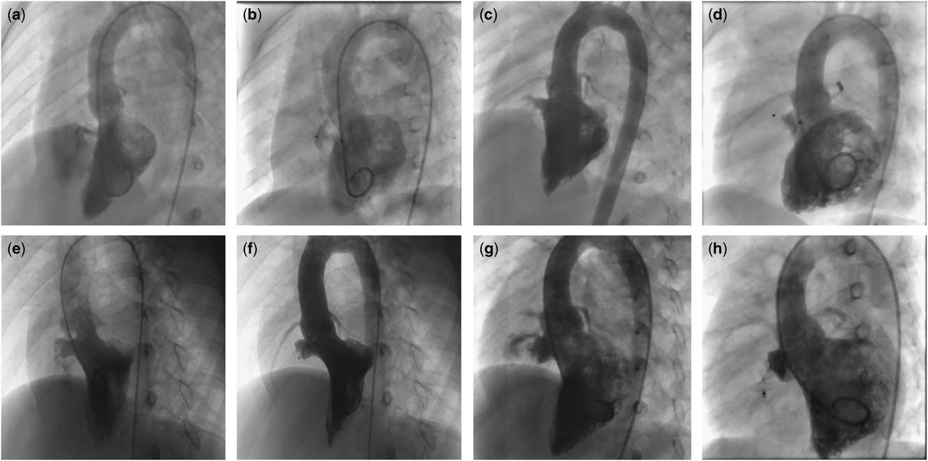
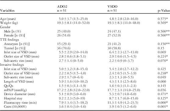
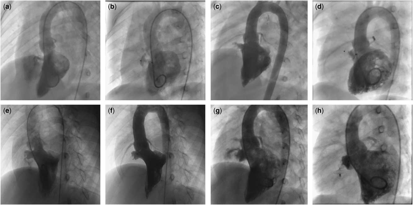
A cohort of 102 patients, of whom 49 were male, was enrolled in this study. Among them, 50 patients were implanted with Amplatzer duct occluder 2 (see Fig 1), and 52 patients were implanted with domestic concentric ventricular septal occluder. The mean age of all the patients was 4.9±3.3 years (range: 1.5–25) and the mean weight was 19.3±8.2 kg (range: 11–52). The mean inlet, outlet, and sub-aortic rim of defects was 6.0±2.6 mm (range: 2.0–14.0), 2.8±0.6 mm (range: 1.5–5.1), and 2.1±1.2 mm (range: 0–5.0) according to echocardiography before operation, respectively. In all, 35.3% (36/102) of the patients were complicated with a septal aneurysm, and one patient was associated with patent ductal arteriosus that was closed simultaneously. A total of 102 devices were implanted. The size of implanted devices ranged from 3 to 6 mm. The detailed baseline characteristics of the two groups are listed in Table 1.
Transcatheter closure of various types of perimembranous ventricular septal defects using Amplatzer duct occluder 2. ( a and b ) Left ventriculography shows small tubular-shaped membranous septal defect in a 5-year-old boy, and control ventriculography showed no residual shunts after one 5/4 mm Amplatzer duct occluder 2 was deployed. ( c and d ) Angiography shows perimembranous septal defect with an aneurysm in a 2.4-year-old girl, and one 6/4 mm Amplatzer duct occluder 2 was successfully implanted with excellent control angiography. ( e and f ) Angiography shows septal aneurysmal defect with a single exit in a 10-year-old girl, which was completely closed by one 5/4 mm Amplatzer duct occluder 2. ( g and h ) A large septal aneurysm is observed with centralised exits in a 3-year-old boy, which was completely closed by one 6/4 mm Amplatzer duct occluder 2; control angiography shows excellent results.
Baseline characteristics of the patients between the two groups.

ADO2=Amplatzer duct occluder 2; mPAP=mean pulmonary artery pressure; Qp/Qs=ratio of pulmonary flow and system flow; TTE=transthoracic echocardiogram; VSD=ventricular septal defect; VSDO=ventricular septal defect occluder
Data are expressed as n (%) or mean±SD (range)
* Z values
** χ2 values
The failed patient was a 5-year-old girl with perimembranous ventricular septal defect who exhibited two disperse exits. Initially, we attempted to use two Amplatzer duct occluder 2 devices to close the exits, independently; however, the device was easily pulled into the left ventricle when closing one of the exits measuring about 4 mm. Consequently, we replaced a 12-mm symmetric device to close the inlet orifice successfully according to the base diameter (11 mm).
A retrograde approach without Arterial–Venous loop establishment was attempted in all patients implanted with Amplatzer duct occluder 2; the approach was successful in 40 patients, and 10 patients failed because the delivery sheath could not traverse the defect from the left ventricle. Then, all the 10 patients were changed to conventional antegrade approach and appropriate Amplatzer duct occluder 2 was implanted smoothly after establishing the Arterial–Venous loop. The fluoroscopy time and cost differed significantly between the two groups (see Table 1).
Complications
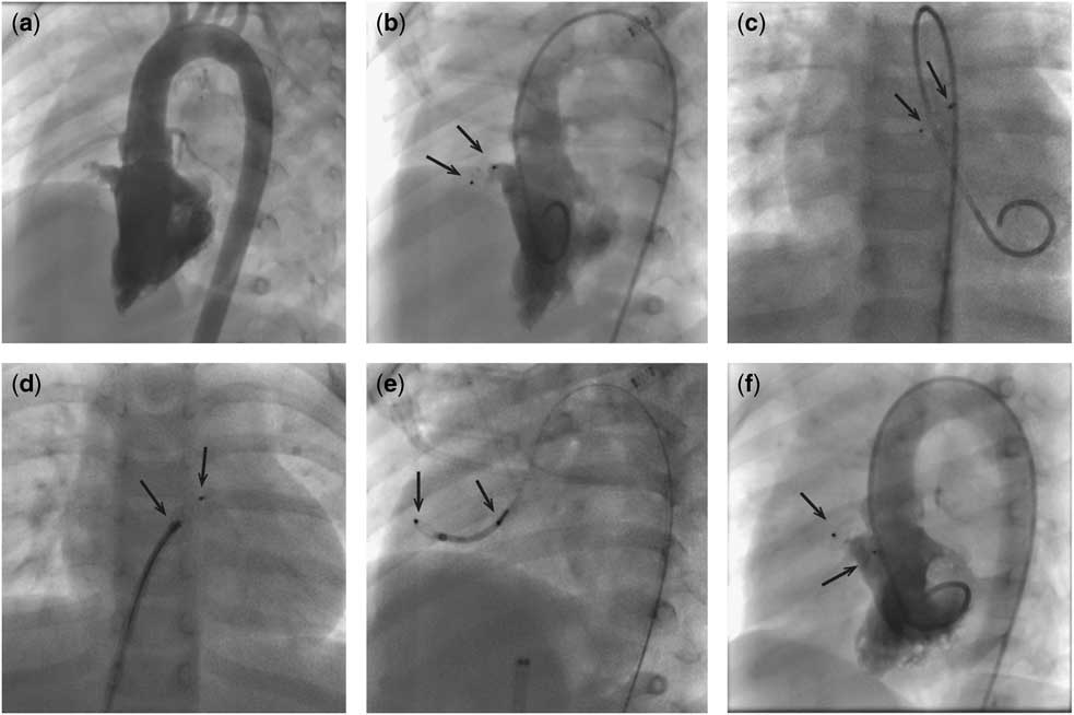
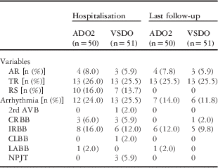
Device embolisation occurred in one patient who presented with perimembranous ventricular septal defect with a large septal aneurysm. We retrogradely deployed one 4/4 mm Amplatzer duct occluder 2 device but failed to pull into the aneurysm sac (right ventricular side); moreover, the angiography by hand and transthoracic echocardiogram both failed to confirm the precise position. After release, the device dislodged into the right ventricular outflow tract, and then it was retrieved by a goose-neck snare, and repeatedly deployed into the aneurysm sac (Fig 2). No death and severe arrhythmia, such as complete atrioventricular block needing a pacemaker, occurred. No vascular complications such as thrombosis, dissection, or an aneurysm occurred. Minor complications including small residual shunt, valve insufficiency, and minor arrhythmia were observed. None of these complications need to be cured. The overall arrhythmia incidence had no significant statistical difference between the two groups. Right branch block was the most common type in both groups; however, no atrioventricular block and left branch block were observed in the Amplatzer duct occluder 2 group. The main complications are listed in Table 2. No significant differences were observed in the residual shunt, aortic regurgitation, and tricuspid regurgitation between the two groups.
Embolisation of Amplatzer duct occluder 2 owing to inappropriate deployment position. Left ventricular angiography showed perimembranous ventricular septal defect with aneurysm in a 2.4-year-old girl ( a ). The base diameter of the aneurysm measured 9.2 mm with several small exits on the right side. A 4/4 mm Amplatzer duct occluder 2 was retrogradely implanted but failed to be deployed into the aneurysm cavity correctly ( b ). The device dislodged into the main pulmonary artery after release ( c ). The device was pulled out from the femoral vein by a snare ( d ). The device was repeatedly implanted via retrograde approach ( e ). Angiography showed the device being deployed into right position with excellent closure result ( f ).

Comparison of complications between Amplatzer duct occluder 2 (ADO2) and ventricular septal defect occluder (VSDO) groups during hospitalisation and last follow-up.

2rd AVB=second degree of atrioventricular block; AR=aortic regurgitation; CLBB=complete left bundle branch block; CRBB=complete right branch block; IRBB=incomplete right branch block; LABB=left-anterior branch block; NPJT=non-paroxysmal junctional tachycardia; RS=residual shunt; TR=tricuspid regurgitation
Follow-up results
Electrocardiogram, transthoracic echocardiogram, and chest X-ray examination were conducted on the day after the procedure, and repeated at 1, 3, 6, and 12 months, subsequently. The follow-up data were collected and analysed. No new onset or aggravated complications were observed during a median follow-up of 26.2 months. The instant closure rate was 80% (40/50) by angiograpy in the Amplazter duct occluder group, and increased to 84% during discharge, and then 92, 96, and 100% at 1, 3, and 6 months of follow-up by echocardiography, respectively. Tricuspid regurgitation and aortic regurgitation remained unaltered until this manuscript was drafted. In addition, one patient in the membranous ventricular septal occluder group developed transient second-degree atrioventricular block, which automatically restored to normal in 3 days. Three patients developed non-paroxysmal junctional tachycardia and all recovered in 1 week. The outcomes of various complications at last follow-up are listed in Table 2.
Discussion
Compared with the other simple CHD, transcatheter closure of perimembranous ventricular septal defect is more challenging owing to variable anatomical morphology and complex manipulation process. A conventional intervention closure technique of ventricular septal defect is needed to establish the Arterial–Venous loop, which involves complex steps. Occasionally, the delivery sheath is difficult to pass through the small defect or be positioned into the left ventricular apex. The procedure often fails for those small defects through which an extremely small delivery sheath cannot pass.
Amplatzer duct occluder 2 is a double-disk appliance made by nitinol mesh wire without the filling of the fabric materials, designed to occlude small type patent ductus arteriosus. It has more flexible profile than the conventional device, and the delivery sheath is small – only four or five French is needed. The size of the occluder is 3–6 mm in diameter with 4- or 6-mm waist length, and can be extended up to 8 and 12 mm in maximum length, respectively. Owing to the softer profile and small delivery sheath, Amplatzer duct occluder could be deployed via retrograde approach (arterial side) or antegrade approach (venous side).
Our preliminary findings confirmed that the transcatheter closure of perimemebranous ventricular septal defect using Amplatzer duct occluder 2 was safe and effective with low medical cost and relatively simple manipulation in selected patients.
We preferred to deploy Amplatzer duct occluder 2 retrogradely because of simple manipulation and less material consumption. The retrograde approach was successful in 40 patients of this group but failed in 10 patients as the delivery sheath failed to cross the defect from the left ventricle side. Thus, we changed the traditional approach establishing the Arterial–Venous loop following which the delivery sheath was introduced from the venous side to complete the closure subsequently using Amplatzer duct occuder 2. The implantation of the delivery sheath of Amplatzer duct occluder 2 was extremely flexible. In this study, we compared the fluoroscopy time and costs of both techniques. The exposure time of the retrograde approach was shorter and the therapy was cost-effective as compared with that of the antegrade approach. The differences in the two methods were statistically significant (p<0.05). The exposure time and costs of the amplatzer duct occluder 2 group were also less than those of the ventricular septal defect occluder group (p<0.05); the average spared cost of the Amplatzer duct occluder 2 group was about ¥3000, which primarily aroused from goose-neck snare, which was an important consideration for limited budgets in a developing country. Diminished exposure time might also decrease the chances of mechanical damages and blood-loss.
Most of the patients in this series are small type defect with mild haemodynamic significance, which means no significant pulmonary hypertension or heart failure; however, all of them had heart murmur more than 2/6 grades. Patients with small defect exhibit the risk of developing infective endocarditis cl. Soufflet et al followed up a cohort of patients with small type ventricular septal defect for 6 years; approximately 4% of the patients developed infective endocarditis. Moreover, the persistent heart murmur may cause a sense of inferiority, which harms the psychological development of the child. On the other hand, the interventional closure was proved to be safe, effective, and less invasive. Therefore, we advised the patients with small type ventricular septal defect to undergo interventional closure before school entrance.
The most common complication of the transcatheter closure of perimembranous ventricular septal defect is heart block,Reference Zuo, Xie and Yi 10 – Reference Zhou, Pan, Guan and Ge 12 with the incidence of complete atrioventricular block about 0.3%. Although the precise mechanism is not very clear, one of the potential reasons might involve mechanical damage to conduction systems caused by hard profile of the conventional occluders. Long procedural time may also increase the chance of damage to conduction tissues. In this series, although the right branch block was common in patients implanted with Amplatzer duct occluder 2, no severe arrhythmias such as atrioventricular block or left bundle branch were observed. The Amplatzer duct occluder 2 seemed safer than the conventional devices in arrhythmia prevalence after the procedure owing to its softer profile, which remained to be confirmed by large sample studies.
Residual shunts
Residual shunts are quite common in device closure of perimembranous ventricular septal defect, especially for an aneurysm with multiple defects.Reference Yang, Tai, Khin and Quek 13 Amplatzer duct occluder 2 is specifically designed to occlude the small type arterial duct (<5.5 mm as per the manual’s instructions). Therefore, residual shunt should be especially concerned when it was off-label used in perimembranous ventricular septal defect. There were 10 patients in the Amplatzer duct occluder group who developed instant minor residual shunts by angiography; only eight remained at discharge and all of them resolved within the 6-month follow-up. The residual shunts presented benign courses with no haemodynamics significance and no mechanical haemolysis occurred. No significant statistical difference was observed as compared with the conventional device group. What sized defect is indicated for Amplatzer duct occluder 2 was still not clear. We once failed to close a defect sized about 4 mm in diameter with the 6/4 mm Amplatzer duct occluder which was easily pulled out of the defect before detachment. We conclude that Amplatzer duct occluder 2 was not indicated for a defect more than 4 mm.
Valve regurgitation
The local anatomy of perimembranous ventricular septal defect is sophisticated and adjacent to the tricuspid and aortic valve. The device implantation may affect the surrounding valve, causing valve insufficiency. As the disk is 4 mm larger than the waist, sufficient distance between the defect and the aortic valve is essential to avoid aortic regurgitation. The ideal distance from the defect to the aortic valve is >4 mm. A previous study suggested that at least 3 mm distance was safe for Amplatzer duct occluder 2 deployment.Reference Kanaan, Ewert, Berger, Assa and Schubert 14 However, in the current cohort, 2 mm distance was also safe for Amplatzer duct occluder 2 deployment without causing aortic insufficiency. The soft property of Amplatzer duct occluder 2 appeared to have little effect on the aortic valve. This might be due to the variable measurement methods among different centres. Nevertheless, a careful examination by an experienced echocardiologist before the procedure is essential for the selection of eligible patients. A minimum of 2 mm distance from the defect to the aortic valve is indicated to deploy the Amplatzer duct occluder 2 device in our studies. Before the release of the device, transthoracic echocardiography was performed regularly to confirm that the aortic valve was not affected. In addition to aortic valve insufficiency, the tricuspid valve is also crucial as the large protruding right disk might interfere with the septal valve movement. Recently, late Amplatzer duct occluder 2-related tricuspid regurgitation had been reported.Reference Zhao, Yu and Gao 7 In the present series, there is no statistically significant difference in the incidence of aortic and tricuspid regurgitation between the two groups.
In our experiences, the main conditions on perimembranous ventricular septal defect closure with Amplatzer duct occluder 2 are as follows: first, small tubular or tunnel-shaped defect with the effective shunt diameter is <4 mm; second; there is sufficient distance from the defect to the aortic valve (>2 mm) to avoid causing aortic regurgitation; third, perimembranous ventricular septal defect with a aneurysm had centralised small exits on the right side that can also be indicated; and fourth, the long length (⩾5 mm) of the tunnel or aneurysm of defects is especially suitable. Although two types of devices with waist lengths of 4mm and 6 mm are available, the 4 mm-waist-length type device was optimal for perimembranous ventricular septal defect. Short waist length design may decrease chances to interfere with tricuspid valve or obstruct right ventricular outflow tract. All the patients in our series received the 4 mm-length waist device successfully. Finally, the delivery sheath tends to bend following withdrawal of the guide wire, in which case,we prefer pulling back the sheath slightly and push the device forward at the same time so as to pass through the kink site. In most cases, the device can pass the kinked part without the need for a replacement with another sheath.
Conclusions
Amplatzer duct occluder 2 has advantages of simple manipulation and less medical costs compared with conventional device in transcatheter closure of small type perimembranous ventricular septal defect.
Acknowledgements
None.
Financial support
This work was supported by Major Research Project of Shandong Province (Science and Technology Key Project): Integrated management of CHD before and after delivery (2016GSF201101).
Conflicts of Interest
None.
Ethical Standards
The study protocol was approved by the Ethics Committee of Shandong Provincial Hospital Affiliated to Shandong University. Written informed consents were obtained from the parents or guardians of the children.