Introduction
Antrochoanal polyps are the commonest variety of choanal polyps, and present most commonly with unilateral nasal obstruction.Reference Robson, Barker and Whittet1–Reference Franche, Granzotto, de Borba, Hermes, Saleh Cde and de Souza4 Several researchers have reported epistaxis as a rare presentation.Reference Robson, Barker and Whittet1, Reference Gendeh, Long and Misiran3–Reference Hardy6 However, how this oedematous connective tissue polyp causes epistaxis is unclear. Robson et al. have described the histological appearance of an antrochoanal polyp presenting with severe epistaxis.Reference Robson, Barker and Whittet1 The specimen showed localised areas of haemorrhage with the formation of abundant granulation tissue; an appearance similar to angiomatous choanal polyps reported by others.Reference Robson, Barker and Whittet1, Reference Batsakis and Sneige2, Reference Som, Cohen, Sacher, Choi and Bryan7–Reference De Vuysere, Hermans and Marchal9 The diagnosis and pathogenesis of angiomatous polyps are not clearly understood, with few published reports. Batsakis and Sneige ascribed it (pathogenesis of angiomatous polyps) to vascular compromise of choanal polyps, due to their origin within a confined space, passage through constrictive ostia, and dependent position within the nasal cavity, choanae or nasopharynx.Reference Batsakis and Sneige2 They stated that ‘this vascular compromise sets up the following sequence: dilatation and stasis of feeder vessels; edema; infarct; neo-vascularization; repeat occlusion and infarct, and the end result is either total necrosis or, more often, an angiomatous polyp’.Reference Batsakis and Sneige2
Over an eight-year period, we recorded the gross and histological appearances of antrochoanal polyps presenting with recurrent, ipsilateral epistaxis, and compared them to those of antrochoanal polyps without a history of bleeding, in order to inform the diagnosis and proper management of these unusual cases.
Materials and methods
All patients presenting with antrochoanal polyps to the out-patient clinic of the Sohag University Hospital ENT department were prospectively included in the study. These patients underwent history-taking, ENT examination, endoscopic examination with 0° and 30° telescopes, computed tomography (CT) scanning of the nose and paranasal sinuses, and endoscopic examination under general anaesthesia, in order to detect the following gross diagnostic criteria for antrochoanal polyp: (1) unilateral, polypoid nasal mass of maxillary sinus origin and with an intrasinus component; (2) a well defined, narrow pedicle protruding through the sinus ostium; (3) the course of the polyp is directed backwards towards the choana between the middle turbinate and the lateral nasal wall, into the nasopharynx; (4) a smooth, glistening surface with a grey-white colour; (5) a characteristic soft rubbery texture; and (6) on CT, a soft tissue shadow involving the maxillary sinus and posterior part of the nasal fossa and nasopharynx, with intact bony confines of the sinus.
All cases were managed with endoscopic sinus surgery involving middle meatal antrostomy, under general anaesthesia, to remove the nasal and intrasinus parts of the polyp.
In patients presenting with ipsilateral epistaxis, multiple biopsies were obtained from resected polyps. Control biopsies were also obtained from a comparable number of ordinary antrochoanal polyps from patients presenting without epistaxis. All biopsy specimens were fixed in 10 per cent formalin, processed into paraffin blocks, sectioned (4 µm thick) and stained with haematoxylin and eosin, according to standard procedures. The histological features observed on light microscopy were recorded. In addition, the number of predominant cells in the corium, just beneath the epithelium, was calculated in five different fields (at ×400 magnification) for each specimen.
Statistical comparison of the epistaxis and non-epistaxis groups was performed using the Student t-test. Calculations were performed using the Statistical Package for the Social Sciences for Windows version 10.0 software package. Statistical significance was defined as p < 0.05.
Results
Over an eight-year period between January 2000 and December 2007, we encountered 84 patients with lesions that met the gross diagnostic criteria for antrochoanal polyp. The age range was eight to 60 years, with a mean age of 22.1 years, and the male/female ratio was 49/35. Recurrent, ipsilateral epistaxis was the presenting symptom in 10 patients (10/84; 11.9 per cent), who were interpreted initially as having a neoplastic vascular nasal mass. However, endoscopic examination, CT scanning of the nose and paranasal sinuses, and endoscopic examination under general anaesthesia indicated that these masses satisfied the gross diagnostic criteria of antrochoanal polyp; in addition, these lesions had a reddish, vascular surface in parts (Figure 1). Endoscopic sinus surgery with middle meatal antrostomy was an adequate procedure in these cases. Polyp removal resulted in little more bleeding than usual, and this bleeding was easily controlled.
Freshly resected angiomatous antrochoanal polyps, displaying a reddish, vascular surface in some parts and a pale surface in others. One polyp's angiomatous part is bisected (c) to demonstrate a highly vascular cut surface. Note the well defined, narrow pedicle (P) in all specimens.

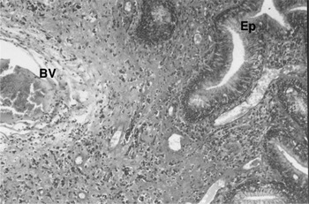
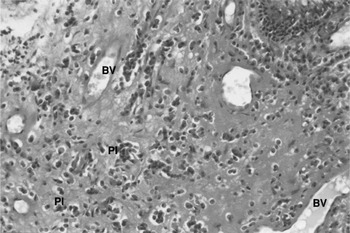
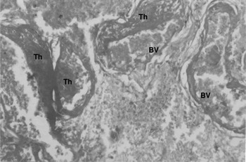
Histological examination of polyp biopsy specimens from patients presenting with epistaxis showed a covering of partially ciliated, pseudostratified, columnar epithelium (Figure 2). The lamina propria was highly vascular, with multiple dilated blood capillaries and engorged veins (Figures 2 to 5), confirming the diagnosis of angiomatous antrochoanal polyp. Marked cellular infiltration with plasma cells, macrophages, lymphocytes and eosinophils was observed, with a predominance of plasma cells (Figure 3) with their characteristic eccentric nucleus and basophilic cytoplasm (Figure 4). Thrombi at different stages of development appeared in some fields of the examined specimens, with a large number of extravasated red blood cells (Figure 5). No infarcts were noted in any of the examined specimens.
Photomicrograph of an angiomatous antrochoanal polyp showing pseudostratified, partially ciliated, columnar epithelium (Ep) and highly vascular stroma. Note the large blood vessel (BV) (H&E; ×100).

Photomicrograph of an angiomatous antrochoanal polyp showing highly vascular stroma with multiple dilated capillaries (BV). The stromal cells are mostly plasma cells (Pl) (H&E; ×200).

High-power photomicrograph of an angiomatous antrochoanal polyp showing highly vascular stroma (BV) and plasma cells (Pl) with their characteristic eccentric nucleus and basophilic cytoplasm (H&E; ×1000).

Photomicrograph of an angiomatous antrochoanal polyp showing numerous dilated and engorged veins (BV). Note the thrombi (Th) inside most of the vessels, at different stages of organisation (H&E; ×400).

The remaining patients (74/84; 88.1 per cent) had conventional antrochoanal polyps, and no history of epistaxis. Histological examination of polyp biopsy specimens from these patients showed a covering of respiratory epithelium, with areas of ulceration and/or squamous metaplasia, and an oedematous connective tissue core infiltrated with a few plasma cells (Figure 6).
Photomicrograph of an ordinary antrochoanal polyp showing a covering of partially ulcerated, pseudostratified, columnar, ciliated epithelium (Ep) with an oedematous connective tissue core (Ed) and a few plasma cells (Pl) (H&E; ×200).

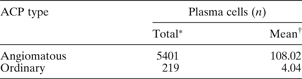
The increased number of plasma cells in the angiomatous antrochoanal polyps, compared with the ordinary antrochoanal polyps, was highly statistically significant (p < 0.00) (Table I).
Plasma cells in angiomatous and ordinary antrochoanal polyps
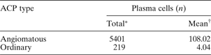
*Over 50 high-power fields (five in each of 10 specimens). †Per high-power field; p < 0.00. ACP = antrochoanal polyp
Discussion
On rare occasions, antrochoanal polyps present with epistaxis, which may confuse the diagnosis and arouse suspicion of a vascular neoplasm, with the consequent risk of mismanagement.Reference Robson, Barker and Whittet1, Reference Hardy6 In our study, 10 patients (11.9 per cent) presented with recurrent, ipsilateral epistaxis; all had antrochoanal polyps of the angiomatous type. Not a single patient with an ordinary antrochoanal polyp presented with epistaxis. It would thus appear that only angiomatous antrochoanal polyps are associated with epistaxis. Bleeding can be explained by the rupture of engorged vessels on the surface of this vascular polyp. The large number of extravasated red blood cells seen in the examined specimens confirms this rupture. The diagnosis of angiomatous antrochoanal polyp is based on gross diagnostic criteria, detected on endoscopic examination and aided by CT scanning. These polyps have the same characteristic gross findings as ordinary antrochoanal polyps, with the addition of a reddish, vascular surface in some areas, an empty pterygopalatine fossa on CT, and epistaxis as a presenting feature. Histological examination confirms the diagnosis, without the need for angiography. Correct diagnosis helps to avoid inappropriate surgery for a misdiagnosed vascular neoplasm. Endoscopic sinus surgery is sufficient treatment for angiomatous antrochoanal polyps, and bleeding can be easily controlled.
The pathogenesis of angiomatous antrochoanal polyp is not clearly understood. Batsakis and Sneige ascribed it to vascular compromise, but this theory is not supported by anatomical, clinical or histological findings.Reference Batsakis and Sneige2 Despite the fact that the maxillary sinus is a confined space; yet, the intrasinus part of ACP does not completely fill the sinus cavity in most cases, so not amenable for constriction within the sinus. In most cases, the sinus ostium is larger than normal, and the polyp pedicle is narrow and not liable to constriction. Moreover, many antrochoanal polyps grow to a large size and become more dependent, with no angiomatous changes. Indeed, vascular compromise, due to occlusion or compression of feeder vessels in the polyp, should lead to necrosis rather than angiomatous change. This view is supported by Ole-Lengine and Manni, who reported a strangulated, black, antrochoanal polyp consisting of two parts – a blackish, choanal part luxated out of the nostril on blowing, and a pinkish, antral part – joined together at the site of strangulation.Reference Ole-Lengine and Manni10 In our study, examined specimens showed no signs of infarction, even those with evident thrombosis.
• Antrochoanal polyp is the commonest variety of choanal polyp, and presents most commonly with unilateral nasal obstruction
• This study investigated the gross and histological appearance of antrochoanal polyps associated with recurrent epistaxis, compared with antrochoanal polyps with a more typical presentation
• Antrochoanal polyps presenting with epistaxis had distinctive histological features, with a highly vascular stroma, and were referred to as angiomatous antrochoanal polyps
• Such polyps had a significantly increased number of plasma cells, which may be the underlying cause of their distinctive histological changes
The most obvious histological features of the observed angiomatous antrochoanal polyps were a highly vascular stroma and a rich inflammatory cellular infiltration (mostly plasma cells); this contrasted with the oedematous core and sparse cellular infiltration seen in ordinary antrochoanal polyps. The difference between the number of plasma cells in both polyp types was highly statistically significant (p < 0.00). This is an interesting finding. To investigate this finding, we searched the Pubmed, Medline, Ovid, Sciencedirect and WinSPIRS databases, from 1996 to 2012, using the keywords ‘predominant AND plasma cells AND antrochoanal polyp OR nasal polyps’. We could find no previous report of a predominance of plasma cells in the cellular infiltrate of a nasal polyp, either for antrochoanal polyps or nasal polyposis. Wittekindt et al. and Ito et al. have linked plasma cells to angiogenesis and vascular endothelial proliferation within nasal polyps, through expression of vascular endothelial growth factor and of vascular proliferating factor and vascular endothelial growth factor mRNA.Reference Wittekindt, Hess, Bloch, Sultanie and Michel11, Reference Ito, Hirota, Mizuno, Kawasaki, Takemura and Nishiura12 This suggests that the significantly increased number of plasma cells detected in our patients' angiomatous polyps may be the underlying cause of the histological changes seen in this type of polyp.
The distinct clinical presentation and gross and histological features of angiomatous antrochoanal polyps indicate that they are a distinct polyp type and not a derivative of ordinary antrochoanal polyps.
Conclusion
It appears that only the angiomatous variant of antrochoanal polyp presents with epistaxis. This polyp is a distinct type and not a derivative of the ordinary antrochoanal polyp. Detection of the characteristic gross findings of an angiomatous antrochoanal polyp will help prevent misdiagnosis as a vascular neoplasm and inappropriate surgery. Angiography is unnecessary; histopathological analysis confirms the diagnosis.
The significantly increased number of plasma cells seen in angiomatous antrochoanal polyps may be the underlying cause of the histological changes observed in this polyp.
Acknowledgement
The authors sincerely acknowledge Mr Liam M Flood FRCS, ENT consultant, James Cook University Hospital, Middlesbrough, UK, for manuscript review and helpful comments.