Introduction
Obesity is a multifactorial disease in which both genetic predisposition and poor regulation of energy play an important role in its development.Reference Chan and Woo1 Several studies have shown that obesity favors the development of other diseases, such as type 2 diabetes,Reference Pi-Sunyer2 cardiovascular diseases,Reference Kotsis, Jordan and Micic3 musculoskeletal disorders,Reference Collins, Herzog and MacDonald4 and some types of cancer.Reference Argolo, Hudis and Iyengar5
In recent years, poor diets and a reduction in physical activity have been shown to induce a big increase in obesity prevalence worldwide.Reference Cohen6,Reference Jiménez-Pavón, Kelly and Reilly7 Likewise, obesity in pregnant women exposes fetuses to an obesogenic intrauterine environment.Reference Zambrano, Ibáñez and Martínez-Samayoa8 Maternal obesity increases maternal and offspring complications and descendants present an increased risk of becoming obese during childhood and/or adulthood.Reference Taveras, Rifas-Shiman and Belfort9 Besides, maternal obesity predisposes the growing fetus to the development of metabolic diseases.Reference Khan, Dekou and Hanson10 Diverse programming effects induce specific metabolic derangements that have been demonstrated across species. Altered expression profiles of growth, proliferation, and circadian rhythm factors; altered antioxidant enzyme actions; and the induction of increased atherogenesis, blood pressure, vascular activity, and heart rate have been demonstrated.Reference Wright, Rifas-Shiman and Rich-Edwards11,Reference Eberle, Merki and Yamashita12 Similarly, in rodents, the change in the mother’s gestational metabolism can affect placental and embryonic development, yielding an increased inflow of nutrients to the fetus and altering future development.Reference Jones, Woollett and Barbour13
Regarding other animal models, it has been reported that male offspring from obese mothers present a significant increase in serum glucose, insulin, leptin, very low-density lipoprotein, apolipoprotein B100, and lipid levels, as well as a permanent reduction in hepatic β-oxidation and an increase in hepatic lipogenesis.Reference Zambrano and Nathanielsz14
However, it has been described that the use of various natural phytochemicals, present in food, help in the prevention of various chronic disorders, such as cancer, cardiovascular diseases, and inflammatory and metabolic diseases, including obesity.Reference Meydani and Hasan15 Within these phytochemicals, (-)-epicatechin (Epi), a flavonoid present in Camellia sinensis, peanuts, apples, grapes, and in cocoa,Reference Harnly, Doherty and Beecher16 has shown to exert beneficial effects on health by activating Endothelial Nitric Oxide synthase (eNOS),Reference Ramirez-Sanchez, Maya and Ceballos17 reducing blood pressure,Reference Schroeter, Heiss and Balzer18 improving oxidative capacity, and increasing resistance to fatigue.Reference Nogueira, Ramirez-Sanchez and Perkins19 Besides, it has an antioxidant role by increasing the level of antioxidant enzymesReference Ramirez-Sanchez, De los Santos and Gonzalez-Basurto20 as well as improving heart structureReference De Los Santos, Palma-Flores and Zentella-Dehesa21 and cardiac function.Reference De Los Santos, García-Pérez and Hernández-Reséndiz22
The use of Epi in high-fat diet (HFD) obesity models has been shown to decrease body weight gain,Reference Hoek-van den Hil, van Schothorst and van der Stelt23 hyperglycemia, and hypertriglyceridemia.Reference Gutiérrez-Salmeán, Ortiz-Vilchis and Vacasey del24 Epi also has beneficial effects by preventing adipose tissue inflammation and insulin resistance.Reference Sano, Nagayasu and Suzuki25–Reference Bettaieb, Cremonini and Kang28 At present, to our knowledge, no study has explored whether Epi has this same impact in the male offspring from maternal obesity rats induced by HFD. Therefore, the purpose of our study was to determine whether Epi can reduce the amount of visceral adipose tissue and improve the metabolic profile in the male offspring of a maternal obesity model.
Methods
Model
The animal research protocol followed in the present study was approved by the Animal Experimentation Ethics Committee of the Instituto Nacional de Ciencias Médicas y Nutrición Salvador Zubirán (INCMNSZ) (CINVA: UIO-1892-17/19-1) and by the Research and Ethic Commissions of the Universidad Nacional Autónoma de México. All procedures were performed in accordance with the Guidelines for the Care and Use of Laboratory Animals of the Institute of Laboratory Animal Resources (https://www.nal.usda.gov/awic/animal-welfare-act), approved in Mexico by the National Academy of Medicine and in accordance with the Mexican Official Standard Norm NOM-062-ZOO-1999.
Standardization of phenotype of females (F0) was used to produce F1 offspring.
Female albino Wistar rats bred in the bioterium of the INCMNSZ were maintained at 22°C–23°C under controlled lighting (lights on from 07:00 to 19:00 h) and were fed with chow diet, containing 23.9% protein, 5.0% fat, 57.9% carbohydrates, 5.1% fiber, 7.0% minerals, and 1.0% vitamins (w/w), with energy 4.07 kcal/g (Purina 5001, Labdiet, St. Louis, Missouri, USA). At the age of 14–16 weeks, that is, postnatal day (PND) 98–112, when they weighed between 200 and 240 g, females were randomly bred with males from other mothers.
The study was performed in two stages. The first stage was performed at weaning (PND 21), in which one female F0 pup from each litter was randomly assigned to either a control (C, n = 8) group that received the laboratory chow diet or to a maternal obesity (MO, n = 8) group fed with a HFD containing 23.5% protein, 20.0% animal lard, 5.0% fat, 20.2% polysaccharide, 20.2% simple sugars, 5.0% fiber, 5.0% mineral mix, and 1.0% vitamin mix (w/w), providing energy 4.9 kcal/g. A total of eight rats were included in each group.
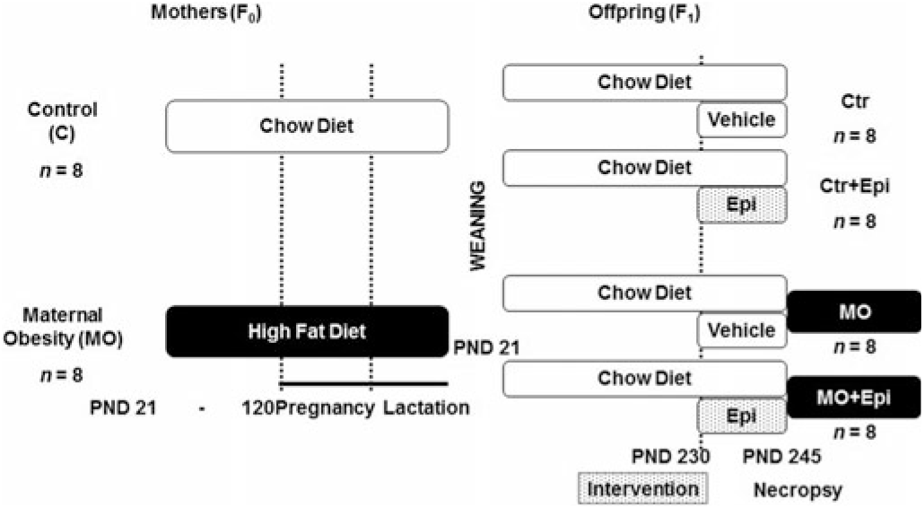
The eight F0 female rats were placed with proven male breeders on PND 120, and they conceived during the next cycle. To minimize consumption of the HFD by males during the mating period, they were placed with females at night and removed during the morning. Lactating mothers were maintained on their pregnancy diet (Fig. 1).
Timeline on Epi intervention in male offspring of the MO at PND 230 (F1). Eight female rats (F0) per group were fed with control or obesogenic diets at weaning and during pregnancy and lactation. At weaning, male offspring (F1) ate control diet throughout the study. At PND 230, 16 males per group (C and MO) from different litters were randomly allocated, to be treated with water or with Epi, twice per day for two weeks (Ctr, C + Epi, MO, MO + Epi, n = 8 rats per group).

Stage two, F1 offspring
Litter size and pup weight were recorded at birth. The anogenital distance was measured to identify males and females.Reference Zambrano, Bautista and Deás29 To ensure F1 offspring homogeneity, on PND 2, all litters studied were adjusted to 10 pups with equal numbers of males and females. Offspring were weaned at PND 21, housed four rats per cage, and fed chow diet throughout the study. Only male offspring were studied. At PND 230, male pups were weighed, and 16 animals per group (2 from the same litter) were randomly selected to the control group (Ctr group = 496.72 ± 58.24 g, n = 8) or to the control group treated with Epi (Ctr + Epi group = 495.13 ± 55.04 g, n = 8) or to the maternal obesity group (MO group = 492.62 ± 70.53 g, n = 8) or to the maternal obesity group treated with Epi (MO + Epi group = 496.21 ± 73.83 g, n = 8) (Fig. 1).
(-)-Epicatechin intervention
At PND 230, male offspring were provided with either vehicle (water) or 1 mg/kg body mass of Epi (Sigma-Aldrich, St Louis, Missouri, USA) by oral gavage twice daily (morning and evening) for 15 days as previously described.Reference De Los Santos, Palma-Flores and Zentella-Dehesa21,Reference De Los Santos, García-Pérez and Hernández-Reséndiz22 No adverse effects were recorded as a result of this dietary intervention.
Food intake
Rats from the same experimental group were housed two per cage. Food intake was measured daily throughout the interventions and was determined as the difference between the food supplied and the amount of food left in the cage after 24 h. The amount consumed was averaged between both rats.
Offspring blood samples and retrieval of organs
At PND 245, after 6 h of fasting, rats were anesthetized with isoflurane (based on the instructions for the use of the isoflurane inhaler of the brand: Stoelting Co, Cat. No. 50207 according to animal weight) and euthanized. Blood was collected, by cardiac puncture, to obtain serum. To calculate the adiposity index, sternal, peripancreatic, retroperitoneal, mesenteric, and gonadal fat were dissected and weighed individually. This index was calculated by dividing the total adipose tissue (g) by body weight (g). Visceral adipose tissues were stored at –70°C for further analysis.
Quantification of adipocyte size
Paraffin sections of retroperitoneal fat were stained with hematoxylin and eosin as described by Fischer et al.,Reference Fischer, Jacobson, Rose and Zeller30 and analyzed with an Axiophot 2 microscope (Carl Zeiss AG, Oberkochen, Germany), Cannon PowerShot A75 camera (Canon Inc, Lake Success, New York, USA). For each animal sample, 30 cells of five nonadjacent areas were evaluated. About 1200 cell areas per group (150 per animal, n = 8) were measured manually by delimiting the adipocyte cross-sectional area using AxioVision® software for digital imaging processing (Zeiss® copyright 2006–2010, Stuttgart, Germany).Reference De Los Santos, García-Pérez and Hernández-Reséndiz22
Biochemical analyses
Serum glucose, triglycerides, and cholesterol of the male offspring were determined enzymatically with a Synchron CX auto analyzer (Beckman Coulter, Fullerton, California, USA). Intra- and inter-assay coefficients of variation (CV) were < 2% and < 3%, respectively.
Serum insulin concentrations were determined using a commercially available enzyme-linked immunosorbent assay (ELISA) kit (EZRMI-13K, Merck, Darmstadt, Germany). The intra- and inter-assay CVs were < 4% and < 6%, respectively; furthermore, the insulin resistance index (IRI) was assessed by the homeostasis model assessment (HOMA) calculated from the formula IRI = glucose (mmol l−1) × insulin (μU ml−1)/22.5.Reference Zambrano, Bautista and Deás29,Reference Vega, Reyes-Castro and Rodríguez-González31
Finally, serum leptin was determined by ELISA (KRC2281, Thermo Fisher Scientific, Waltham, Massachusetts, U.S.). The intra- and inter-assay CVs were < 5% and < 7%, respectively.
Statistical analyses
Values are expressed as mean ± SD. All data were analyzed with the GraphPad Prism 6.0 software (GraphPad Software, San Diego, California, USA). Sample size and power were calculated according to Dell et al.Reference Dell, Holleran and Ramakrishnan32
Data distribution was tested using the Kolmogorov-Smirnov test. Kruskal-Wallis with the post hoc Conover method was used to determine fat cell size differences, and ANOVA with the post hoc Tukey HSD Test analysis for pair comparison was used to determine differences between groups for weight, adiposity index, fat depots, and metabolic parameters; P value < 0.05 was accepted as statistically significant.
Results
Biochemical parameters
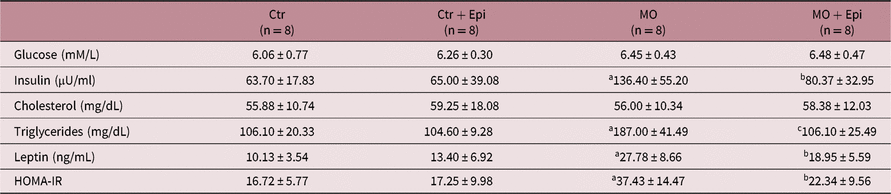
There were no differences in serum glucose nor in cholesterol in any of the four male offspring groups. Nevertheless, male offspring of MO group presented elevated insulin and HOMA-IR as well as elevated serum concentrations of triglycerides and leptin when compared with the male offspring of both Ctr and Ctr + Epi groups (Table 1). However, in the male offspring of MO + Epi group, insulin, HOMA-IR, and leptin decreased significantly (P < 0.05), as did triglycerides (P < 0.01), when compared with the male offspring of the MO group (Table 1).
Effects of (-)-Epicatechin on biochemical parameters of the male offspring from maternal obesity rats induced by high-fat diet

Ctr = control; Ctr + Epi = control group treatment with Epi; MO = male offspring from maternal obesity rats induced by high-fat diet; MO + Epi = male offspring from maternal obesity rats induced by high-fat diet treatment with Epi. Data are expressed as mean ± SD *P was obtained from One-way ANOVA with post hoc Tukey HSD Test. aP < 0.01 vs C, bP < 0.05 vs MO, cP < 0.01 vs MO
We did not find any differences in insulin, HOMA-IR, triglycerides, and leptin among the male offspring of Ctr group and the offspring of Ctr + Epi group (Table 1).
Body weight, food intake, visceral fat and adiposity index
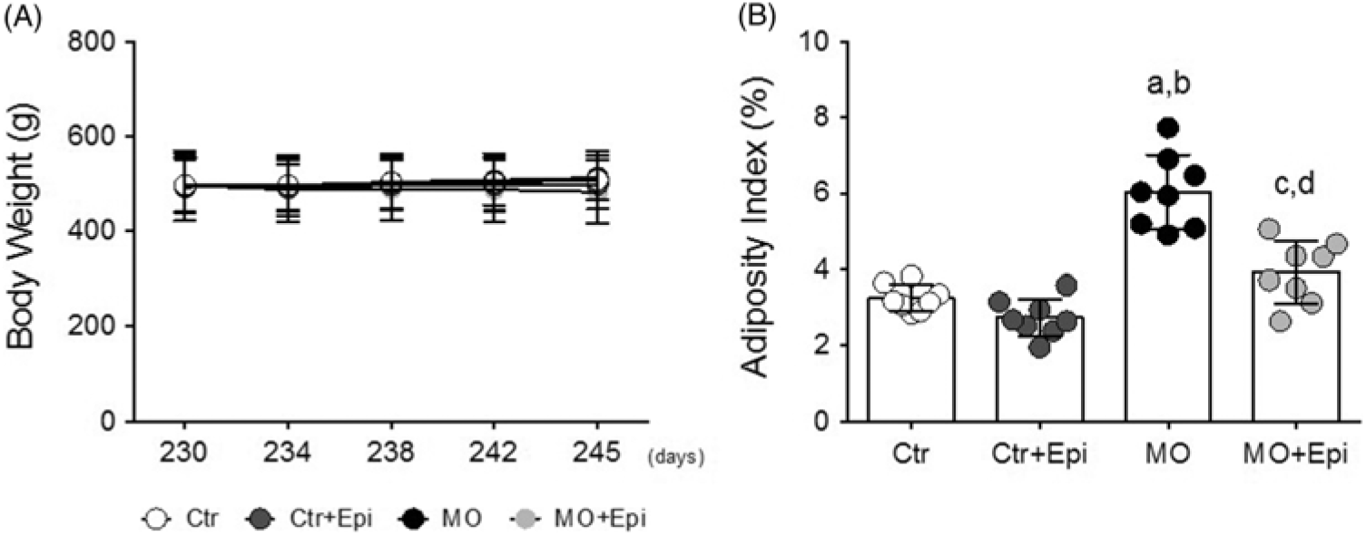
There were no differences in body weight in any of the male offspring groups at PND 245 (Fig. 2A). Chow diet consumption, using averaged data, was similar in the four groups (Ctr 24.10 ± 2.7, Ctr + Epi 24.24 ± 2.4, MO 24.34 ± 2.0 and MO + Epi 23.87 ± 1.8 g per day per group). Epi intervention did not affect food intake.
A) Body weight along the 15 days of treatment. There were no differences in body weight in any of the male offspring groups. B) Adiposity index. Differences of the adiposity index between the male offspring of the maternal obesity model (MO) group compared to the male offspring of controls (Ctr) and the male offspring of the control group treated with Epi (Ctr + Epi). Besides, male offspring of the MO + Epi group had a lower adiposity index when compared to the male offspring of the MO group. Data are expressed as mean ± SD and were analyzed by ANOVA, with post hoc Tukey HSD for pair comparison. aP < 0.01 vs Ctr; bP < 0.01 vs Ctr + Epi; cP < 0.05 vs Ctr + Epi; dP < 0.01 vs MO, n = 8 rats per group.

We observed a significantly higher amount of visceral fat tissue in male offspring of MO group when compared with the male offspring of both Ctr group and Ctr + Epi group, reflecting a higher adiposity index in the male offspring of MO group (P < 0.01) (Fig. 2B). Although we considered sternal fat for calculating the adiposity index, this tissue did not show any differences between MO and Ctr groups (data not shown).
Interestingly, male offspring of MO + Epi group presented 11.61 g less of visceral fat when compared with the male offspring of the MO group. These differences were statistically significant, representing a lower adiposity index (P < 0.01; Fig. 2B), although this index in the MO + Epi male offspring group was higher when compared with the male offspring of Ctr + Epi group (P < 0.05). We did not find differences in the adiposity index between male offspring of Ctr group and male offspring of Ctr + Epi group (Fig. 2B).
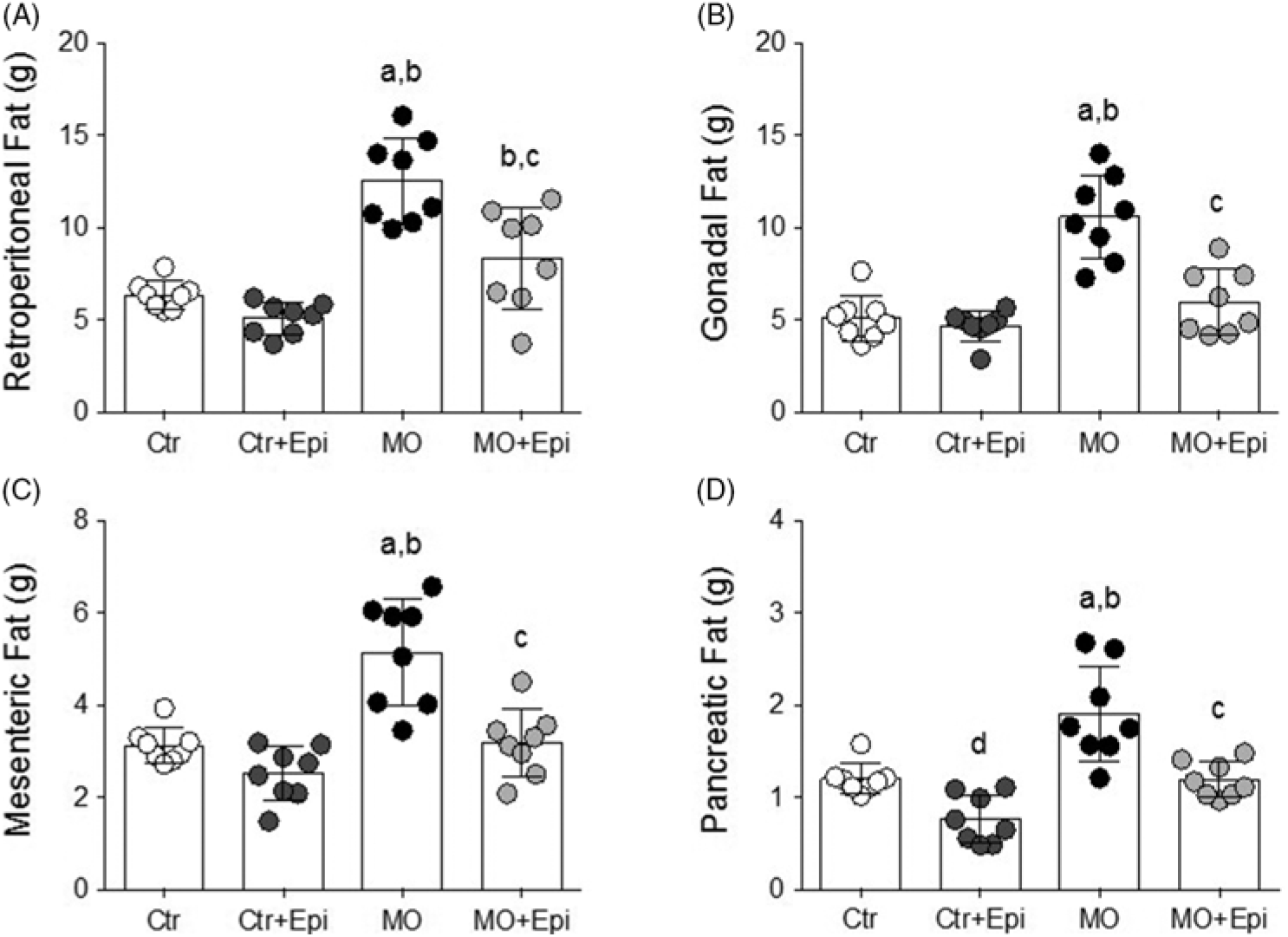
Besides, we found at least a 50% increase in deposits of visceral fat (retroperitoneal, gonadal, peripancreatic, and mesenteric) in male offspring of MO group when compared with the male offspring of both Ctr group and Ctr + Epi group (P < 0.01; Fig. 3A–D). Excepting sternal fat, all other deposits of visceral fat of the MO + Epi male offspring group were significantly decreased when compared with the male offspring of MO group (P < 0.01; Fig. 3A–D). Moreover, these results were similar to what we observed in male offspring of Ctr group and in the male offspring of Ctr + Epi group other than for retroperitoneal visceral fat for which male offspring of MO + Epi group had higher values compared with the male offspring of Ctr + Epi group (P < 0.01; Fig. 3A).
Regarding the male offspring of control groups, we observed that Epi treatment induced a significant reduction only in peripancreatic fat in the male offspring of Ctr + Epi group when compared with the male offspring of Ctr group (P < 0.05; Fig. 3D).
Weights of different fat deposits in male offspring of the MO group at 245 postnatal day (F1). A) Retroperitoneal fat, B) Gonadal fat, C) Mesenteric fat and D) Pancreatic fat. All deposits of visceral fat in the male offspring of the MO group were higher when compared to the male offspring of the control (Ctr) groups treated and not treated with Epi (Ctr + Epi). Besides, male offspring of the MO group treated with Epi (MO + Epi) presented a decrease of all deposits of visceral fat when compared to the male offspring of the MO group. Data are expressed as mean ± SD and were analyzed by ANOVA, with post hoc Tukey HSD for pair comparison. aP < 0.01 vs Ctrl, bP < 0.01 vs Ctr + Epi, cP < 0.01 vs MO, dP < 0.05 vs Ctr, n = 8 rats per group.

Epi reduces body fat and fat cell size
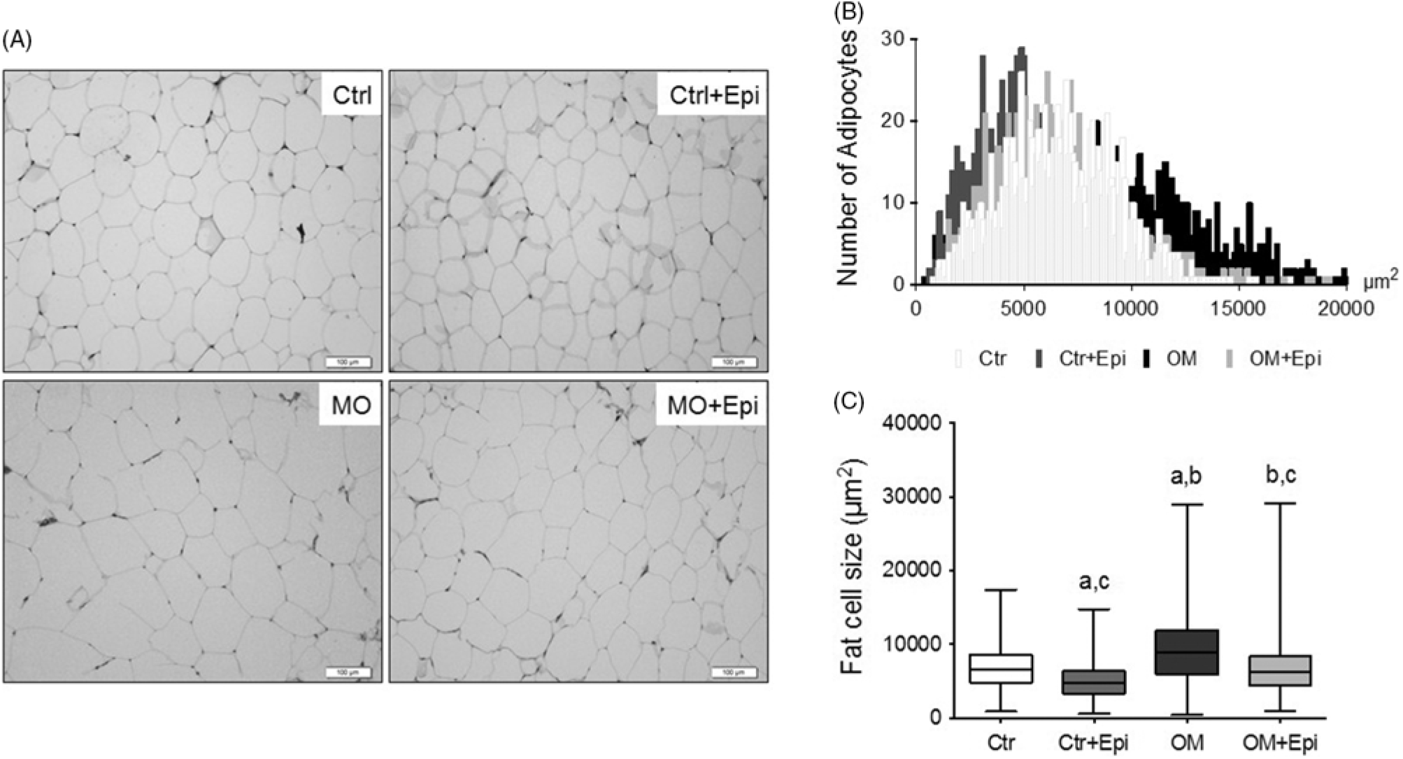
Figure 4 shows characteristics of the adipose tissue of the four groups of male offspring. We found that male offspring of MO group had a greater amount of body fat, as well as larger retroperitoneal fat cell size, when compared with the male offspring of both Ctr and Ctr + Epi groups (P < 0.01; Fig. 4A–C). Remarkably, the male offspring of the MO + Epi group had these parameters diminished when compared with the male offspring of MO group (P < 0.01), but the amount of body fat and size of the retroperitoneal adipocytes of male offspring of MO + Epi group was higher than in the male offspring of the Ctr + Epi group (P < 0.01).
Adipose tissue characteristics of the male offspring at postnatal day 245. A) Example of hematoxylin and eosin staining of fat cells. Scale bars = 100 μm. B) Histogram of the area shows the distribution of fat cells C). Comparison data of box plot; boxes extend from maximum to minimum, with a horizontal line at the median. Data were analyzed by Kruskal-Wallis, with post hoc Conover method. aP < 0.01 vs Ctr, bP < 0.01 vs Ctr + Epi, cP < 0.01 vs MO, n = 1200 cells per group.

Furthermore, after evaluating the area of 150 retroperitoneal adipocytes per rat, a significant difference was observed in the median between male offspring groups treated with Epi (Ctr + Epi and MO + Epi) with respect to the male offspring of the Ctr and MO groups (P < 0.001). Interestingly, the median presented in male offspring of MO + Epi group was similar to the median of the male offspring of the Ctr group (Fig. 4C).
Discussion
In animal models, it has been shown that maternal obesity during gestation is an important determinant of offspring body weight.Reference Nathanielsz, Ford and Long33 In several studies, it has been reported that F1 males from F0 obese mothers exhibit increases in body weight, fat, adiposity index, serum triglycerides, leptin, insulin, and insulin resistance.Reference Nathanielsz, Ford and Long33–Reference Vega, Reyes-Castro and Bautista35 This occurs even when F1 males from F0 obese mothers had been fed with a normal laboratory diet.Reference Rodríguez-González, Vega and Boeck36
It is well known that adverse environments in utero, including maternal overnutrition, may disturb the process of cell proliferation and differentiation, leading to changes in organogenesis and developmental programming. Subsequent changes in phenotype can enhance susceptibility to certain diseases in adult offspring,Reference Parlee and MacDougald37,Reference Williams, Seki and Vuguin38 such as type 2 diabetes,Reference Portha, Chavey and Movassat39 hypertension,Reference Ingelfinger and Nuyt40 and obesity.Reference Sarr, Yang and Regnault41,Reference Desai, Beall and Ross42 Besides, it has been reported that Epi, a flavonoid abundant in cocoaReference Heiss, Dejam and Kleinbongard43 is associated with a decreased rate of weight gain in a rat model of obesity induced by HFD.Reference Gutiérrez-Salmeán, Ortiz-Vilchis and Vacasey del24
Due to the existence of multiple negative effects of adverse programming on life-course health, in the present study we analyzed whether Epi can modify biochemical parameters and examined the amount of adipose tissue in the male offspring of maternal rats that had obesity induced by HFD. We decided to use rats as an animal model, as our group has extensive experience in the handling of this model,Reference Zambrano, Bautista and Deás29,Reference Vega, Reyes-Castro and Rodríguez-González31,Reference Zambrano, Martínez-Samayoa and Rodríguez-González34,Reference Vega, Reyes-Castro and Bautista35 and because previous studies have shown that Epi has similar effects in metabolic parameters in humans and in rats.Reference Gutiérrez-Salmeán, Meaney and Lanaspa44
Santos et al.Reference Santos, Rodríguez-González and Ibáñez45 demonstrated that exercise intervention in the offspring of maternal obesity did not decrease body weight but partially improved the adiposity index and gonadal fat. In relation to our results, we observed a similar effect on adipose tissue, as we found a significant lower adiposity index and a large decrease in all visceral adipose tissue depots in the group of male offspring of maternal rats with obesity induced by HFD treated with Epi (MO + Epi treated group). In this regard, Nogueira et al.,Reference Nogueira, Ramirez-Sanchez and Perkins19 showed that Epi increases the capacity for aerobic muscle metabolism, and this molecule has been proposed as a new class of low-toxin exercise mimetics.Reference Fan and Evans46 The latter could be because Epi can induce mitochondrial biogenesis in vitroReference Moreno-Ulloa, Cid and Rubio-Gayosso47 and in vivo,Reference Nogueira, Ramirez-Sanchez and Perkins19 as in a HFD-induced obesity model, Epi was able to increase protein levels of genes related to mitochondrial biogenesis related.Reference Gutiérrez-Salmeán, Ortiz-Vilchis and Vacasey del24 Besides, Varela et al.Reference Varela, Rodriguez and Romero-Valdovinos48 have indicated the possible mechanisms by which Epi induces such effects on adipose tissue as well as its impact on mitochondrial function and fatty acid oxidation. These authors suggested that Epi induces fat browning by promoting mitochondrial biogenesis, enhancing indicators of mitochondrial structure and function, increasing fatty acid metabolism, and upregulating the expression of brown adipose tissue-specific proteins.
Moreover, Gutiérrez-Salmeán et al.Reference Gutiérrez-Salmeán, Ortiz-Vilchis and Vacasey del24 administered 1 mg/kg of Epi for 2 weeks to an rat model with HFD-induced obesity and found that this intervention did not change the weekly weight gain among the normal diet animals; however, Epi significantly reduced the rate of weekly weight gain in the obese HFD-fed rats. Nonetheless, we did not find body weight reduction in the MO + Epi group. This could be because we analyzed the male offspring of maternal rats with obesity induced by HFD and not the mothers with obesity induced with HFD.
Interestingly, the differences we found were in visceral fat accumulation and in the metabolism profile in the MO group. According to these results, we suggest that the effect of the MO + Epi intervention is directly on the fat tissue as we did not observe this effect in the control groups. This is important as visceral fat has been associated with numerous metabolic abnormalities.Reference Pi-Sunyer2,Reference Montague and O’Rahilly49 It well known that different adipose depots have intrinsic differences in adipocyte characteristics and differences in immune cell composition.Reference Kintscher, Hartge and Hess50,Reference Weisberg, McCann and Desai51 Emerging evidence indicates that mesenteric fat is an independent determinant of metabolic syndrome;Reference Liu, Chan and Chan52 besides, mesenteric and gonadal fat are important for the development of hepatic insulin resistance and steatosis.Reference Choe, Huh and Hwang53 Moreover, peripancreatic adipose tissue is associated with increased inflammation in the mouse model of pancreatic cancer.Reference Hertzer, Xu and Moro54 In addition, retroperitoneal fat constantly releases free fatty acids into the circulation, which contributes to hyperlipidemia, atherogenesis, and hyperinsulinemia.Reference Jensen55
Several studies performed in animal models of HFD-induced obesity treated with Epi had shown that this flavonoid decreases the levels of glucose, insulin, and triglycerides.Reference Gutiérrez-Salmeán, Ortiz-Vilchis and Vacasey del24,Reference Cremonini, Bettaieb and Haj26,Reference Varela, Rodriguez and Romero-Valdovinos48 This could be explained by the ability of Epi to restore protein levels of those molecules known to play critical roles in regulating adipose and skeletal muscle metabolismReference Gutiérrez-Salmeán, Ortiz-Vilchis and Vacasey del24 through the amelioration of adipose tissue inflammation.Reference Sano, Nagayasu and Suzuki25,Reference Bettaieb, Cremonini and Kang28 Similarly, Epi could improve insulin sensitivity, in part through downregulation of the inhibitory molecules c-Jun N-terminal kinase (JNK), IkB kinase (IKK), protein kinase C (PKC), and protein tyrosine phosphatase 1B (PTP1B).Reference Cremonini, Bettaieb and Haj26 In our results, we found that in the male offspring from maternal rats with HFD-induced obesity and treated with Epi (MO + Epi), serum triglycerides, leptin, and insulin, as well as the HOMA-IR were significantly reduced when compared with the male offspring from maternal rats with HFD-induced obesity not treated with Epi. These findings agree with previous reports, although in our study, cholesterol and glucose remained unchanged.Reference De Los Santos, García-Pérez and Hernández-Reséndiz22–Reference Cremonini, Bettaieb and Haj26,Reference Varela, Rodriguez and Romero-Valdovinos48
Adverse effects in the metabolic function of the offspring from obese maternal rats could be influenced by epigenetic changes that modify the expression of various genes.Reference Wright, Rifas-Shiman and Rich-Edwards11–Reference Zambrano and Nathanielsz14 In the future, we should consider analyzing histone modifications as a possible explanation of our results, because it has been shown that Epi is capable of inducing posttranslational changes of histone acetyltransferase (HAT) p300, as well as of histone deacetylase-4 (HDAC4) in human monocyte cell line-1 (THP-1) under different concentrations of glucose.Reference Cordero-Herrera, Chen, Ramos and Devaraj56
In contrast, Zambrano et al.,Reference Zambrano, Martínez-Samayoa and Rodríguez-González34 in a study of dietary intervention performed in a model of maternal obesity, analyzed the outcome of male offspring and observed that although dietary intervention in the mothers significantly reduced the mass of fat deposit and the size of the adipose cell, these were significantly higher than in controls. Differing from this outcome, we found that in the male offspring from maternal rats with HFD-induced obesity treated with Epi, fat deposits and adipocytes size decreased, being similar to those observed in the male offspring of the Ctr group. In addition, we also observed that treatment with Epi significantly decreased the size of the adipocytes and peripancreatic deposit in the male offspring of the Ctr + Epi group, when compared with the Ctr group.
Conclusions
(-)-Epicatechin (Epi) administration can reverse the negative effects that maternal obesity has on the male offspring. This could be because Epi reduces the amount of visceral fat and improves the metabolic profile.
Our findings are similar to those reported in diet-induced obesity models, showing that treatment with Epi has a similar effect in our obesity model, where epigenetic programming and predisposition to the development of obesity are different. Further studies will be needed to explore whether Epi treatment in this model of obesity constitutes a natural adjuvant in the treatment of obesity.
Author ORCIDs
Patricia Canto 0000-0002-1891-480X
Acknowledgments
This work was partially supported by the Newton Fund RCUK- CONACYT-2015 (Research Conuncils UK – Consejo Nacional De Ciencia Y Tecnología, México) and by Apoyo al Fortalecimiento y Desarrollo de la Infrastructura Científica y Tecnológica (CONACyT, México; Grant: 250786).
Sergio De los Santos received a postdoctoral fellowship from Programa de Becas Posdoctorales de la Dirección General de Asuntos del Personal Académico (DGAPA), División de Investigación, Faculta de Medicina, Universidad Nacional Autónoma de México.
Conflict of Interest
None.
Ethical Standards
The animal research protocol followed in the present study was approved by the Animal Experimentation Ethics Committee of the Instituto Nacional de Ciencias Médicas y Nutrición Salvador Zubirán (INCMNSZ) (CINVA: UIO-1892-17/19-1) and by the Research and Ethic Commissions of the Facultad de Medicina, Universidad Nacional Autónoma de México. All procedures were performed in accordance with the Guidelines for the Care and Use of Laboratory Animals of the Institute of Laboratory Animal Resources (http://www.nal.usda.gov/awic/animal-welfare-act), approved in Mexico by the National Academy of Medicine and in accordance with the Mexican Official Standard Norm NOM-062-ZOO-1999.
Declarations of interest
None.