INTRODUCTION
Regenerative phenomena are typical developmental processes which, although not representing an accelerated version of ontogenetic events (Candia Carnevali & Bonasoro, Reference Candia Carnevali and Bonasoro2001; Candia Carnevali, Reference Candia Carnevali2006), involve analogous problems in all animal models and often superficially resemble embryonic processes in terms of basic mechanisms and events such as cell proliferation, identity and positioning (Davidson et al., Reference Davidson, Cameron and Ransick1998). It is therefore not surprising that the nervous system plays a crucial role by directing the correct progression of regenerative events in most animal models. It is well established that it can supply regulatory mitogenic or morphogenic factors that regulate the basic developmental processes of regeneration. Among these molecules, serotonin has been cited often as a multifunctional neurohumoral agent (Franquinet, Reference Franquinet1979; Lauder, Reference Lauder1988; Whitaker-Azmitia et al., Reference Whitaker-Azmitia, Druse, Walker and Lauder1996; Buznikov et al., Reference Buznikov, Woyne Lambert and Lauder2001). During regenerative events in planarians, serotonin is a critical regulatory factor which stimulates, via adenylate cyclase, RNA and DNA synthesis (Franquinet & La Moigne, Reference Franquinet and La Moigne1979). Moreover, flatworms exposed to antiserotonergic drugs display a retarded regeneration rate (Saitoh et al., Reference Saitoh, Yuruzume and Nakata1996).
Echinoderms are well known for their capacity to regenerate many body parts and organs following traumatic or self-induced mutilation (autotomy) (Wilkie, Reference Wilkie2001). Moreover, their regenerative processes are also nerve-dependent (for review see Thorndyke & Candia Carnevali, Reference Thorndyke and Candia Carnevali2001), and serotonin is one of the main neural regulatory factors identified so far (Bonasoro et al., Reference Bonasoro, Candia Carnevali, Thorndyke, Welsch, Emson, Smith and Campbell1995; Candia Carnevali et al., Reference Candia Carnevali, Bonasoro, Invernizzi, Lucca, Welsch and Thorndyke1996, Reference Candia Carnevali, Bonasoro, Thorndyke, Patruno and Barker2001). Previous results obtained in the crinoid Antedon mediterranea (Lamarck, 1816), a common featherstar endemic in the Mediterranean Sea (Figure 1), showed that the process of arm regeneration is characterized by changes in the tissue localization and concentration of serotonin (Bonasoro et al., Reference Bonasoro, Candia Carnevali, Thorndyke, Welsch, Emson, Smith and Campbell1995; Candia Carnevali et al., Reference Candia Carnevali, Bruno, Denis Donini, Melone, Kiortsis, Koussoulakos and Wallace1989, Reference Candia Carnevali, Bonasoro, Invernizzi, Lucca, Welsch and Thorndyke1996, Reference Candia Carnevali, Bonasoro, Trezzi, Giardina, Candia Carnevali and Bonasoro1999, Reference Candia Carnevali, Bonasoro, Thorndyke, Patruno and Barker2001). In particular, immunocytochemical techniques revealed variations in serotonin distribution during the different regenerative phases, in comparison with the standard pattern of positive reactions displayed by the cellular elements of the different nervous system components (brachial nerve and basiepithelial plexuses) in non-regenerating arms. It is notable that during regeneration labelling was found both in neural structures, where it was appreciably enhanced, and at the level of non-neural elements, such as migrating amoebocytes, coelothelial cells and differentiating myocytes. Quantitative HPLC data confirmed the involvement of serotonin in regenerative events and showed fluctuations in serotonin concentration during the crucial phases. These data showed a clear variability in absolute values, an initial peak being detectable in the early regenerative stages (24–72 hours post amputation (p.a.)) when most proliferative phenomena occur, and a further concentration increase being measurable at the onset of advanced regenerative stages (72 hours–4 weeks) characterized by early cell and tissue differentiation (Candia Carnevali et al., Reference Candia Carnevali, Bonasoro, Thorndyke, Patruno and Barker2001).
The featherstar Antedon mediterranea (Lamarck, 1816): different stages of the regeneration process are in progress in different arms (A and B: SEM images).

The immunocytochemical and biochemical results obtained so far thus suggest that serotonin is involved in crinoid regenerative processes. In the present work we employed neuropharmacological compounds that interfere with serotonin uptake or metabolism. Treatment of other animal models with serotonin receptor antagonists or with serotonin synthesis inhibitors induces appreciable anomalies in both the embryonic and regenerative development (Franquinet, Reference Franquinet1979; Palén et al., Reference Palén, Thörneby and Emanuelsson1979; Lauder, Reference Lauder1988; Saitoh et al., Reference Saitoh, Yuruzume and Nakata1996; Buznikov et al., Reference Buznikov, Woyne Lambert and Lauder2001). The aim of the present investigation was to explore the possible role of serotonin in crinoid arm regeneration using the serotonin synthesis inhibitor, parachlorophenylalanine, and the serotonin receptor antagonist, methiothepin.
MATERIALS AND METHODS
Animals
Adult specimens of Antedon mediterranea (Figure 1) were collected by SCUBA divers from the Tyrrhenian Sea (Giglio Island, 42°21′57″96N 10°54′7″20E) and immediately transferred to our laboratory in Milan. The experimental animals were maintained at 14–16°C in artificial seawater tanks (Instant Ocean; salinity 37‰) provided with chemical and biological filters and water circulation and aeration systems, and were fed once a week with drops of an artificial diet (Tetramin). The following parameters were monitored daily: temperature, water pH, nitrite and nitrate levels, animal mortality and health (general aspects, behaviour and vital reactions). Experiments were started after animals had been acclimatized for a few days.
All the experiments were carried out according to the current laws of Italy.
Experimental amputation and regeneration
Arm regeneration was experimentally induced as described previously (see Candia Carnevali et al., Reference Candia Carnevali, Lucca and Bonasoro1993): each specimen was subjected to the amputation of two different arms by cutting at the level of autotomy planes (syzygies) about half way down the length of the arm. The experimental animals were then left to regenerate for a predetermined time and treated according to different protocols. Exposure to anti-serotonergic compounds (see below) in 4 l tanks started immediately after experimental amputation.
The regenerating samples of early stages were prepared for light microscopy as described in detail by Candia Carnevali et al. (Reference Candia Carnevali, Lucca and Bonasoro1993, Reference Candia Carnevali, Bonasoro, Lucca and Thorndyke1995). Briefly, collected samples were fixed with 2% glutaraldehyde in 0.1 M cacodylate buffer for 2 hours, washed overnight in the same buffer and postfixed with 1% osmium tetroxide in 0.1 M cacodylate buffer. After standard dehydration in ethanol and propylene oxide, samples were embedded in Epon–Araldite 812. Semithin sections (about 1 µm thick) were cut with a Reichert Ultracut E, stained with crystal violet-basic fuchsin and observed under a Jenaval light microscope. Some samples were processed for specific immunocytochemical (ICC) methods for monitoring cell proliferation (BrdU labelling; see below) using previously established protocols (Candia Carnevali et al., Reference Candia Carnevali, Bonasoro, Lucca and Thorndyke1995, Reference Candia Carnevali, Bonasoro and Biale1997).
Regenerating specimens at advanced stages were observed under a stereomicroscope (Leica Wild M3C Planapo) and photographed using a Leica digilux zoom 18 102 E-type digital camera.
BrdU labelling
Cell proliferation was monitored with the thymidine analogue, 5-bromo-deoxyuridine (BrdU), localized by a monoclonal mouse antibody against BrdU (Amersham: cell proliferation kit). Animals were immersed in a BrdU and FldU (5-fluoro-2′-deoxyuridine; an inhibitor of thymidine syhthetase) solution (0.05% in seawater; BrdU:FdU = 10:1) for the final 2 hours prior to regenerate collection and processing for optical microscopy. The standard BrdU-immunocytochemistry protocol for paraffin was modified for use on semithin Epon–Araldite sections, as described previously (Candia Carnevali et al., Reference Candia Carnevali, Bonasoro, Lucca and Thorndyke1995, Reference Candia Carnevali, Bonasoro and Biale1997). After a brief treatment (2 minutes) with a resin-remover mixture (10 ml methanol, 5 ml propylene oxide and 2 g KOH), sections were rinsed with methanol (2 minutes), then with PBS (3 × 5 minutes) and incubated overnight at 4°C with anti-BrdU antibody diluted (1:100) in an aqueous solution containing nuclease (Amersham: cell proliferation kit). Pre-treatment for 20 minutes with 0.3% H2O2 in PBS was performed to exclude potential endogenous peroxidase activity. After washing in PBS (3 × 10 minutes) samples were incubated (3 hours) with a peroxidase anti-mouse IgG (1:70; Amersham: cell proliferation kit) at room temperature and, after further washing in PBS (3 × 10 minutes), incubated (5 minutes) with 0.05% 3,3′-diaminobenzidine (DAB) in PBS and then washed in distilled water. In order to amplify the peroxidase reaction product, a cobalt and nickel intensifier supplied with the kit was employed. Control reactions omitted the primary antibody.
Exposure experiments
Two different anti-serotonergic drugs were tested:
– parachlorophenylalanine (pCPA) (SIGMA): a potent inhibitor of tryptophan-hydroxylase, one of the key enzymes involved in serotonin biosynthesis;
– methiothepin (as the mesylate salt; SIGMA): a serotonin receptor antagonist which has a high affinity for most invertebrate serotonin receptors (Tierney, Reference Tierney2001).
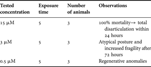
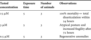
Optimal concentrations and treatment times were determined in initial tests by examining the effect of the above agents on samples that had been regenerating for one week. The developmental stage after one week is well defined and characterized in terms of growth and differentiation in standard regenerating samples (Candia Carnevali & Bonasoro, Reference Candia Carnevali and Bonasoro2001; Candia Carnevali, Reference Candia Carnevali2006). During this period the regenerating animals were daily exposed to different drug concentrations for prefixed times as shown in Tables 1 & 2. Daily treatment for a few hours was preferred to continuous exposures in order to minimize neurotoxic effects, since the selected drugs are likely to affect all serotonin-dependent nervous functions, i.e. both neurotransmission and any possible broader spectrum neuroendocrine functions.
Effect of different pCPA concentrations. Exposure times are given as hours/day.

Effect of different methiothepin concentrations. Exposure times are given as hours/day.
In order to avoid differences in physicochemical parameters, the exposure solutions for all the experimental groups were prepared by dissolving appropriate amounts of the substances in autoclaved seawater drawn from the maintenance aquarium. During the daily neuropharmacological treatment the animals were temporarily transferred to exposure tanks (4 l) provided with an aeration system. At the end of the daily treatment/exposure time they were returned to their original maintenance aquarium, which was subdivided into compartments in order to guarantee the same maintenance conditions for all the experimental groups.
After these initial tests and once the optimal concentrations and exposure times had been established, two sets of definitive experiments were performed on different regenerative phases.
Early regenerative phase (48–72 hours)
In the initial tests, samples at this stage remained healthy only in 0.5 µM methiothepin for 5 hours/day (Tables 1 & 2). This treatment was therefore repeated using seven experimental animals and regenerating arms were collected at 48 and 72 hours p.a. and processed as described above. Since in the initial tests pCPA treatment resulted in high mortality, extreme fragility or lack of observable morphological alterations (Table 1), no further experiments on the early regenerative stages were carried out with this compound.
Advanced regenerative phase (1 week)
The second set of experiments was performed as follows: amputated animals, randomly subdivided in three different groups of ten individuals each, were daily exposed to: (1) 0.5 µM methiothepin for 5 hours/day; (2) 0.2 µM pCPA for 5 hours/day; and (3) artificial seawater for 5 hours/day (control group). The intermediate pCPA concentration was selected on the basis of the results of the initial tests (see Table 1), which showed that 0.5 µM was too toxic and 0.1 µM had no detectable effect. At the end of the daily exposure, animals from all the experimental groups were placed again in the maintenance aquarium. After treatment for 1 week, regenerating samples were collected, processed and photographed as described above. Photographs were used for measuring regenerate lengths by employing the computer program ArcView 3.2 (GIS). The homogeneity of variance of the obtained data was verified by a Levene test and a one-way ANOVA test was performed.
RESULTS
The results of the initial tests are summarized in Tables 1 & 2. The highest concentrations of pCPA (1 mM and 0.1 mM) were extremely toxic, causing the death of all animals within the first 24 hours of treatment. Lower pCPA concentrations (1 µM and 0.5 µM) also appeared to cause general damage. In all specimens exposed to these concentrations the jointed appendages (arms, pinnules and cirri) displayed extreme fragility, i.e. they fragmented at preformed autotomy planes. On the other hand, at a lower concentration (0.1 µM) and exposure time (3 hours), there were no observable morphological anomalies.
Although a lethal effect was observed with the highest methiothepin concentration (15 µM; 5 hours/day), the specimens exposed to the lowest concentration (0.5 µM; 5 hours/day) had regenerative abnormalities, yet showed no increase in mortality or fragility (Table 2). We therefore selected the latter concentration and exposure time for the subsequent tests with methiothepin. The intermediate methiothepin concentration (3 µM) induced the same tendency to break at autotomy planes as did pCPA.
Early regenerative phase (48–72 hours p.a.)
Figure 2 gives a schematic view of a regenerating arm (in sagittal section) at an early regenerative phase (48–72 hours p.a.). In standard conditions these stages are characterized by proliferation and migration phenomena which contribute to new arm formation (Candia Carnevali et al., Reference Candia Carnevali, Bonasoro, Lucca and Thorndyke1995, Reference Candia Carnevali, Bonasoro and Biale1997).
Schematic reconstruction showing the main anatomical components of an arm at early regenerative phase (72 hours post amputation). AG, ambulacral groove; B, blastema; CC, coelomic canals, L, ligament; M, muscle; N, nerve.

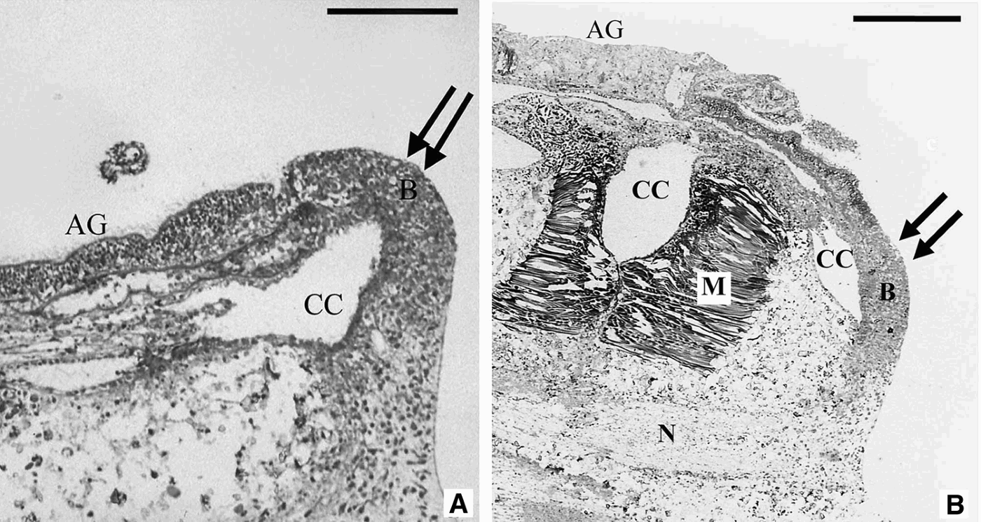
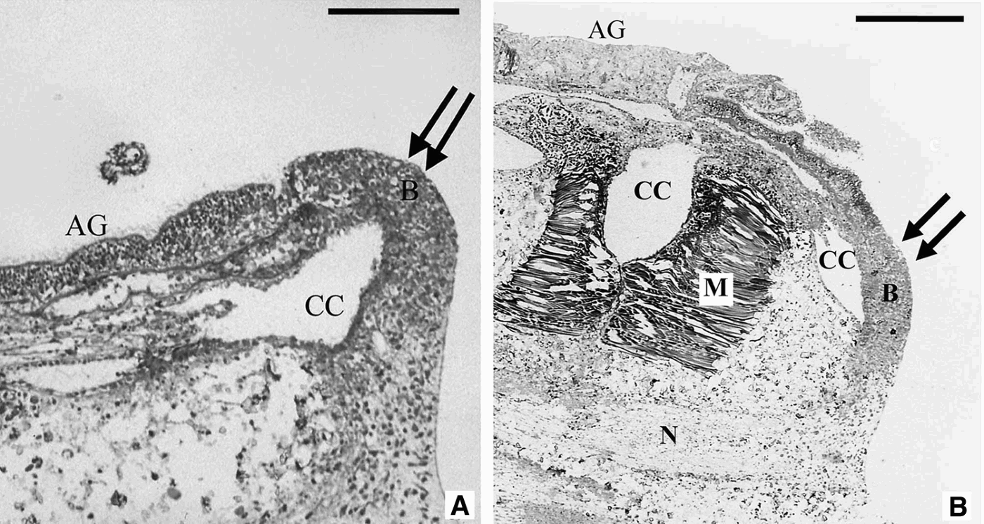
Exposure to 0.5 µM methiothepin produced obvious anomalies in microscopic anatomy indicating dysfunctions in early regenerative development. The morphological analysis of treated samples showed significant malformations at the cellular and tissue levels in both the regenerate and the stump. The regenerate showed a general delay in regenerative growth starting from the 48 hour-stage. The blastemal bud, which is usually easily recognizable in normal regenerating arms at this stage (Figure 4), was replaced by a flattened and poorly defined blastemal area. In addition, at the level of the amputation region, the re-growing coelomic canal was abnormally flattened and bent aborally, following the general flat shape of the regenerate. The apparent growth inhibition displayed by the exposed samples was confirmed by BrdU–ICC results. As observed in Figure 5A, in standard regenerating samples the BrdU reaction was particularly evident at the level of the regenerative bud, where both coelothelial and blastemal cells were labelled, the latter representing presumptive stem elements. At the level of the stump (Figure 5C), in addition to labelling of the coelothelium, a strong BrdU reaction was shown by scattered cells of the brachial nerve, particularly non-neural satellite elements in the cortex (amoebocytes). In contrast, in methiothepin treated samples, the blastemal region was less immunoreactive (Figure 5B). Reduced labelling was even more evident in the stump where only a very small number of elements were immunoreactive (Figure 5D).
Schematic reconstruction showing the main anatomical components of an arm at advanced regenerative phase (1 week). B, blastema; L, ligament; M, muscle; N, nerve; RM, regenerating muscle.

Light microscopy view of the effects of methiothepin post-amputation (p.a.) treatment (0.5 µM; 5 hours/day) on regenerative growth at early regenerative stages. Histological sagittal sections. (A) Standard regenerate at 72 hours p.a. showing a protruding regenerative bud (double arrow). Bar = 100 µm; (B) sagittal section of a treated regenerating sample (72 hours p.a.). The blastema (double arrow) is scarcely developed and rather flat in comparison with standard regenerating bud. Bar = 200 µm. B, blastema; CC, coelomic canals; M, muscle; N, nerve.
Light microscopy details of the effects of post-amputation (p.a.) methiothepin treatment (0.5 µM; 5 hours/day) on cell proliferation. ICC–BrdU method. Sagittal sections of regenerating samples at 72 hours p.a. (A) Normal regenerating blastema showing many immunolabelled blastemal cells. Bar = 70 µm; (B) blastema of a treated sample. Only few cells (arrow) are immunoreactive for BrdU. Bar = 60 µm; (C) coelomic canals of a normal regenerate. Arrow = labelled epithelial cells. Bar = 20 µm; (D) coelomic canals of treated sample: labelling (arrow) is localized in few cells. Bar = 60 µm. CC, coelomic canal; M, muscle. (Note: in order to highlight the immunolabelling, the semithin-sections were not counter-stained and the image contrast was slightly enhanced using the computer program Adobe Photoshop 7.0.)

At the level of the stump, treated samples also displayed an unusual rearrangement of differentiated tissues and cells, specifically involving the muscles, in which marked dedifferentiation and/or transdifferentiation processes were frequently detectable. Muscle bundles in treated samples lost their normal compact structure (Figure 6A) and their individual myocytes underwent intensive reorganization and progressive dedifferentiation. These myocytes showed progressive disorganization of their contractile apparatus, particularly in terms of filament arrangement (Figure 6B, C & D). This phenomenon was very evident in the periphery of the muscle bundles, particularly in the areas close to the coelomic canals. Observation of serial sections indicated that the de-differentiating myocytes migrated towards the coelomic cavity (Figure 6B, D).
Light microscopy details of the effects of methiothepin on the muscles of the stump. Sagittal sections. (A) Muscle bundle of a normal regenerating arm at 72 hours showing the classic compact arrangement of the fibres. Bar = 80 µm; (B) muscle bundle of a treated regenerating sample at 72 hours post amputation (p.a.) showing an anomalous histological pattern. The muscle fibres have lost their defined structure and numerous dedifferentiating myocytes (arrow) appear to detach from the bundle and move towards the coelomic cavity. Bar = 60 µm; (C) muscle of a treated sample at 48 hours p.a.: some myocytes (arrow) seem to be migrating toward the blastema. Bar = 100 µm; (D) detail of (C); (E) ectopic mass of dedifferentiating myocytes (arrow) in the muscle bundle of a treated sample. Bar = 100 µm. B, blastema; CC, coelomic canals; M, muscle.

Advanced regenerative phase (1 week p.a.)
Figure 3 shows a schematic view of a standard regenerating arm (in sagittal section) at an advanced regenerative phase (1 week p.a.). Both methiothepin and pCPA produced a statistically significant decrease in regenerate growth (expressed in terms of regenerate length) in comparison with the control group (ANOVA test: P < 0.001; Tukey and Bonferroni post-hoc tests: P < 0.001 for both compounds). Moreover, methiothepin inhibited growth more strongly than pCPA (P < 0.05) (Figure 7).
DISCUSSION
In echinoderms, and most other animals, interactions between specific neurochemicals and cellular elements are necessary for the successful course of most developmental phenomena, including regenerative processes. Several authors have suggested that many common neurotransmitters, such as serotonin, are broad-spectrum messengers responsible for promoting and controlling embryonic and regenerative developmental processes in many animal models (Franquinet, Reference Franquinet1979; Lauder, Reference Lauder1988; Whitaker-Azmitia et al., Reference Whitaker-Azmitia, Druse, Walker and Lauder1996; Buznikov et al., Reference Buznikov, Woyne Lambert and Lauder2001; Moiseiwitsch et al., Reference Moiseiwitsch, Lambert, Lauder, Kalverboer and Gramsbergen2001). In agreement with this hypothesis, our previous (Candia Carnevali et al., Reference Candia Carnevali, Bruno, Denis Donini, Melone, Kiortsis, Koussoulakos and Wallace1989, Reference Candia Carnevali, Bonasoro, Invernizzi, Lucca, Welsch and Thorndyke1996; Thorndyke & Candia Carnevali, Reference Thorndyke and Candia Carnevali2001) and present results suggest that serotonin is also involved in the overall process of arm regeneration in the crinoid Antedon mediterranea, influencing morphogenesis, differentiation and growth. In addition, our present and past data are consistent with the idea that in echinoderms serotonin functions both as a specific neurotransmitter and as a neurohumoral factor influencing basic developmental processes such as cell proliferation and differentiation (Candia Carnevali et al., Reference Candia Carnevali, Bonasoro, Thorndyke, Patruno and Barker2001; Thorndyke & Candia Carnevali, Reference Thorndyke and Candia Carnevali2001). The involvement of serotonin in arm regeneration was indicated previously by the finding of significant differences in its distribution, localization and concentration between normal and regenerating samples at different regenerative stages (Candia Carnevali et al., Reference Candia Carnevali, Bruno, Denis Donini, Melone, Kiortsis, Koussoulakos and Wallace1989, Reference Candia Carnevali, Bonasoro, Invernizzi, Lucca, Welsch and Thorndyke1996; Thorndyke & Candia Carnevali, Reference Thorndyke and Candia Carnevali2001). Our present results showed that repeated treatment with neuropharmacological compounds interfering with serotonin synthesis or action induced appreciable anomalies in regenerative development. Although more tests are needed to definitively confirm the specificity of the antiserotonergic treatments employed, our present results provide further evidence to support the view that serotonin-regulated processes are crucial to the correct course of regeneration. Different cellular processes appear to be affected by these neuropharmacological treatments, suggesting that serotonin is involved at different levels and with different functions. This is in agreement with the well known multifunctional role proposed for serotonin in early development (Whitaker-Azmitia et al., Reference Whitaker-Azmitia, Druse, Walker and Lauder1996), where this molecule has been reported as a mitogen, morphogen and migration-inducing substance (Gustafson & Toneby, Reference Gustafson and Toneby1970; Moiseiwitsch & Lauder, Reference Moiseiwitsch and Lauder1995a, Reference Moiseiwitsch and Lauderb; Fanburg & Lee, Reference Fanburg and Lee1997; Moiseiwitsch, Reference Moiseiwitsch2000; Bhasin et al., Reference Bhasin, Kernick, Luo, Seidel, Weiss and Lauder2004).
Our results suggest that serotonin influences cell proliferation and muscle differentiation/maintenance.
Proliferation
Serotonin involvement in cell division can be inferred from the remarkable decrease in the number of proliferating cells at the blastemal and coelothelial levels following 48 hours of antiserotonergic treatment (Figure 5). An inhibited proliferation rate could explain the reduced regenerative regrowth observable in all the exposed samples at advanced stages (Figure 7). These data are consistent with the often reported role of serotonin in promoting mitosis in a wide range of cultured mammalian cell types (Nemecek et al., Reference Nemecek, Goughlin, Handley and Moskowits1984; Zachary et al., Reference Zachary, Woll and Rozengurt1987; Fanburg & Lee, Reference Fanburg and Lee1997).
Effects of para-chlorophenylalanine (pCPA: 0.2 µM; 5 hours/day) and methiothepin (0.5 µM; 5 hours/day) on regenerative growth. Advanced regenerative stages. Both neuropharmacological treatments caused highly significant reductions on regenerate length after 1 week of treatment (**P < 0.001, ANOVA and Tukey post hoc tests) in comparison with controls (CTL), but the receptor antagonist methiothepin displayed more evident effects than pCPA (P < 0.05).

Although the in vivo approach employed here does not help to identify the specific signalling pathway of serotonin or its interactions with other regulatory factors, the reduced proliferation rate observed after antiserotonergic treatment indicates that it probably influences cell division during regenerative phenomena.
Muscle differentiation/maintenance
Our previous work on A. mediterranea arm regeneration showed that there is a prominent localization of serotonin at the level of the differentiating muscle bundles and individual muscle fibres in the new arm (Bonasoro et al., Reference Bonasoro, Candia Carnevali, Thorndyke, Welsch, Emson, Smith and Campbell1995; Candia Carnevali et al., Reference Candia Carnevali, Bonasoro, Invernizzi, Lucca, Welsch and Thorndyke1996, Reference Candia Carnevali, Bonasoro, Thorndyke, Patruno and Barker2001; Thorndyke & Candia Carnevali, Reference Thorndyke and Candia Carnevali2001). This localization led us to suggest that serotonergic innervation might be relevant for the development and maintenance of the motor components (Candia Carnevali et al., Reference Candia Carnevali, Bonasoro, Trezzi, Giardina, Candia Carnevali and Bonasoro1999). The present work appears to provide further support for this idea. In contrast to the rather good histological preservation and apparent morphological integrity of most structures/tissues of the arm stump (e.g. epithelial tissues, brachial nerve, skeletal components, etc.), the muscles appeared to be selectively targeted by the neuropharmacological treatment. In fact, the receptor antagonist methiotephin produced an intensive and extensive rearrangement of myocytes in the muscle bundles, particularly in the oral portion adjacent to the coelomic canal (Figure 6B, D). These muscular elements had lost their typical elongate structure, though retaining their normal cytochemical properties, as indicated by heavy staining. These cytological features have been described at the ultrastructural level (Candia Carnevali & Bonasoro, Reference Candia Carnevali and Bonasoro2001; Candia Carnevali et al., Reference Candia Carnevali, Bonasoro, Thorndyke, Patruno and Barker2001; Candia Carnevali, 2005) and are also seen less frequently in normal muscle bundles implying that they represent a basal rate of physiological cell turn-over. The anomalous increase in the number of muscle fibres apparently in progressive stages of dedifferentiation, which was detected after methiothepin exposure, suggests a loss of balance in the intrinsic mechanism of muscle maintenance. Serotonin may act as a specific signal for motor components, playing an important role in both their development and maintenance. As stated above, in the regenerating arm this molecule is associated specifically with differentiating myocytes (as demonstrated by its widespread and intense localization on the surface of these elements during the advanced regenerative phase: Candia Carnevali et al., Reference Candia Carnevali, Bonasoro, Invernizzi, Lucca, Welsch and Thorndyke1996, Reference Candia Carnevali, Bonasoro, Thorndyke, Patruno and Barker2001) and its presence might be critical for the correct anatomical and functional development of the regenerating muscles. In the stump, decreased serotonin leads to an extensive disorganization of myocytes and their consequent dispersion into the pool of migratory cells recruited for regeneration (Figure 6B, C & D). In other words, the presumptive neurotrophic function of serotonin in muscles may not be restricted to development, but may also be important for adult muscles: as long as the serotonergic signal is present, muscle cells may retain their normal morphology and motor functions; whereas a deficiency in serotonin may lead to a progressive rearrangement of individual muscle fibres and detachment of peripheral dedifferentiating myocytes from the bundle. It is perhaps relevant that serotonin has been reported to modulate CAM (cellular adhesion molecules) expression, therefore influencing cell adhesion and, consequently, interactions between cells (Peter et al., Reference Peter, Aronoff, Wu and Schacher1994; Zhu et al., Reference Zhu, Wu and Schacher1994).
Current knowledge of crinoid molecular biology is very limited. However, we anticipate that future research based on a genetic approach to the selective blocking of serotonin receptors will help to elucidate the specific role of this mono-amine. In addition, an in vitro model is currently being developed (A. mediterranea cell culture; Parma et al., Reference Parma, Di Benedetto, Denis Donini, Cossu and Candia Carnevali2006), which could help to clarify the mechanisms of action of serotonin on crinoid cells. Such information, integrated with in vivo results, will provide a fuller picture of the role of serotonin in A. mediterranea arm regeneration.
ACKNOWLEDGEMENTS
The authors wish to thank Professor Paolo Mantegazza and Professor Alberto Panerai for their valuable advice and profitable contribution to the neuropharmacological approach. This work received financial support from the MIUR COFIN2003 Research Project (Coordinator: M.D. Candia Carnevali).