INTRODUCTION
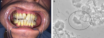
The complex composition and organization of periodontium (Menicanin et al. Reference Menicanin, Hynes, Han, Gronthos and Bartold2015; Mark Welch et al. Reference Mark Welch, Rossetti, Rieken, Dewhirst and Borisy2016) can be affected by a disruption of the homoeostasis between the oral microbiome and the host, which can lead to two major diseases: gingivitis and periodontitis. Gingivitis is a reversible inflammatory disease of the superficial periodontium most commonly occurring in response to a lack of hygiene (Burt, Reference Burt2005). In a permissive host, both infection and inflammation may synergistically extend to deeper periodontium, leading to an irreversible loss of tooth-supporting tissues (periodontitis). Clinical signs include gingival inflammation and bleeding (Fig. 1A), alveolar bone loss leading to periodontal pocket formation, gingival recession and, in later phases, tooth mobility and subsequent loss.
(A) Clinical intra-oral view of a patient with periodontal disease (general gingival inflammation along with a significant amount of calculus and loss of tooth-supporting tissues), where Trichomonas species were detected, as exhibited in (B). (B) Trichomonas sp. during observation of dental plaque (taken inside the sulcus area) using phase-contrast microscopy (×1000).

For several decades, research in periodontology has focused on the characterization of bacterial communities thought to be involved in periodontal diseases. This ‘reductionist approach’ (Baker et al. Reference Baker, Bor, Agnello, Shi and He2017), has failed to demonstrate that a shift from a healthy to a periopathogenic bacterial microflora represents the onset/aetiopathogeny of the tooth-supporting tissue diseases. Thus, the future of periodontal therapies should not target the periopathogenic bacteria alone, but rather consider the whole oral microflora, and also include parasites, and how it interacts with local immunity and the micro-environment homoeostasis.
The impact of parasites in periodontium pathophysiology is still debated and little information is available regarding the conditions favourable for their occurrence. Among the parasites found in dental plaque, Trichomonas tena x, an anaerobic motile-flagellated protozoan, may play a role in the pathophysiology of periodontal diseases. This 12–20 µm long and 5–6 µm wide organism is either ellipsoidal or ovoid in shape and has four anterior flagella of unequal lengths (Fig. 1B). Trichomonas tena x is a protozoan classified in the same genus as Trichomonas vaginalis (Roberts and Schmidt, Reference Roberts and Schmidt2009). The pathogenicity of T. vaginalis is due to multiple virulence factors and has been extensively investigated: it possesses the ability to adhere to a target cell with the mediation of a range of iron-dependent surface adhesins (Muñoz et al. Reference Muñoz, Pérez, Orrego, Osorio, Gutiérrez, Sagua, Castillo, Martínez-Oyanedel, Arroyo, Meza-Cervantez, da Silveira, Midlej, Benchimol, Cordero, Morales, Araya and González2012), has a cytotoxic effect (Bastida-Corcuera et al. Reference Bastida-Corcuera, Okumura, Colocoussi and Johnson2005), causes apoptosis of epithelial cells through release of cysteine proteases (Kummer et al. Reference Kummer, Hayes, Gilbert, Beach, Lucas and Singh2008) and induces a weakening of the junctional complex within the epithelial monolayer (da Costa et al. Reference da Costa, de Souza, Benchimol, Alderete and Morgado-Diaz2005). However, the putative pathogenic role of T. tenax is less documented. This parasite has nevertheless been reported to be involved in some extra-oral bacterial diseases (Duboucher et al. Reference Duboucher, Farto-Bensasson, Chéron, Peltier, Beaufils and Périé2000; Leterrier et al. Reference Leterrier, Morio, Renard, Poirier, Miegeville and Chambreuil2012; Lopez-Escamilla et al. Reference Lopez-Escamilla, Sanchez-Aguillon, Alatorre-Fernandez, Aguilar-Zapata, Arroyo-Escalante, Arellano, Moncada-Barron, Romero-Valdovinos, Martinez-Hernandez, Rodriguez-Zulueta and Maravilla2013) such as pleural effusion or lymph node infections. The presence of T. tena x within periodontal pockets was reported earlier, but its involvement in periodontitis pathophysiology has not been demonstrated yet (Feki et al. Reference Feki, Molet, Haag and Kremer1981; Ghabanchi et al. Reference Ghabanchi, Zibaei, Afkar and Sarbazie2010; Özçelık et al. Reference Özçelık, Gedık, Gedık and Malatyali2010).
The role of T. tenax in periodontopathies is discussed through an analysis of clinical and experimental studies. The data presented here may constitute a crucial contribution to a holistic approach to the interactions between host and oral microbiome as targets for innovative therapies in periodontitis. The objective of this concise review was to collect data about T. tenax, its prevalence in oral infections and its pathogenicity, and search for a treatment consensus.
MATERIAL AND METHODS
Aim
This review aims to collect data about the prevalence and the potential pathogenic role of T. tenax during periodontal diseases.
Searching for relevant studies
We searched the following databases: Ovid MEDLINE In-Process; the databases of MEDLINE (1950 to present) via Pubmed; Biosis (1997 to present) via OvidSP®; Google Scholar (the first 500 results, sorted by relevance); LILACS via the Virtual Health Library search form (1982 to present). The search strategy is provided in Appendix 1. Reference lists of publications were inspected to detect any additional relevant data.
Inclusion criteria
Based on information provided by titles and abstracts, full-text articles were retrieved if studying T. tenax was an objective of the work. Prevalence studies in animals were not considered. For human epidemiological studies to be taken into account, oral conditions had to be described. The last search was conducted on 20 August 2016. Two blinded authors performed the screening (MM and PM).
RESULTS
We screened 1030 studies and 92 full-text articles were retrieved on the basis of titles and abstracts. Finally, 47 studies about prevalence and eight studies about pathogenicity were included.
Although most studies did not report an adequate control group (no similarity of population with the test group), the prevalence of oral protozoan infections was found to be largely greater in patients with periodontal diseases than in controls (healthy periodontium). The presence of T. tenax in periodontal diseases started to be investigated in the 1960s and more importance was attached to the research during 1980s. Most of the authors performed the parasite detection by direct microscopy (Fig. 1B), using staining (e.g. Giemsa or trichrome staining) or culturing to identify T. tenax in dental plaque samples. Only four studies used the polymerase chain reaction (PCR) assay to highlight this pathogen (Athari et al. Reference Athari, Soghandi, Haghighi and Kazemi2007; Kurnatowska et al. Reference Kurnatowska, Dudko and Kurnatowski2004a ; Turkowicz et al. Reference Turkowicz, Tomaszewska and Cielecka2004; Mehr et al. Reference Mehr, Zarandi and Anush2015).
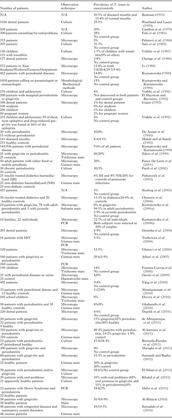
Data from 47 studies (detailed in Table 1) showed that T. tenax prevalence in diseased periodontium was heterogeneous, ranging from 0 to 94·1%. In the eight studies focusing on children and students, the prevalence was quite low, ranging from 0 to 4%, except in one study reporting a T. tenax prevalence in young people of about 14% (Fuentes Cuevas et al. Reference Fuentes Cuevas, Ramos, De, Castillo Contreras and Hernández-Sierra2008). Among subjects with periodontal diseases, three studies (Kurnatowska et al. Reference Kurnatowska, Dudko and Kurnatowski2004a ; Al-hamiary et al. Reference Al-hamiary, Kezar and Al-Khafaji2011; de Albuquerque et al. Reference de Albuquerque, de Melo, de Santana, Ribeiro and Silva2011) reported a higher T. tenax prevalence in periodontitis than in gingivitis patients. Conversely, two other studies did not report such differences (Sumaiah and Rasha, Reference Sumaiah and Rasha2012; Khalid et al. Reference Khalid, Kim, Duan, Hoang, Elashoff and Kim2014). Althought the presence of T. tenax seems to be correlated to the severity of periodontal diseases (de Albuquerque et al. Reference de Albuquerque, de Melo, de Santana, Ribeiro and Silva2011), no data were found about a quantitative correlation between pocket depth and parasite abundance. This may due to the lack of accuracy of the identification by light microscopy, that would be replaced by PCR, as already used for bacteria identification. Moreover, information about pocket measurement are often unavailable.
Details of included studies about prevalence of T. tenax occurrence in gum diseases.

N/A, no data; PCR, polymerase chain reaction.
Systemic diseases may also have impacted T. tenax prevalence in subjects displaying periodontopathies. Interestingly, this ratio was stronger in non-diabetics than in type 1 diabetic subjects (Piekarczyk et al. Reference Piekarczyk, Fiedor, Chomicz, Szubinska, Starościak, Piekarczyk, Zawadzki, Zebrowska and Dudziński2003; Chomicz et al. Reference Chomicz, Szubińska, Piekarczyk, Wojtowicz, Piekarczyk, Starościak and Fiedor2004). In one study on HIV+ patients, the authors reported a 5·5% T. tenax prevalence in diseased periodontium (no control group) (Turkowicz et al. Reference Turkowicz, Tomaszewska and Cielecka2004). Finally, T. tenax prevalence was higher in patients with Down Syndrom and periodontitis (18·8%) than in healthy individuals (3%) (Mehr et al. Reference Mehr, Zarandi and Anush2015).
DISCUSSION
Overall, the prevalence of T. tenax is higher in diseased (gingivitis and periodontitis) than in healthy periodontium. The high heterogeneity of T. tenax periodontal prevalence reported in the literature may be correlated with the diversity of the population screened (age, systemic diseases), and the methods used for diagnosis (insufficient use of a molecular, e.g. identification PCR method).
Sociodemographic and medical characteristics of the screened population
A wide time window was used in this study, and many countries were included. The hygiene habits, economic limitations and psycho-social perception of the disease may thus vary strongly and therefore influence the data collection. This points out the need to develop new epidemiological studies in this scientific field of investigation.
Considering the pediatric populations, the prevalence of T. tenax in periodontal tissues is higher in adolescents than in young children (Vráblic et al. Reference Vráblic, Tomová and Catár1992). In adult populations, gender does not seem to correlate with T. tenax prevalence (Cambon et al. Reference Cambon, Petavy, Guillot, Glanddier, Deguillaume and Coulet1979; Palmieri et al. Reference Palmieri, Halverson, Sudjadi, Purnomo and Masbar1984), while age may impact the T. tenax occurrence. Indeed, it has been suggested that periodontal tissues of patients over 40 years old are more infected by T. tenax (Cambon et al. Reference Cambon, Petavy, Guillot, Glanddier, Deguillaume and Coulet1979; Kurnatowska and Kurnatowska, Reference Kurnatowska and Kurnatowska1990) than those of younger subjects. Those results may be correlated with the increased prevalence of periodontitis with age, particularly from 40 to 50 years. The effect of age and systemic diseases on sustaining immunity homoeostasis and the decrease of stromal cells’ renewal potential may explain this fact (Lemaitre et al. Reference Lemaitre, Monsarrat, Blasco-Baque, Loubières, Burcelin, Casteilla, Planat-Bénard and Kémoun2017). This notion of local immunity alteration in periodontal tissue due to general pathology (Blasco-Baque et al. Reference Blasco-Baque, Serino, Vergnes, Riant, Loubieres, Arnal, Gourdy, Sixou, Burcelin and Kemoun2012) is crucial because it can explain the difference of prevalence between studies among heterogeneous populations. For example, DS could create conditions for the development of periodontal inflammation, so patients with DS would be more infected by T. tenax than healthy patients (Mehr et al. Reference Mehr, Zarandi and Anush2015).
Is T. tenax a pathogen and/or a marker of periodontal diseases?
For decades, diseases of tooth-supporting tissue were considered as ‘microbial’ diseases, meaning that bacteria may induce, by themselves and in anyone, the onset of the illness. However, there is currently no consensus about the ‘primitive’ aetiological agent of periodontal diseases. Low oral hygiene and plaque/calculus accumulation are often correlated with the grade of damage, especially gingival inflammation for gingivitis, and number and depth of periodontal pockets in case of periodontitis (Bartold and Van Dyke, Reference Bartold and Van Dyke2013). Nevertheless, there is accumulated evidence that periodontal diseases develop mainly in permissive hosts, who have local dysimmunity (Monsarrat et al. Reference Monsarrat, Blaizot, Kémoun, Ravaud, Nabet, Sixou and Vergnes2016). Despite the fact that more experiments are needed, it can be hypothesized that one or several events may upset the balance of host–microbiome homoeostasis. Thus, a local dysimmunity may favour the development, selection and pathogenicity expression of periopathogens (including bacteria and protists), leading to a shift from ‘eubiosis’ to ‘dysbiosis’ (change of the pressure of the environment). The crucial point is that, once the host–microbiome homoeostasis is unbalanced, a vicious cycle of pathogen activity – inflammation – tissue destructions, may be set up and become self-perpetuating.
It has been shown that protozoa can both induce tissue damage and benefit from this modified micro-environement to grow and exacerbate the injuries (see the next section). Because it is suggested here that the existence of T. tenax infection is correlated with periodontal diseases and its amount with their severity, we hypothesized that T. tenax may not only have an intrinsic periopathogenicity, but also reflect the activity of tooth-supporting tissue inflammation, as is known to occur for periopathogens such as Porphyromonas gingivalis or Fusobaterium nucleatum, for example (Bartold and Van Dyke, Reference Bartold and Van Dyke2013). Case–control and/or cohort studies paired with biological analyses are necessary for a better understanding of these processes.
Trichomonas tenax involvement in periodontal disease pathophysiology
No animal study on the pathophysiology of T. tenax virulence regarding periodontal diseases. was found. However, several mechanisms may explain the deleterious effects of the T. tenax parasite towards periodontal tissues.
First, the pathogenicity of T. tenax may be inferred from a comparison with the urogenital parasite T. vaginalis, the first human non-viral sexually transmitted disease worldwide (Kumar et al. Reference Kumar, Chandra, Reddy, Reddy, Reddy and Naveen2015; Alcaide et al. Reference Alcaide, Feaster, Duan, Cohen, Diaz, Castro, Golden, Henn, Colfax and Metsch2016). A genetic comparison between T. vaginalis and T. tenax, suggested that the two parasites were variants (Kucknoor et al. Reference Kucknoor, Mundodi and Alderete2009). Despite more experiment are needed to identify mechanisms of T. tenax pathogenicity, it may be hypothized that T. vaginalis may have colonized oral cavity during evolution, and developed survival strategies by genotype changes to acquire phenotype suitable to the new ecosystem, as T. tenax. T. vaginalis often occurs in tandem with bacterial vaginitis caused by an imbalance of the bacterial flora (Fichorova et al. Reference Fichorova, Buck, Yamamoto, Fashemi, Dawood, Fashemi, Hayes, Beach, Takagi, Delaney, Nibert, Singh and Onderdonk2013), escaping the host immune response via a complex strategy (Menezes and Tasca, Reference Menezes and Tasca2016). Recent studies have emphasized the ability of parasites to induce changes in some features of microbial communities (Bhattacharjee et al. Reference Bhattacharjee, Kalbfuss and Prazeres da Costa2017). The presence of T. tenax in the deep periodontal pocket may substantiate this role in periodontal dysbiosis. Indeed, along with inflammation and the deepening of the periodontal pockets, the periodontal environement becomes more anaerobic, resulting in a bacterial shift from a Gram-positive to a Gram-negative flora. This decrease in the partial pressure in oxygen may explain why the depth of periodontal pocket may be a critical factor for the anaerobic T. tenax colonization and growth.
Pathogenic bacteria involved in periodontal host colonization and immune subversion use complement and toll-like receptor (TLR) signalling pathways (Bartold and Van Dyke, Reference Bartold and Van Dyke2013; Hajishengallis, Reference Hajishengallis2015). Like bacteria, parasites are recognized by TLR (Gazzinelli and Denkers, Reference Gazzinelli and Denkers2006). Because stimulation of the immune system via TLR4 by T. vaginalis has been reported (Zariffard et al. Reference Zariffard, Harwani, Novak, Graham, Ji and Spear2004), such a mechanism within the periodontium was also hypothesized for T. tenax. Moreover, patients infected by T. vaginalis harbored a predominant tissue recruitment of neutrophils (Escario et al. Reference Escario, Gómez Barrio, Simons Diez and Escario2010) similarly to the massive neutrophil recruitment found in human periodontitis (Dutzan et al. Reference Dutzan, Konkel, Greenwell-Wild and Moutsopoulos2016).
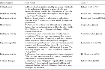
Second, these pathogens produce fibronectin-like proteins, responsible for tissue adhesion (Ribaux et al. Reference Ribaux, Magloire, Joffre and Morrier1983). Given this pathogenic property, host-tissue disruption and lysis may be induced by T. tenax secretion of peptidases (Bózner and Demes, Reference Bózner and Demes1991a ; El Sibaei et al. Reference El Sibaei, Abdel-Fattah, Ahmed and Abou-Seri2012) such as cathepsin B-like proteinases for matricial type 1 collagen and gelatin hydrolyses (Yamamoto et al. Reference Yamamoto, Asaga, Nagao, Igarashi and Goto2000) or haemolysins for erythrolysis (Nagao et al. Reference Nagao, Yamamoto, Igarashi, Goto and Sasa2000) (Table 2). Recently Ribeiro et al. analysed the in vitro relationship of T. tenax with mammalian cells (Ribeiro et al. Reference Ribeiro, Santos and Benchimol2015). They confirmed the ability of the flagella to adhere to periodontal epithelial cells. After only 6 h, T. tenax in co-culture caused significant damage (disrupting some of the cells in the tissue, inducing membrane damage and cell apoptosis). In contrast, T. vaginalis has been found in the oral cavity (Crucitti et al. Reference Crucitti, Jespers, Mulenga, Khondowe, Vandepitte and Buvé2010), but failed to elicit in vitro damage on periodontal cells (Ribeiro et al. Reference Ribeiro, Santos and Benchimol2015).
Details of included studies on the pathogenicity of Trichomonas tenax

Methods of parasite identification
Another potential factor of prevalence discrepancies in literature reports was the method of parasite identification. The method most frequently used was direct microscopy, with or without staining (e.g. Giemsa). This method is time consuming and insufficient to discriminate parasite species, because T. tenax is morphologically close to T. vaginalis. Nevertheless, such a method is used at chair-side for diagnosis and treatment management (Fig. 1) but it could give rise to more false negative results (Marty et al. Reference Marty, Bonner and Vaysse2015). In vitro culture is also a method for detecting the presence of the protozoa (Table 1), but its ability to detect T. tenax was found to be similar to that of direct observation (El Sibaei et al. Reference El Sibaei, Abdel-Fattah, Ahmed and Abou-Seri2012). Molecular methods, e.g. PCR, appear to be the more reliable, as already performed for T. tenax (Kikuta et al. Reference Kikuta, Yamamoto, Fukura and Goto1997) and Entamoeba gingivalis detection (Bonner et al. Reference Bonner, Amard, Bar-Pinatel, Charpentier, Chatard, Desmuyck, Ihler, Rochet, Roux de La Tribouille, Saladin, Verdy, Gironès, Fresno and Santi-Rocca2014). Unfortunately, only four published studies used such a method to assess T. tenax identification in periodontal tissues. Direct microscopy is a simple procedure used for immediate diagnosis, but the molecular method is more specific and allows a lower detection threshold to be used, meeting the prevalence research requirement (Patil et al. Reference Patil, Nagamoti and Metgud2012).
Antiparasitic treatments
Oral trichomonads are sensitive to anti-parasitic treatment. Zvetkova et al. investigated the clinical improvement and the titration of antibodies after anti-parasitic treatment with tinidazole in patients infected by oral trichomonads. After 3 days of treatment, oral clinical signs had disappeared, and titration of antibodies was normalized by months 6–12 after treatment (Zvetkova et al. Reference Zvetkova, Isev and Stoilova1988). Tinidazole is also efficient against anaerobic bacteria involved in periodontal disease (Alou et al. Reference Alou, Giménez, Manso, Sevillano, Torrico, González, Granizo, Bascones, Prieto, Maestre and Aguilar2009). However, additional studies are needed to support these results.
Concluding remarks
The involvement of the whole oral communities, including parasites, fungi and bacteria, has been suggested to be critical in the pathophysiology of periodontitis (Lamont and Hajishengallis, Reference Lamont and Hajishengallis2015). However, many questions remain. Among them, the periopathogeny of protozoans, and especially T. tenax, has to be deciphered for a rational development of new efficient periodontal therapy strategies (Costalonga and Herzberg, Reference Costalonga and Herzberg2014). Animal experimentation, using pertinent physiopathology models of periodontitis (Lemaitre et al. Reference Lemaitre, Monsarrat, Blasco-Baque, Loubières, Burcelin, Casteilla, Planat-Bénard and Kémoun2017) may be implemented to investigate the ability of T. tenax to cause and/or exacerbate the disease.
Dental practitioners should take time to analyse the intra-sulcular dental plaque of patients with periodontitis by direct microscopy. The discovery of T. tenax may be a marker of a specific clinical type of periodontitis and modify the therapeutic management and prognosis (Marty et al. Reference Marty, Bonner and Vaysse2015).
FINANCIAL SUPPORT
This study was supported by funding from the Midi-Pyrenees region, France, Paul Sabatier University and the research platform of Toulouse Dental Faculty (PLTRO).
CONFLICT OF INTEREST
None.
APPENDIX 1
Search strategy
MEDLINE via Pubmed
#1: Stomatognathic Diseases [mh:exp]
#2: buccal [tw] OR oral [tw] OR mouth [tw] OR periodont* [tw] OR gingiva* [tw] NOT ‘oral contraceptives’ [tw] NOT ‘oral contraceptive’ [tw]
#3: Trichomonas [mh:exp]
#4: trichomona* [tw]
#5: #1 OR #2
#6: #3 OR #4
#7: #5 AND #6
Biosis, Ovid MEDLINE® In-Process and Other Non-Indexed Citations via OvidSP®
1: (buccal or oral or mouth).tw.
2: (trichomon*).tw.
3: 1 and 2
LILACS
[(trichomon$ OR ‘TRICHOMONAS’) AND (‘STOMATOGNATHIC DISEASES’ OR buccal OR oral OR mouth)][Words]
Google Scholar
‘trichomonas tenax’