The yeast Malassezia pachydermatis, a common colonizer of dogs and cats, can result in healthcare-associated infections, primarily in neonates.Reference Chen, Chiu and Chi1–Reference Ilahi, Hadrich and Goudjil3 The first reported outbreak of M. pachydermatis bloodstream infections (BSIs) occurred in the 1990s in a neonatal intensive care unit (NICU).Reference Welbel, McNeil and Pramanik4 A subsequent report of a BSI outbreak in another NICU implicated indirect patient-to-patient transmission via the hands of healthcare workers (HCWs) who became colonized with M. pachydermatis after contact with pet dogs.Reference Chang, Miller and Watkins5 Using the traditional molecular technique restriction fragment length polymorphism (RFLP), these authors reported identical DNA patterns among specimens from patients, HCWs, and pet dogs of HCWs.
Fluconazole is commonly used in NICUs to prevent Candida infections in very-low-birthweight preterm infants.Reference Silva-Rios, Camargos, Correa and Romanelli6 Infections with fluconazole-resistant M. pachydermatis have been reported in preterm infants receiving fluconazole prophylaxis.Reference Chen, Chiu and Chi1,Reference Al-Sweih, Ahmad, Joseph, Khan and Khan7
We investigated a suspected outbreak of M. pachydermatis to determine epidemiologic linkages, and we performed whole-genome sequencing (WGS) to assess genetic relatedness of isolates. WGS is a powerful technology that can assist in public health investigations by providing a high degree of resolution for microbial subtyping but has not been previously used to investigate an M. pachydermatis outbreak.
Methods
We examined cases of M. pachydermatis infection and colonization during December 2015–September 2016 in an 84-bed, level 3 NICU. A case was defined as isolation of M. pachydermatis from sterile sites or skin of a patient. After the second case of M. pachydermatitis was identified, infection prevention practices were reviewed with the staff to mitigate further transmission of M. pachydermatis. Clinical data were extracted from medical records.
Susceptibility testing was performed at the University of Texas Health Science Center at San Antonio. We performed WGS on case isolates and on historical M. pachydermatis isolates stored at the US Centers for Disease Control and Prevention (CDC) using the Illumina MiSeq (Illumina, San Diego, CA). Paired-end sequences that had at least 50X coverage were used for downstream analyses. Read data were aligned to an assembly generated by PacBio sequencing (PacBio, Menlo Park, CA). Single-nucleotide polymorphism (SNP) variants were identified using SAMtools and were filtered using the publicly available SNP analysis pipeline NASP to remove positions that had <10X coverage, <90% variant allele calls, or those identified by Nucmer as being within duplicated regions in the reference.Reference Sahl, Lemmer and Travis8 Phylogenetic analyses were performed on SNP matrices using MEGA, and bootstrapping was performed with 1,000 iterations.
Results
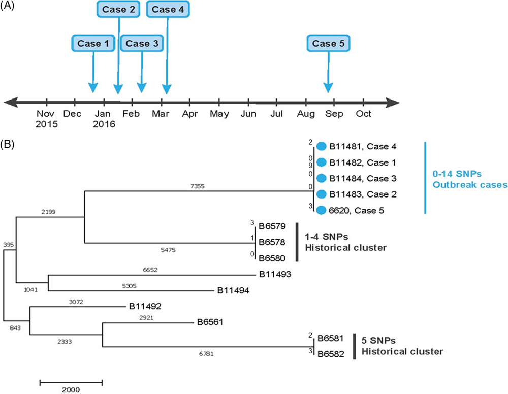
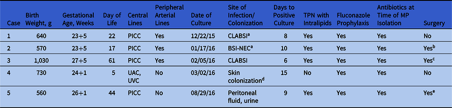
Overall, 5 cases were identified in neonates hospitalized in NICU A; the first 4 cases (cases 1–4) occurred from December 2015 to March 2016 and the last occurred in August 2016 (case 5) (Fig. 1A and Table 1). For cases 1–4, M. pachydermatis was recovered from specimens taken in NICU A. For case 5, the culture was obtained at a different hospital (NICU B) 23 days after transfer (44th day of life) from NICU A. Using National Healthcare Safety Network surveillance definitions, 2 cases were categorized as central-line–associated BSIs (cases 1 and 3): 1 patient had a secondary BSI due to necrotizing enterocolitis (case 2); 1 patient had skin colonization (case 4); and 1 patient had peritonitis, infected urinoma, and positive urine culture (case 5).
(A) Outbreak timeline: Cases 1–4 were identified in neonatal intensive care unit (NICU) A and case 5 was identified in NICU B. (B) Maximum parsimony tree showing genetic relationships among Malassezia pachydermatous isolates based on whole-genome sequencing. Outbreak isolates are marked with blue circles, and isolates from historical clusters are marked with vertical lines.

Clinical Characteristics by Case

Note. TPN, total parenteral nutrition; PICC, peripherally inserted central catheter; CLABSI, central-line–associated bloodstream infection; BSI-NEC, secondary bloodstream infection attributed to necrotizing enterocolitis (NEC); UAC, umbilical arterial catheter; UVC, umbilical venous catheter.
a Surveillance definitions per National Healthcare Safety Network (NHSN).
b Thoracotomy chest tube for pneumothorax; spontaneous intestinal perforation with repair.
c Laparotomy for gastric perforation; necrotizing enterocolitis; extended right colectomy.
d Methicillin-resistant Staphylococcus aureus (MRSA) bacteremia with diffuse MRSA and aspergillus skin infection. M. pachydermitis felt to be colonizer.
e Laparotomy for suspected necrotizing enterocolitis.
On susceptibility testing, all 5 isolates had minimum inhibitory concentration (MIC) values of ≤0.03–0.06 for amphotericin B, >8 for micafungin, and >64 for fluconazole.
All 5 patients had stayed in the same 29-bed NICU pod. Birth weights ranged from 560 to 1,010 g, with gestational ages between 23+5 and 27+5 weeks. Positive cultures were obtained between ages 5 and 44 days. All patients had central venous catheters, were exposed to broad-spectrum antibiotics, and received fluconazole prophylaxis (Table 1). Also, 4 patients received total parenteral nutrition and 3 had comorbid conditions. No patients died.
The isolation of M. pachydermatis in this NICU and the epidemiologic clustering raised concern that an outbreak was present. Consequently, the following infection prevention strategies were implemented: (1) the importance of optimal catheter insertion and maintenance practices was reinforced; (2) NICU staff were provided with education on M. pachydermitis epidemiology; (3) proper frequency and quality of hand hygiene practices were ensured; and (4) enhanced environmental cleaning was initiated. A wider point-prevalence survey for colonization in the NICU, culturing of HCWs, and environmental sampling was not performed because it was uncertain at the time whether molecular subtyping was available. However, subsequently, using WGS, CDC confirmed that an outbreak was present.
The 5 case isolates were highly related (maximum pairwise difference of 14 SNPs), whereas case isolates were not closely related to any of the 9 historical isolates (>10,000 SNPs) (Fig. 1B). Isolates from the first 4 cases were nearly identical (≤2 SNPs), and the fifth isolate differed by ≤14 SNPs. Among the historical CDC isolates, 2 clusters were detected, with ≤4 and ≤5 SNPs among them. A review of the records revealed epidemiologic links among these isolates.
Discussion
We describe an outbreak of M. pachydermatis in a NICU that was confirmed with epidemiologic clustering and high-relatedness by WGS. Although the source of the outbreak was not identified, given that all neonates stayed in the same 29-bed pod, gaps in optimal hand hygiene practices, as previous studies have shown, likely played a role. The contribution of the environment to indirect transmission remains to be determined. WGS characterizes genetic diversity among isolates at a higher resolution than traditional methods such as RFLP. Case 5 differed from the others in several ways. Whereas isolates from cases 1–4 were nearly identical (maximum pairwise difference, 2 SNPs), they differed by 12–14 SNPs from the case 5 isolate. Case 5 was identified 5 months after the case 1–4 cluster at a different hospital, suggesting a more distant relationship. However, several factors suggest that this case was related to the outbreak. First, a retrospective review of laboratory reports at NICU B did not identify additional M. pachydermatis isolates. Second, before transferring to NICU B, where the isolate was recovered, the patient had been hospitalized in NICU A in the same pod as the other patients where the patient could have become colonized. Third, the number of SNP differences (12–14 SNPs) was still very small compared with the differences with historical isolates (>10,000 SNPs) and might simply reflect the greater time between cases.
Although there are no standardized MIC cutoffs for M. pachydermatis, all isolates had elevated MICs to fluconazole. Additionally, because M. pachydermatis is a basidiomycete and has B1,6-glucan in the cell wall rather than B1,3-glucan, it is intrinsically resistant to echinocandins. Because all 5 case patients had received fluconazole prophylaxis, our findings, together with previous reports of fluconazole-resistant M. pachydermatis, should alert clinicians to the possibility of an infection with this organism in neonates receiving fluconazole prophylaxis. The susceptibility testing profile of M. pachydermatis observed to date,Reference Leong, Buttafuoco, Glatz and Bosshard9,Reference Cafarchia, Iatta, Immediato, Puttilli and Otranto10 suggests that patients with M. pachydermatis infections should be treated with amphotericin B.
In conclusion, in response to a suspected outbreak of M. pachydermatis in the NICU, we ensured adherence to infection prevention measures, implemented enhanced environmental hygiene, and reviewed the epidemiology with HCWs to mitigate further transmission. The presence of an outbreak was confirmed using WGS. We anticipate increasing use of WGS for fungal disease outbreak investigations as the technology become increasingly affordable and accessible.
Acknowledgments
The use of product names in this manuscript does not imply their endorsement by the US Department of Health and Human Services. The finding and conclusions in this article are those of the authors and do not necessarily represent the views of the Centers for Disease Control and Prevention.
Financial support
No financial support was provided relevant to this article.
Conflicts of interest
All authors report no conflicts of interest relevant to this article.