Implications
The commercial breeding of deer for antler trophies has a turnover of many millions of Euros worldwide. Our results demonstrate that Cu supplementation tended to increase the cortical thickness of adult red deer antlers, which may indicate increases in their weight, value as trophies and resistance to fracture. As the fastest growing bone, antlers may exhibit the effects of supplementation with Cu more clearly than internal bones. Thus, these results may also have implications for human bones. Our results suggest that medical research into the potential benefits of Cu supplementation injections in improving bone resistance in elderly humans could be interesting.
Introduction
Antler is a unique form of bone and by far the fastest growing mammalian tissue with an average increase of 0.67 cm/day in Iberian red deer (based on Gómez et al., Reference Gómez, García, Luna, Kierdorf, Kierdorf, Gallego and Landete-Castillejos2013), demanding a high mineral transfer from the skeleton. Blood plays an essential role in maintaining the flow of minerals and enzymes required for antler growth, which may eventually constitute as much as 28% of the weight of a deer skeleton (Gómez et al., Reference Gómez, Ceacero, Landete-Castillejos, Gaspar-López, García and Gallego2012). In addition to Ca, other minerals, such as Mn, have essential roles in incorporating circulating bone Ca into the growing antlers (Landete-Castillejos et al., Reference Landete-Castillejos, Estévez, Martínez, Ceacero, García and Gallego2010; Cappelli et al., Reference Cappelli, García, Ceacero, Gómez, Luna, Gallego, Gambín and Landete-Castillejos2015). Even when the levels of other minerals and diet quality are appropriate, deficiency of a single mineral may lead to bones having low Ca content (Strause et al., Reference Strause, Hegenauer, Saltman, Cone and Resnick1986). Copper may be another such mineral, as it is involved in the development and maintenance of collagen, elastin, bones and connective tissue. In particular, Cu plays an essential role in the normal maturation of collagen, specifically the synthesis of lysine-derived cross-links (Hyun et al., Reference Hyun, Barrett-Connor and Milne2004). Therefore, Cu deficiency may directly damage bone cell formation (by increasing chondrogenesis and delaying cartilage mineralization) with negative effects on the regulation of bone turnover, bone maturation or skeletal growth (Suttle, Reference Suttle1972), and alter the hormonal regulation of Ca and vitamin D3 metabolism (Sadeghi et al., Reference Sadeghi, Oveisi, Jannat, Hajimahmoodi, Behzad, Behfar, Sadeghi and Saadatmand2014). In fact, a decrease in the formation of amino acid cross-links is thought to cause the increased bone fragility associated with Cu deficiency (Opsahl et al., Reference Opsahl, Zeronian, Ellison, Lewis, Rucker and Riggins1982); therefore, Cu deficiency may increase the risk of fractures (Kierdorf et al., Reference Kierdorf, Kierdorf and Boyde2000). Insufficient Cu constitutes one of the most common and severe trace mineral deficiencies in deer (Grace and Wilson, Reference Grace and Wilson2002) and these animals frequently require Cu supplementation.
The mineral composition of antlers is influenced by diet and affects their mechanical properties (Estévez et al., Reference Estévez, Landete-Castillejos, Martínez, García, Ceacero, Gaspar-López, Calatayud and Gallego2009; Landete-Castillejos et al., Reference Landete-Castillejos, Estévez, Martínez, Ceacero, García and Gallego2010). However, even when deer are fed a balanced diet, the presence of antagonists (such as S, Mo, Zn or Fe) among the raw materials commonly used as feed for deer can decrease the absorption and digestibility of Cu (Jacob et al., Reference Jacob, Skala, Omaye and Turnlund1987), leading to increased bone fragility and a consequent risk of fractures. Copper supplementation delivered by injection bypasses the gastrointestinal tract and, thereby, dietary antagonists, and has the potential to improve the properties of bone and increase its ash and mineral content. In a study of the effects of trace minerals on the mechanical properties of antlers, Landete-Castillejos et al. (Reference Landete-Castillejos, Estévez, Martínez, Ceacero, García and Gallego2010) suggested that reduced Mn content in diet was responsible for generalized antler breakage and a 27% reduction in impact energy, in addition to a wide range of other effects. That study also reported a lower level of Cu of antlers with Mn deficiency. Recently, Cappelli et al. (Reference Cappelli, García, Ceacero, Gómez, Luna, Gallego, Gambín and Landete-Castillejos2015) presented very similar data demonstrating the effect of Mn deficiency on various characteristics of antler and, again, an effect on antler Cu content. However, no study has yet assessed the effects of supplementation with Cu injections on the mechanical and structural properties, and mineral composition, of antlers or other types of bone.
The effects of Cu injections on bone characteristics and mineral content may be modified depending on the age of deer, as yearlings and adults respond differently to supplementation with other minerals, such as Mn (Cappelli et al., 2015). The degree of skeleton demineralization and, by implication, the physiological efforts required to grow each part of the antler, increases as an antler grows (Baxter et al., Reference Baxter, Andrews and Barrell1999; Landete-Castillejos et al., Reference Landete-Castillejos, Currey, Estévez, Fierro, Calatayud, Ceacero, García and Gallego2007a and Reference Landete-Castillejos, Currey, Estévez, Gaspar-López, García and Gallego2007b). In addition, yearlings are under greater growth constraint, as they require Ca and other minerals for both skeleton and antler growth (Gaspar-López et al., Reference Gaspar-López, Landete-Castillejos, Estévez, Ceacero, Gallego and García2008; Ceacero et al., Reference Ceacero, Landete-Castillejos, García, Estévez and Gallego2010). Thus, we studied the effect of Cu supplementation by injection on the mechanical and structural properties and mineral content of antler bone tissue from both yearling and adult red deer (Cervus elaphus hispanicus).
Material and methods
Ethical statement
This study was carried out in accordance with Spanish legislation regarding the use of animals in research (Boletín Oficial del Estado, 2013) and with the approval of the Ethical Committee in Animal Experimentation at the Universidad de Castilla-La Mancha (UCLM, Permit Number: 1002.04). Because antlers are dead when they are hard and clean of velvet (Currey et al., Reference Currey, Landete-Castillejos, Estévez, Ceacero, Olguín, García and Gallego2009), their removal causes no pain; therefore, no anaesthesia is required. Nevertheless, a low dose of xylazine (0.3 mg/kg BW) was used as tranquilizer to reduce stress.
Experimental design
This study was performed at the UCLM experimental farm in Albacete, south-eastern Spain (38°57′10′′N, 1°47′00′′W, altitude 690 m). Copper was administered by subcutaneous injection of Glypondin (König S.A., Argentina) solution (1 cm3/30 kg live BW) containing 0.83 mg of Cu per kg BW in the treatment group. Injections were administered every 42 days during the whole antler growth period. The control group was injected with a physiological saline solution. Each group comprised animals of different ages: there were 10 yearlings and seven adults in the control group, and 11 yearlings and seven adults in the treatment group. As average, yearlings were 1.5 years old (range from 17 to 18 months) and adults were 4 years (range from 2 to 6 years).
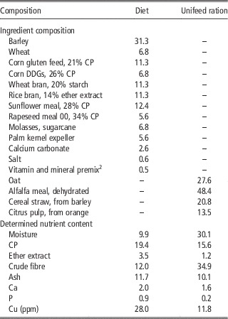
Feed and water were offered ad libitum throughout the trial. The feeding program (diet and unifeed ration) was the same for all deer and met or exceeded the nutrient requirements for deer (National Research Council (NRC), 2007). The ingredients and determined nutrient content (Association of Official Analytical Chemists (AOAC), 2000) of the diet and unifeed ration are shown in Table 1. Diet and unifeed ration mineral contents were analysed by inductively coupled optical emission spectrometry (ICP-OES) at the Ionomic Laboratory (IL), Centro de Edafología y Biología Aplicada del Segura-Consejo Superior de Investigaciones Científicas (CEBAS-CSIC, Murcia, Spain). Unifeed rations were homogenized and cut into small portions in a tractor-driven commercial mixer.
Ingredient composition (% dry matterFootnote 1 ) and determined nutrient content (% dry matterFootnote 1 ) of diet and unifeed ration

1 Unless otherwise indicated.
2 Supplied per kg of diet: vitamin A (trans-retinyl acetate), 10 000 IU; vitamin D3 (cholecalciferol), 2000 IU; vitamin E (all-rac-tocopherol-acetate), 15 IU; Mn (MnSO4·H2O), 75 mg; Fe (FeCO3), 50 mg; Zn (ZnSO4·H2O), 115 mg; Cu (CuSO4·5H2O), 7.5 mg; I (KI), 2 mg; Co (2CoCO3·3Co(OH)2·H2O), 0.83 mg; Se (Na2SeO3), 0.22 mg; ethoxiquin, 0.025 mg; butilhidrotoluen (BHT), 0.18 mg; butylhydroxyanisole (BHA), 0.016 mg; sepiolite, 950 mg.
All animals were adapted to routine management and maintained in good health and body condition during the experiment. At the beginning of the trial, deer were divided in two groups matched for BW (mean BWs of yearlings and adults were 72.9 and 147.4 kg, respectively) and body condition (means of 3.5 and 3.8 for yearlings and adults, respectively). Body weight and body condition were measured every 42 days throughout the trial, at the same time that Cu injections were administered. Animals were weighed using a ±50 g electronic balance. Average daily gain (ADG) was calculated from BW data for yearlings and adults. Body condition was measured as indicated by Audige et al. (Reference Audige, Wilson and Morris1998) and Carrión et al. (Reference Carrión, García, Gaspar-López, Landete-Castillejos and Gallego2008). Average daily feed intake of diet and unifeed ration was calculated from the amount used to replenish the feeders daily.
Antler and serum sample collection
The effects of Cu supplementation were analysed at four positions along the antler beam in adults, as indicated by Landete-Castillejos et al. (Reference Landete-Castillejos, Estévez, Martínez, Ceacero, García and Gallego2010): position one, close to the base (burr); position two, the first third of the antler shaft; position three, the second third of the shaft; and position four, close to the base of the crown. Measurements were taken ~2 cm above the burr, 2 cm below the main tip, 2 cm above the main tip and 2 cm below the crown. Antlers from yearlings were sampled following a similar procedure to that for adults; however, only three sampling points (positions one, two and three) were included, due to the smaller size and different structure of the antlers. The last position sampled in the antler indicates more clearly the effects of diet and other factors (Landete-Castillejos et al., Reference Landete-Castillejos, Currey, Estévez, Fierro, Calatayud, Ceacero, García and Gallego2007a, Reference Landete-Castillejos, Currey, Estévez, Gaspar-López, García and Gallego2007b and Reference Landete-Castillejos, Estévez, Ceacero, García and Gallego2007c).
From each position, a 1 cm slice and a cylinder of about 5 to 6 cm (the latter to obtain bars from the cortical wall) were obtained. Bars were identified according to individual deer and level in the main beam. The outer, or periosteal, side was also marked to ensure consistent testing with this side in tension, and to mark the end closer to the antler base.
Blood samples were taken from resting deer after an overnight fast on the first day of the trial and on the 84th day after the first Cu injection. Blood samples were drawn by jugular venepuncture using Vacutest Clot activators and were coagulated in the tubes (Kima S.A., Italy). Serum was separated using a centrifugal separator (Jouan® CR23-22; 2848 g at 4°C for 15 min) and samples were kept in a freezer at −20°C until the analysis.
Mechanical bone properties
First, samples were placed in Hank’s balanced salt buffer solution (BioWhittaker, Belgium) for 48 h to minimize the risk of small amounts of mineral leaching out of the bone (Currey et al., Reference Currey, Landete-Castillejos, Estévez, Ceacero, Olguín, García and Gallego2009). Thereafter, to standardize humidity content, the bars were dried at 20°C, 40% relative humidity, for 72 h. Bars with homogeneous humidity content were tested by three-point bending, with the periosteal side in tension, as described by Cappelli et al. (Reference Cappelli, García, Ceacero, Gómez, Luna, Gallego, Gambín and Landete-Castillejos2015), in a Zwick/Roell 500 N machine (Germany), head speed 32 mm/min, and gauge length 40 mm.
A low-speed circular saw was used for initial cutting of the antler. The surfaces were then abraded using semiautomatic polishing device (LaboPol-21, Struers Inc., Ballerup, Denmark) to create bars of the correct size. Parallel surfaces were obtained and the width and depth of each sample recorded to the nearest 0.01 mm using a digital calliper before testing. The final size was 4.5 mm wide, 2.5 mm deep, with variable length sufficient for the gauge length mentioned above.
The mechanical properties of antler bone samples were measured as previously described (Landete-Castillejos et al., Reference Landete-Castillejos, Currey, Estévez, Fierro, Calatayud, Ceacero, García and Gallego2007a, Reference Landete-Castillejos, Estévez, Ceacero, García and Gallego2007c and Reference Landete-Castillejos, Estévez, Martínez, Ceacero, García and Gallego2010; Currey et al., Reference Currey, Landete-Castillejos, Estévez, Ceacero, Olguín, García and Gallego2009) and included Young’s modulus of elasticity (E), an estimate of stiffness; bending strength (BS), calculated from the maximum load borne and the work to peak force (W), determined by total work done on the specimen up to the greatest load borne divided by the central cross-sectional area.
The impact work (U) testing procedure measures the energy used to break an un-notched specimen by a falling pendulum. This value of U was normalized by dividing by the cross-sectional area of the specimen (Cappelli et al., Reference Cappelli, García, Ceacero, Gómez, Luna, Gallego, Gambín and Landete-Castillejos2015). Tests were carried out in a CEAST-IMPACTOR II (CEAST S.p.A., Pianezza, Italy) with a hammer of 1 J.
Bone structural properties
To calculate the specific gravity or density of cortical bone, samples were placed in a controlled heating chamber for 72 h at 60°C to dry out fully. Each sample was then weighed with a precision balance (±0.01 g) and measured with a precision scale (±0.01 mm). Density was calculated by dividing the weight by the volume (calculated as length×width×depth).
As described by Landete-Castillejos et al. (Reference Landete-Castillejos, Estévez, Martínez, Ceacero, García and Gallego2010), complete transverse cross-sections were scanned on a flatbed scanner (ScanJet 4370 Photo Scanner, HP Inc., Palo Alto, CA, USA) at 2400×2400 dpi into image analysis software (IMAGEJ), and the cortical bone thickness of the cross-sections was measured at six equally spaced points around the shaft. The average cortical thickness was calculated for each cross-section. In addition, the area of the cortical section was measured.
Chemical analysis of antlers and serum samples
The breakage of one antler bar to determine the value of U and the other for the BS test produced four similar sized fragments. One of these fragments was used to determine the mineral content of the antlers. To this end, the edge that was fractured was polished to achieve a regular shape and also in any areas with pen marks. Antler mineral composition, including the contents of ash, Ca, P, Mg, Na, S, K, B, Cu, Fe, Mn, Sr, Zn, Al, Bi, Li and Cr, was analysed. Also, the ratio of Ca to P was calculated. The segment of bar was placed in a tube for mineral analysis. Details of the method used for mineral analysis are described in Cappelli et al. (Reference Cappelli, García, Ceacero, Gómez, Luna, Gallego, Gambín and Landete-Castillejos2015). Another fragment was used to measure ash content; samples were heated for 72 h at 60°C, then weighed using a precision balance (±0.01 g) to determine the dry weight. Subsequently, fragments were placed in a muffle furnace (HTC 1400, Carbolite-Gero Ltd, Derbyshire, UK) for 6 h at 480°C. Ash content was defined as the weight of the ashes thus obtained divided by the dry weight. Serum Cu content was analysed by ICP-OES at IL, CEBAS-CSIC Centre (Murcia, Spain).
Statistical analyses
A GLM test was performed to determine the effects of treatment and age class on each of the studied antler variables. Interaction between treatment and age was not significant for any trait studied. However, preliminary analysis indicated a very clear difference between yearlings and adults. Consequently, all subsequent statistical tests were carried out separately for yearlings and adults.
Deer were weighed and their body condition was measured at the beginning of the trial (before being subjected to Cu treatment). Deer were assigned to the experimental treatments so that the average BW (72.9 v. 72.9 kg for yearlings and 144.4 v. 147.4 kg for adults) and body condition (3.5 v. 3.5 for yearlings and 3.8 v. 3.8 for adults) in the control and Cu groups, respectively, were not different (P>0.10). A GLM procedure (GLMM model) was used to study the effect of Cu supplementation on ADG and body condition at the beginning of the trial and every 42 days, coinciding with each Cu injection, until the end of the experiment.
General linear mixed analysis was performed to study the effect of Cu supplementation on the serum content of this mineral. In addition, antler data were analysed using the GLMM model; the model included the main effects of Cu supplementation and antler position, as well as the interaction between these variables. When the model indicated a significant difference, a Tukey’s test was used to make pairwise comparisons of treatment means. Also, Pearson’s correlation coefficients (r) were calculated to determine the correlation between the increase in serum Cu level between days 0 (beginning of the trial) and 84 (after two injections with Cu) of the trial, and the cortical thickness.
As values derived from samples from the base (which indicate the condition of the animal at the start of antler growth) and top part (which, for the majority of parameters, indicates the physiological effort made to grow the antler, as described by Landete-Castillejos et al., Reference Landete-Castillejos, Currey, Estévez, Fierro, Calatayud, Ceacero, García and Gallego2007a and Reference Landete-Castillejos, Currey, Estévez, Gaspar-López, García and Gallego2007b) of antlers often differed from one another, a GLM analysis was performed to study the proportional difference between the top or last position and the base or position one ((Last position−Position one)/Position one). A decrease in values from the base relative to the top of antlers was indicated by a negative percentage difference. All statistical analyses were carried out using SPSS version 19 (SPSS Inc., Chicago, IL, USA).
Results
Determined analysis of the nutrient diet and unifeed rations indicated that they were similar to calculated values recommended by Fundación Española para el Desarrollo de la Nutrición Animal (2010), demonstrating that the raw materials comprising the diet and unifeed rations were properly mixed.
Effects of Cu supplementation on the average daily gain and body condition of deer
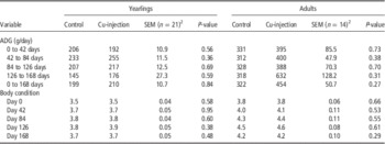
Copper supplementation did not influence ADG or body condition of yearlings or adult deer at any time point (Table 2). Average daily intakes of dry matter diet and unifeed ration were 2.1 and 1.4 kg, respectively.
Effects of Cu supplementation on average daily gain (ADG) and body condition measured every 42 daysFootnote 1 throughout the trial in yearling and adult red deer

1 Coinciding with each Cu injection.
2 The experimental unit was the animal.
Effects of Cu supplementation on the mechanical and structural properties and ash and mineral content of antlers
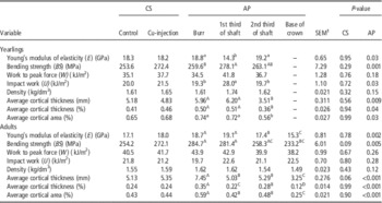
No significant interactions between Cu supplementation and antler position were detected for any traits studied; therefore, only main effects are shown for yearlings and adults. Supplementation with Cu did not influence any antler traits studied in yearlings (Table 3). However, average cortical thickness tended to increase by a mean of 4.1% in adults receiving Cu supplementation (P=0.06). In addition, cortical thickness correlated positively with increased Cu serum before and after treatment in the supplemented group, supporting an effect of Cu in increasing cortical thickness; however, the high correlation coefficients (r=0.73 and 0.80 for yearlings and adults, respectively) did not achieve statistical significance (P>0.10), likely due to the small sample size.
Effects of Cu supplementation (CS) and antler position (AP) on the mechanical and structural properties of antlers from yearling and adult red deer

a,bValues within a row with different superscripts differ significantly at P⩽0.05.
A,B,C,DValues within a row with different superscripts differ significantly at P⩽0.01.
Interaction of Cu supplementation×antler position was not significant (P>0.10) for any trait studied; therefore, only the main effects are presented.
1 The experimental unit was the animal (21 and 14 for yearlings and adults, respectively).
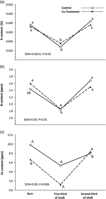
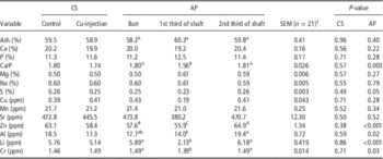
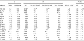
Copper supplementation led to an increase in the serum content of this mineral in yearlings from 0.68 to 0.94 ppm (P=0.02) and in adults from 0.59 to 0.95 ppm (P=0.02) compared with control groups. The magnitude of the observed effect was greater in adults than yearlings (38% v. 28%). Despite the fact that Cu injection led to increased serum Cu content, no effect on the Cu content of antlers was observed in either yearlings or adult deer (Tables 4 and 5). In fact, Cu supplementation had little influence on the ash or mineral content of yearling or adult antlers. The only effect observed in antlers of yearling deer was a significant interaction between Cu supplementation and antler position for K, B and Fe content (Figure 1). In adults, Cu supplementation tended to increase the Sr content (P=0.06) and significantly increased the Zn content (P=0.02) of antlers.
Interaction effect between Cu supplementation and antler position (burr, first third of shaft and second third of shaft) on K content (%), B content (ppm) and Fe content (ppm) of antlers from yearling red deer. The experimental unit was the animal, with a total of 21 deer. a,bDifferent alphabetic superscripts denote mean values which are significantly different at P<0.05.A,BDifferent alphabetic superscripts denote mean values which are significantly different at P < 0.01.

Effects of Cu supplementation (CS) and antler position (AP) on ash and mineral content of antlers from yearling red deer

a,bValues within a row with different superscripts differ significantly at P⩽0.05.
A,B,CValues within a row with different superscripts differ significantly at P⩽0.01.
The interaction of Cu supplementation × antler position was not significant (P>0.10) for any trait studied; therefore, only main effects are presented.
1 The experimental unit was the animal.
Effects of Cu supplementation (CS) and antler position (AP) on ash and mineral content of antlers from adult red deer

a,bValues within a row with different superscripts differ significantly at P⩽0.05.
A,BValues within a row with different superscripts differ significantly at P⩽0.01.
The interaction of Cu supplementation×antler position was not significant (P>0.10) for any trait studied; therefore, only main effects are presented.
1 The experimental unit was the animal.
Effect of Cu supplementation on the variation in characteristics and mineral composition between the base and top of antlers from yearling and adult deer
In yearlings, the difference between the top and the base of the antler (expressed as a percentage), which indicates depletion of body mineral stores or physiological exhaustion, was not affected by Cu supplementation, other than the characteristics U and Ca/P proportion. In this age group, U increased in the Cu supplemented group, while it decreased in the control group (20.4% v. −9.6%; P=0.04). The ratio of Ca to P tended to decrease in deer administered Cu by injection, whereas this was not the case in control deer (−1.6% v. 1.5%; P=0.06). In adults, the average cortical thickness increased in the Cu supplemented group, while it decreased in the control group (27.8% v. −23.7%; P=0.02). In the same age group, the P content increased in antlers from the control group and decreased in antlers from the Cu supplemented group (6.4% v. −1.4%; P=0.02). Also in adults, the increase of Fe content tended to be higher in control animals than in Cu injected animals (98.5% v. 28.1%; P=0.07).
Discussion
Our results indicate a limited effect of Cu supplementation on antler characteristics and mineral composition. There may be a number of factors underlying these data. First, and importantly, animals were fed a balanced diet that met or exceeded their nutrient requirements (NRC, 2007). Second, the dose of Cu used for supplementation was relatively small, because excess of Cu is toxic for some ruminants, including sheep (Winge and Mehra, Reference Winge and Mehra1990) and deer (Fairley, Reference Fairley2008). This context limited the effects of Cu supplementation and prompted us to perform blood tests 84 days after the first Cu injection to check if Cu levels in serum were increased by the supplementation. The results demonstrated that Cu levels in serum were higher in the supplemented than the control group, as expected. In general, supplementation with minerals produces an increase of the serum mineral content; an exception is Ca, the serum homeostasis of which is the most tightly physiologically controlled among minerals (Favus et al., Reference Favus, Bushinsky and Lemann2006).
Wilson and Grace (Reference Wilson and Grace2001) proposed reference criteria for deer serum Cu content: values <0.32 ppm were considered to indicate deficiency; values between 0.32 and 0.50 ppm were considered marginal; and values >0.50 ppm were considered adequate. In the current trial, the average serum Cu content from the yearling and adult control groups were 0.68 and 0.59 ppm, respectively. Therefore, according to the criteria of Wilson and Grace (Reference Wilson and Grace2001), even deer in the control group had adequate serum Cu levels in this study. In early studies, Strause et al. (Reference Strause, Hegenauer, Saltman, Cone and Resnick1986) showed that either Mn or Cu deficiency led to reduced Ca and P content in the bones of rats, and the animals did not appear to suffer from any other problems. In the current trial, there was no general effect of Cu supplementation on Ca or P levels or the Ca/P ratio in either yearling or adult deer. It is possible that, if this study had been conducted using deer suffering from Cu deficiency, the results would have been very different from those reported here.
Different patterns of serum Cu levels were observed in yearling and adult deer during this study. The percentage increase of serum Cu content was higher in adults than in yearlings (38% v. 28%). One reason for this may be that the blood Cu content in the control yearling group was much higher than that of adult controls. The fact that the percentage increase of Cu content in adult serum was higher than that in yearlings may account for the greater effect of Cu supplementation on the antlers of adults than yearlings. These results indicate that Cu may be more highly mobilized in adults than yearlings. Given that the same dose of Cu was administered to each group, one possible explanation for such an effect could be that the higher metabolism of yearlings and their greater need for minerals (and for nutrients in general) may have led to a faster use of the injected Cu in these animals, and a consequent reduction in their serum levels. In general, plasma mineral levels vary with deer age (Kučer et al., Reference Kučer, Kuleš, Barić Rafaj, Tončić, Vicković, Štoković, Potočnjak and Šoštarić2013). In fact, in a previous Mn supplementation experiment carried out by our group (Cappelli et al., Reference Cappelli, García, Ceacero, Gómez, Luna, Gallego, Gambín and Landete-Castillejos2015), adults showed a wide range of effects, whereas yearlings only exhibited increases in Mn and Fe antler content. However, in the current study, the variation of serum Cu level with age was higher than expected. Our data include more blood samples taken at different dates to assess the influence of Cu supplementation on the evolution of serum mineral content, comparing both yearling and adult deer.
Although Cu injections did not influence antler characteristics as extensively as expected, supplementation did influence some of the traits investigated. The most interesting effects of Cu supplementation were the trend towards increased cortical thickness and the attenuation of reduction in cortical thickness from the base to the top of antlers, both of which were observed in adults. The increase in cortical thickness may increase the structural resistance of bone to fracture. The mechanical performance of structures depends on two types of factors (Landete-Castillejos et al., Reference Landete-Castillejos, García and Gallego2012): those depending on structure, such as cortical thickness, diameter or area, and those derived from the mechanical quality of the material, such as E, BS, W or U. In fact, diameter and cortical thickness account for 55% of the variation in the mechanical performance of bones (Davidson et al., Reference Davidson, Siminoski, Adachi, Hanley, Goltzman, Hodsman, Josse, Kaiser, Olszynski, Papaioannou, Ste-Marie, Kendler, Tenenhouse and Brown2006). Moreover, given that an increase in cortical thickness influences antler weight, supplementation with Cu, even when animals are receiving a balanced diet, may affect the trophy value of antlers, as weight is an influential measure in the International Council for Game and Wildlife Conservation method of trophy valuation.
In the current paper, which is the first to investigate mineral content in both serum and antlers, the increase in serum Cu content did not result in an increase in the content of this mineral in antlers, demonstrating the limitations of using serum Cu content to assess bone Cu status. These results may have several contributing factors. It is possible that increases in serum Cu are not reflected in antlers. Alternatively, serum Cu may be captured by the liver, or other organs with higher priority for uptake than bone tissue. Indeed, the liver is an organ where Cu is stored, while the amounts of this mineral in bone are very small (Suttle, Reference Suttle2010).
In conclusion, even when animals are receiving a balanced diet and the level of Cu supplementation is rather low in order to avoid the risk of death by Cu toxicity, supplementation with this mineral appears to affect the mechanical properties of antlers, and induce some changes in their mineral composition. One of the most interesting effects is the increase of the cortical thickness in Cu supplemented adult deer. Such an effect may increase the trophy value or the velvet production of antlers (depending on the purpose for which deer are bred). Similar results have previously been observed in humans, in which dietary Cu supplements reduced the rate of bone loss (Lowe et al., Reference Lowe, Fraser and Jackson2002). However, administration of Cu supplementation by injection, rather than in the diet, may increase its potential benefits in improving bone resistance in elderly humans.
Acknowledgements
The authors wish to thank The Charlesworth Group for professional language editing. This work was supported by Project No. PEII-2014-004-P (Regional Consejería de Educación y Deportes de Castilla-La Mancha) and INCYDEN Project RTC-2016-5327-2 (Ministerio de Economía y Competitividad).