Introduction
Schistosomiasis is regarded as one of the most significant helminthic infections in the world (King, Reference King2009). The concept of developing a vaccine against schistosomiasis has strong arguments in its favour since the stand-alone treatment approach with praziquantel proved insufficient to eradicate the disease. Nevertheless, only two antigens; Sh28GST or BILHVAX from Schistosoma haematobium (Capron et al., Reference Capron, Riveau, Capron and Trottein2005; Riveau et al., Reference Riveau, Deplanque, Remoue, Schacht, Vodougnon, Capron, Thiry, Martial, Libersa and Capron2012) and Sm14 from S. mansoni (http://clinicaltrials.gov/show/NCT01154049) have reached clinical trials. Looking for new potential antigens and antigen combinations is of paramount importance to obtain a protective vaccine.
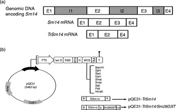
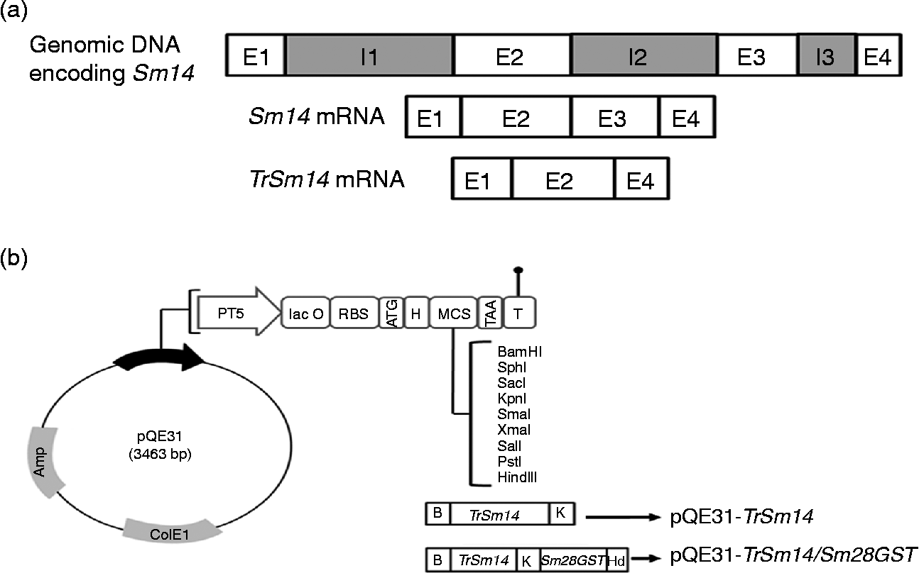
Sm14 is a fatty acid binding protein (FABP) which is encoded in a 402 bp cDNA gene (Moser et al., Reference Moser, Tendler, Griffiths and Klinkert1991). Animal studies revealed that immunization with Sm14 showed variable percentages of adult worm reduction in animal challenge models, such as 25% (Fonseca et al., Reference Fonseca, Brito, Alves and Oliveira2004), 36.9% (Ribeiro et al., Reference Ribeiro, Vieira Cdos, Fernandes, Araujo and Katz2002) and 67% (Tendler et al., Reference Tendler, Brito, Vilar, Serra-Freire, Diogo, Almeida, Delbem, Da Silva, Savino, Garratt, Katz and Simpson1996). Another schistosomal fatty acid binding protein is the S. mansoni Sm14 delta E3 variant. The 297-nucleotide mRNA (GenBank accession number AF492390) is transcribed by alternative splicing of the Sm14 gene, so that exon 3 is completely missing (fig. 1a). This truncated form of Sm14, herein designated TrSm14, was discovered in only two clones from cDNA libraries constructed from two Brazilian S. mansoni strains (Ramos et al., Reference Ramos, Figueredo, Pertinhez, Vilar, do Nascimento, Tendler, Raw, Spisni and Ho2003). However, since only one band, that of the full-length 402-bp Sm14 cDNA, appeared following reverse transcription-polymerase chain reaction (RT-PCR) amplification using specific Sm14 primers, the truncated form was postulated to be a ‘rare misprocessing of the mRNA precursor’ (Ramos et al., Reference Ramos, Figueredo, Pertinhez, Vilar, do Nascimento, Tendler, Raw, Spisni and Ho2003).
(a) Diagramatic representation of Sm14 and TrSm14 exon splicing. E, exon; I, intron. (b) Schematic representation of the construction of pQE31-TrSm14 and pQE31-TrSm14/Sm28GST. Amp, β-lactamase coding sequence; ColE1, ColE1 origin of replication; PT5, T5 promoter; lac O, lac operator element; RBS, ribosomal binding site; H, hexahistidine tag; MCS, multiple cloning site; T, terminator; B, BamHI recognition site; K, KpnI recognition site; Hd, HindIII recognition site.
Since data about the alternatively spliced TrSm14 are scarce in the literature, we investigated this antigen in an Egyptian S. mansoni strain. In the current study, efforts were made to detect TrSm14 and to express it in Escherichia coli, either singly or fused to the schistosomal antigen Sm28GST, for potential research and medical uses.
Materials and methods
Construction of plasmid vectors
Adult worms, both sexes, of an Egyptian strain of S. mansoni were perfused, as described previously (Smithers & Terry, Reference Smithers and Terry1965), from the livers of hamsters 7 weeks after infection with S. mansoni cercariae. RNA was extracted from adult worms following a sequential two-step protocol using BIOZOL® reagent (Bioer, China) and Oligotex® mRNA Mini Kit (Qiagen GmbH, Hilden, Germany), respectively. First-strand cDNA was subsequently synthesized using RevertAid H Minus First Strand cDNA Synthesis Kit (ThermoScientific Inc., Vilnius, Lithuania). The gene encoding S. mansoni fatty acid binding protein Sm14 (Accession no. XM_002580386; 402 bp) was PCR-amplified from cDNA using the primers 5′-TAGTTTCTTGGGAAAGTGGAA-3′ and 5′-TTAGGATAGTCGTTTATAATTGC-3′. This resulted in two close bands upon electrophoresis which were separately gel extracted. The smaller band, corresponding to TrSm14, was used as a template to amplify pure TrSm14 by PCR using the primers 5′-TGAAGGATCCGATGTCTAGTTTCTTGGGAAAGTGGAA-3′ and 5′-ACGCGGTACCGGATAGTCGTTTATAATTGC-3′ which contain BamHI and KpnI restriction sites, respectively. Similarly, Sm28GST was amplified from cDNA using the primers 5′-ATGGCTGGCGAGCATATCAA-3′ and 5′-TTAGAAGGGAGTTGCAGG-3′. This Sm28GST PCR product acted as a template for a second PCR amplification of Sm28GST using the primers 5′-TATAGGTACCATGGCTGGCGAGCATATCAA-3′ and 5′-ATTAAAGCTTTTAGAAGGGAGTTGCAGG-3′, containing KpnI and HindIII sites, respectively, for subsequent construction of the fusion gene TrSm14/Sm28GST.
For construction of pQE31-TrSm14, the TrSm14 gene was double digested using BamHI and KpnI and ligated to a similarly digested pQE31 vector using T4 DNA ligase. For construction of pQE31-TrSm14/Sm28GST, both TrSm14 and Sm28GST PCR products were cut using KpnI and ligated together, where the ligation reaction mixture was used as a template to amplify TrSm14/Sm28GST fusion gene using TrSm14 forward primer and Sm28GST reverse primer with appropriate flanking restriction sites. The resulting TrSm14/Sm28GST fusion PCR product was cut with BamHI and HindIII and ligated to a similarly treated pQE31 plasmid. Plasmids pQE31-TrSm14 and pQE31-TrSm14/Sm28GST (fig. 1b) were ultimately transformed into E. coli M15 (pREP4) by electroporation. All genes were sequenced and identity was confirmed.
Protein expression and detection
Engineered E. coli M15 (pREP4) strains were induced with isopropyl β-d-thiogalactopyranoside (IPTG) for production of corresponding antigens. His-tagged antigens were purified from induced cultures using metal-affinity nickel-nitrilotriacetic acid (Ni-NTA) chromatography (Qiagen) under denaturing conditions, following the manufacturer's instructions. Purified proteins were checked by sodium dodecyl sulphate-polyacrylamide gel electrophoresis (SDS-PAGE) and Western blot using pooled serum from three S. mansoni-infected mice. In addition, serum from S. mansoni-infected patients was used to test reactivity with expressed antigens using a standard enzyme-linked immunosorbent (ELISA) assay (Cardoso et al., Reference Cardoso, Pacifico, Mortara and Oliveira2006).
Results and discussion
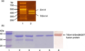
PCR amplification of Sm14 from first-strand cDNA showed two equally intense, close bands corresponding to Sm14 (402 bp) and an alternatively spliced Sm14 isoform, herein designated TrSm14 (297 bp) (fig. 2a). On the contrary, only one band, that of the full-length 402-bp Sm14 cDNA, appeared upon RT-PCR amplification in all studies carried out to date using specific Sm14 primers (Moser et al., Reference Moser, Tendler, Griffiths and Klinkert1991; Brito et al., Reference Brito, Oliveira, Oliveira, Street, Riengrojpitak, Wilson, Simpson and Correa-Oliveira2002; Ramos et al., Reference Ramos, Figueredo, Pertinhez, Vilar, do Nascimento, Tendler, Raw, Spisni and Ho2003; Angelucci et al., Reference Angelucci, Johnson, Baiocco, Miele, Brunori, Valle, Vigorosi, Troiani, Liberti, Cioli, Klinkert and Bellelli2004). Consequently, TrSm14 was formerly misinterpreted as a ‘rare misprocessing of the mRNA precursor’ (Ramos et al., Reference Ramos, Figueredo, Pertinhez, Vilar, do Nascimento, Tendler, Raw, Spisni and Ho2003). However, we obtained a double band consistently in different independent RT-PCR reactions, reflecting a good level of expression of TrSm14 mRNA. This may be ascribed to strain differences, since this study is the first attempt of RT-PCR and cloning of TrSm14 from an Egyptian S. mansoni strain. The sequence of the smaller truncated PCR product, TrSm14, showed 100% identity with the alternatively spliced S. mansoni Sm14 delta E3 variant (Ramos et al., Reference Ramos, Figueredo, Pertinhez, Vilar, do Nascimento, Tendler, Raw, Spisni and Ho2003).
(a) Agarose gel picture of PCR amplification of Sm14 from cDNA of Egyptian S. mansoni strain. Lanes: 1, DNA ladder (sizes indicated in bp); 2, two bands corresponding to Sm14 (402 bp) and TrSm14 (297 bp). (b) Lanes 1–5 show SDS-PAGE of the purified TrSm14/Sm28GST fusion protein. Lanes: 1, protein marker; 2 and 3, two elutions using Qiagen buffer E (pH 4.5); 4 and 5, two elutions using Qiagen buffer D (pH 5.9). Lane 6 represents the Western blot of the fusion protein using pooled serum from S. mansoni-infected mice as a primary antibody.
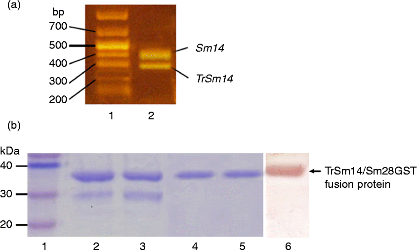
We successfully cloned the genes of TrSm14 and TrSm14/Sm28GST fusion in pQE31 plasmids where an N-terminus His tag was added to expressed proteins (fig. 1b). Escherichia coli M15 (pREP4) (pQE31-TrSm14/Sm28GST) produced the fusion protein (fig. 2b) with an average yield of 1 mg per litre of culture upon induction with 0.3 mm IPTG at 25°C for 16 h. TrSm14/Sm28GST fusion protein reacted positively with pooled serum from S. mansoni-infected mice upon Western blotting (fig. 2b). On the other hand, production of TrSm14 by E. coli M15 (pREP4) (pQE31-TrSm14) was not observed under various induction conditions. This might be due to its small size and possible instability. Using the ELISA assay, serum from infected patients reacted positively with the fusion protein as well as with recombinant Sm28GST antigen. It is noteworthy that pooled serum from S. mansoni-infected mice was also reactive with purified recombinant Sm28GST upon Western blotting (data not shown). Consequently, the apparent Western blot reactivity of the serum with the TrSm14/Sm28GST fusion might be due to reactivity with either of the single antigen components or with both antigens. Since TrSm14 could not be expressed alone, confirming the specific serum reactivity against the TrSm14 part of the fusion was tested indirectly by proving the cross-antigenicity between TrSm14 and Sm14 antigens. We performed an ELISA assay with anti-Sm14 serum obtained from mice formerly immunized with recombinant Sm14 and polyinosinic-polycytidylic acid (poly (I:C)) as adjuvant. Briefly, ELISA plates were coated with recombinant Sm14 and TrSm14/Sm28GST fusion on different wells and the anti-Sm14 serum was used as primary antibody. High absorbance readings were observed in wells coated with either Sm14 or the fusion protein. These readings were significantly different from the low readings of the negative control wells involving serum from non-immunized mice. This provides experimental proof for the cross-antigenicity of Sm14 and TrSm14.
It has been reported that Sm14 is a twisted V-shaped barrel that comprises a large internal cavity with a volume of 300 Å3 in which fatty acids are bound (Angelucci et al., Reference Angelucci, Johnson, Baiocco, Miele, Brunori, Valle, Vigorosi, Troiani, Liberti, Cioli, Klinkert and Bellelli2004). In silico analysis revealed that the missing exon in TrSm14 could have a direct effect on its fatty acid binding specificity (unpublished data, Prof. M. Brunori and Dr F. Angelucci, University of Rome ‘La Sapienza’, pers. comm.). Consequently, the role/function of TrSm14 needs to be determined through further functional and structural studies
Previous in vivo immunization experiments demonstrated that Sm14 has two main immunogenic peptide epitopes: peptide 1 (residues 85–94) and peptide 2 (118–125) (Angelucci et al., Reference Angelucci, Johnson, Baiocco, Miele, Brunori, Valle, Vigorosi, Troiani, Liberti, Cioli, Klinkert and Bellelli2004). The missing exon in TrSm14 encodes the amino acid sequence from 81 to 116 in Sm14. While the amino acids of peptide 1 are missing in TrSm14, it still contains the immunogenic epitope of peptide 2.
Our created TrSm14/Sm28GST fusion protein offers several advantages. First, it allowed successful expression of the small-sized unstable TrSm14. Second, rather than fusion of TrSm14 to a foreign protein, Sm28GST is another promising schistosomal antigen (Balloul et al., Reference Balloul, Sondermeyer, Dreyer, Capron, Grzych, Pierce, Carvallo, Lecocq and Capron1987) that is expected to increase the protection level induced by TrSm14 if this fusion protein is tested as a vaccine. Third, in addition to purification using Ni-NTA metal-affinity chromatography, the TrSm14/Sm28GST fusion protein could be purified using glutathione affinity chromatography, owing to the glutathione S-transferase activity of Sm28GST. Indeed, purification of Sm28GST was previously achieved by affinity chromatography on immobilized glutathione (Ivanoff et al., Reference Ivanoff, Phillips, Schacht, Heydari, Capron and Riveau1996).
In conclusion, we detected, for the first time, the alternatively spliced TrSm14 cDNA in an Egyptian strain of S. mansoni. This truncated Sm14 (TrSm14) gene was found to be expressed at a comparable level to the native Sm14 gene, as confirmed by RT-PCR reactions. This suggests a potential role/function of TrSm14 protein. The TrSm14 antigen was successfully expressed and purified as a fusion protein with Sm28GST. Pooled serum from S. mansoni-infected mice reacted positively with the fusion protein, which indicates a possible diagnostic value for detection of anti-schistosomal antibodies. Moreover, this TrSm14/Sm28GST fusion protein, which harbours two promising schistosomal antigens, represents a potential bivalent vaccine against S. mansoni, which is worthy of further investigation.
Acknowledgements
The authors thank Dr Yosra Elnaggar and Michael George for help and support.
Financial support
This work was funded by Alexandria University under the Alexandria University Research Enhancement Program (ALEX-REP), project code HLTH10.
Conflict of interest
None.
Ethical standards
The authors assert that all procedures contributing to this work comply with the ethical standards of the national guidelines on human experimentation and with the Helsinki Declaration of 1975, as revised in 2008, and have been approved by the research ethics committee of Alexandria University. All human serum samples were collected following informed consent from the patients. The authors also assert that all procedures contributing to this work comply with the ethical standards of the national guidelines on the care and use of laboratory animals and have been approved by the Animal Care and Use Committee (ACUC) of Alexandria University.