The hypothesis that ‘the duodenum yields a chemical excitant for the internal secretion of the pancreas’ was proposed over 100 years ago (Refs Reference Creutzfeldt1, Reference Moore, Edie and Abram2). It is now well known that the intestine contains numerous enteroendocrine cells dispersed throughout the epithelial lining from the stomach to the colon, which secrete over 20 different peptides (Ref. Reference Rehfeld3). These gut peptides play a critical role in local gastrointestinal and whole-body physiology, ranging from gut motility and epithelial repair to adiposity, appetite regulation and glucose homeostasis. Two of these hormones, glucagon-like peptide 1 (GLP-1) and glucose-dependent insulinotropic polypeptide (GIP; also known as gastric inhibitory polypeptide), mediate the ‘incretin effect’ – the phenomenon that glucose ingested orally elicits a much greater insulin response than glucose delivered intravenously, even when plasma glucose levels are matched (Refs Reference Elrick4, Reference Creutzfeldt5). Indeed, this indirect pathway is estimated to underlie up to 70% of glucose-stimulated insulin secretion in healthy individuals (Refs Reference Baggio and Drucker6, Reference Holst7). At the level of the pancreatic β-cell, GLP-1 and GIP arriving in the circulation following food ingestion provide a signal that amplifies the secretory response to concomitant changes in the glucose concentration, thereby enhancing insulin release under conditions when it is most needed (Ref. Reference Gromada, Holst and Rorsman8).
Taking advantage of the potent action of GLP-1 on pancreatic β-cells, several GLP-1-based therapies are now entering the market for the treatment of type 2 diabetes. In contrast to many other antidiabetic medications, they have a low incidence of hypoglycaemic side effects, due to the strict glucose-dependence of GLP-1-triggered insulin release. They also cause weight loss, or at least tend to be weight neutral – an additional benefit for the generally overweight diabetic population. A new therapeutic avenue under exploration is to attempt to stimulate endogenous GLP-1 secretion. The biology of the GLP-1-producing L-cells in the gut has therefore become a topic of renewed interest, and dissecting the relative importance of different L-cell signalling pathways is viewed as critical for the design of L-cell-targeted drugs.
Incretin biology
Timing and source of incretin secretion
The enteroendocrine L-cells that synthesise and release GLP-1 are found scattered throughout the small and large intestine, with the highest density in the distal ileum and colon (Ref. Reference Eissele9). GIP, by contrast, is produced by an anatomically distinct cell type known as K-cells, which are predominantly located in the duodenum (Refs Reference Polak10, Reference Buchan11). Perhaps in part reflecting the different locations of K- compared with L-cells, the circulating concentrations of GIP and GLP-1 exhibit distinct profiles following food ingestion. Depending on the meal composition, GLP-1 secretion can exhibit a biphasic profile, with an early phase occurring soon after food intake and lasting for 15–30 min, and a late phase persisting for 1–2 h or longer (Ref. Reference Herrmann12). GIP secretion occurs with a similar short time delay, and may also remain elevated for several hours (Refs Reference Cataland13, Reference Elliott14). In humans, glucose can be detected in the proximal duodenum within 5 min of the ingestion of a liquid glucose load, correlating with the time taken to see the first rapid elevation of GLP-1 and GIP concentrations in the bloodstream (Refs Reference Balks15, Reference Schirra16).
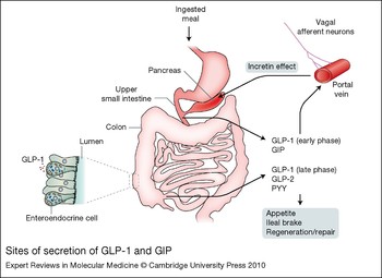
While the early release of GIP is readily explained by the arrival of nutrients in the duodenum with its high local density of K-cells, it has long posed a dilemma that the rapid secretion of GLP-1 occurs before nutrients reach the majority of the L-cells in the ileum and colon. This gave rise to the idea that the rapid phase of GLP-1 secretion must be triggered indirectly by neuronal signals or hormonal factors arising in the proximal intestine and stimulating the distal L-cell population (Refs Reference Hansen and Holst17, Reference Rocca and Brubaker18). However, L-cells have been identified at low density in the duodenum (Ref. Reference Mortensen19) and in higher numbers in the jejunum (Ref. Reference Eissele9). Glucose infusion studies in human subjects showed that GLP-1 secretion was stimulated only when the rate of glucose entry into the duodenum exceeded the duodenal absorptive capacity – that is, when nonabsorbed glucose would leak through to the jejunum, and potentially stimulate L-cells in that region (Ref. Reference Schirra16). In the same studies, GIP secretion was stimulated by even low duodenal glucose infusion rates, consistent with the more proximal location of the majority of K-cells. Interestingly, a proportion of cells in the proximal small intestine reportedly express both GLP-1 and GIP (Refs Reference Mortensen19, Reference Theodorakis20). Taken together with the finding that the rapid phase of GLP-1 secretion is preserved in patients after ileal resections or proctocolectomy, the data suggest that the early release of GLP-1, which accounts for the majority of the incretin effect in normal individuals, is triggered by local luminal nutrient-sensing pathways (Ref. Reference Nauck21) (Fig. 1). Nevertheless, the relative contribution of the ileal and colonic L-cells to the incretin effect remains surprisingly unclear.
Sites of secretion of GLP-1 and GIP. Plasma levels of glucagon-like peptide 1 (GLP-1) and glucose-dependent insulinotropic polypeptide (GIP) increase rapidly after the ingestion of a meal as a result of the direct stimulation of both L- and K-cells located in the duodenum. The later phase of GLP-1 secretion coincides with the time taken for nutrients to reach the L-cells in the lower ileum. As well as entering the circulation and having direct effects on their target tissues, such as the pancreatic β-cell (incretin effect), GLP-1 might also mediate its effects via receptors in close proximity to the enteroendocrine cells, such as those expressed by vagal afferent neurons in the portal vein. Enteroendocrine cells secreting hormones like GLP-1 and GIP are found scattered through the epithelial layer, as shown in the inset (bottom left). L-cells from the distal ileum and colon secrete peptide YY (PYY), GLP-1 and GLP-2. The extent to which GLP-1 from distal L-cells contributes to the incretin effect is unclear, but it also suppresses appetite and slows peristalsis (the ileal brake). PYY acts centrally as a satiety signal, and GLP-2 stimulates regeneration and repair of the intestinal epithelium.

Incretin hormone processing
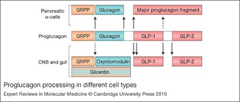
GLP-1 is post-translationally cleaved from the product of the proglucagon gene (GCG), which is expressed not only in the L-cell but also in pancreatic α-cells and subsets of neurons in the brainstem. Alternative processing of the prohormone is largely attributable to the differential expression of prohormone convertase (PC) enzymes. Whereas pancreatic α-cells express PC2 (PCSK2) and generate glucagon, L-cells in the gut express PC1/3 (PCSK1) and produce the bioactive peptides GLP-1, GLP-2 and oxyntomodulin (Ref. Reference Holst7) (Fig. 2). Proglucagon processing in the brainstem is believed to follow a similar pathway to that in the intestine. In fact, GLP-1 exists in two active forms, GLP-1(7–36)amide and GLP-1(7–37), which are produced in varying proportions across species but have equipotent insulinotropic activity (Ref. Reference Orskov, Wettergren and Holst22). The C-terminal moiety of proglucagon is processed almost completely to GLP-1 and GLP-2 in L-cells, but only 20–40% of the N-terminal region is released as the active peptide oxyntomodulin, with the remainder taking the form of glicentin (60–80%) (Ref. Reference Meier and Nauck23). Certain doubts therefore remain about the physiological importance of oxyntomodulin, and its broadly similar effects to those of GLP-1 probably reflect the fact that its action in vivo is largely mediated through the GLP-1 receptor (Refs Reference Baggio24, Reference Druce25). By contrast to proglucagon, the proGIP precursor in K-cells is not believed to contain any bioactive peptide sequences in addition to that of GIP. ProGIP processing in K-cells, like that of proglucagon in L-cells, is attributed to the action of PC1/3 (Ref. Reference Ugleholdt26).
Proglucagon processing in different cell types. Proglucagon processing in pancreatic α-cells generates the major products glucagon, GRPP (glicentin-related pancreatic polypeptide) and the major proglucagon fragment. In the brainstem and intestinal L-cells, by contrast, processing generates glucagon-like peptide 1 (GLP-1), GLP-2, glicentin, oxyntomodulin and GRPP. Removal of the first six amino acids of GLP-1 generates the active forms GLP-1(7–36)amide and GLP-1(7–37). The major processing enzymes are believed to be prohormone convertase 2 (PC2) in the α-cells, and PC1/3 in the L-cells. Solid arrows depict points where cleavage is largely complete; dashed arrows depict sites of incomplete processing (in L-cells ~20–40% of the N-terminal region is released as the active peptide oxyntomodulin). The uncoloured segments of proglucagon between glucagon and GLP-1 and between GLP-1 and GLP-2 correspond to intervening peptides 1 and 2, respectively. For further details see Ref. Reference Rouillé, Martin and Steiner135.

Incretin hormone degradation
Following their secretion, both GLP-1 and GIP are rapidly degraded by the enzyme dipeptidyl peptidase 4 (DPP4), which is ubiquitously expressed on the surface of endothelial and epithelial cells and exists as a soluble form in plasma (Refs Reference Demuth, McIntosh and Pederson27, Reference Hansen28). DPP4 has a preference for substrate peptides containing a proline or alanine residue at position 2 (Ref. Reference Mentlein29) and readily cleaves N-terminal dipeptides from GLP-1 and GIP, forming GLP-1(9–37) or GLP-1(9–36) amide and GIP(3–42), which lack activity at their respective receptors (Ref. Reference Holst7). As a result, it is estimated that only 10–15% of the secreted active form of GLP-1 passes into the systemic circulation, where it has a short half-life of <2 min (Refs Reference Baggio and Drucker6, Reference Hansen28), and only after larger meals does the active GLP-1 level rise sufficiently to be detected in the peripheral circulation. GIP inactivation by DPP4 is slightly slower (t½ ~6 min in humans and 2 min in rodents) (Refs Reference Deacon30, Reference Kieffer, McIntosh and Pederson31), and the peak postprandial GIP levels are correspondingly higher – factors that probably contribute to the effectiveness of GIP as a circulating physiological incretin hormone.
Site of action of incretins
While the best-characterised effect of GLP-1 is its stimulatory action on the pancreatic β-cell, GLP-1 receptors are found in a variety of extrapancreatic sites, including the lungs, kidneys, stomach, heart, central nervous system, major blood vessels (including the portal vein) and vagal neurons (Refs Reference Baggio and Drucker6, Reference Vahl32, Reference Bucinskaite33). The rapid clearance of GLP-1 from the circulation suggests that there may also be primary sites of GLP-1 action located closer to its site of secretion. The nodose ganglion, which contains the cell bodies of afferent vagal neurons innervating the viscera, expresses receptors for GLP-1, GLP-2 and GIP, although only GLP-1 has been shown to reliably elicit changes in the electrical activity of the neurons themselves (Refs Reference Nakagawa34, Reference Kakei35). The findings suggest that GLP-1, like cholecystokinin (CCK), might exert a proportion of its effect by acting on nerve endings located in close proximity to the enteroendocrine cells themselves (Refs Reference Simasko and Ritter36, Reference Simasko37). This idea is supported by a recent report that the appetite-suppressive effect of systemic GLP-1 was inhibited when the GLP-1 receptor antagonist exendin(9–39) was administered peripherally but not centrally (Ref. Reference Williams, Baskin and Schwartz38).
Nutrient detection in enteroendocrine K- and L-cells
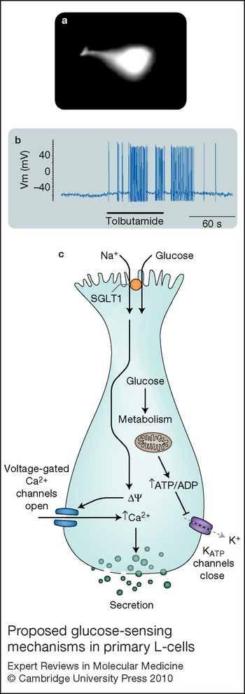
K- and L-cells are open-type enteroendocrine cells residing in the intestinal epithelium, and have long, slender apical processes extending to the gut lumen (Ref. Reference Eissele9) (Fig. 3a). This morphology has long suggested that the cells could directly sense the presence of luminal nutrients. The molecular mechanisms underlying nutrient detection by enteroendocrine cells remain only partially characterised, however, because of the limitations of the experimental systems available to study them.
In vitro models
Three cell line models are widely used to investigate the secretion of GLP-1, GIP and other gut hormones. The GLUTag cell line was originally derived from endocrine colonic tumours taken from a transgenic mouse expressing SV40 large T antigen under the control of the proglucagon promoter (Ref. Reference Drucker39), and has proved to be a reliable model of GLP-1 secretion, responding to a range of physiologically relevant stimuli (Ref. Reference Brubaker, Schloos and Drucker40). The secretin tumour cell line (STC-1), derived from an endocrine tumour of the mouse small intestine, secretes a range of gut hormones including GLP-1, GIP, CCK and secretin (Ref. Reference Rindi41). It has been used as a model to investigate the release of each of these peptides, although it is unclear whether its ability to secrete such a range of hormones is due to a loss of cell differentiation that may also impact on its expression of stimulus–secretion coupling components, or whether this actually reflects the behaviour of distinct populations of enteroendocrine cells that similarly release a range of hormonal products. As there are very few model systems for studying GIP secretion in vitro, subclones of STC-1 cells have been generated with increased GIP expression, such as GTC-1, STC6-14 and GIP-Ins (Refs Reference Ramshur, Rull and Wice42, Reference Cheung43, Reference Kieffer44). NCI-H716 is a GLP-1-secreting human cell line derived from a caecal adenocarcinoma (Ref. Reference Reimer45). It responds to a limited range of secretagogues, but was found to exhibit impaired physiological regulation of the proglucagon promoter (Ref. Reference Cao46). The validity of this cell line as a model of the human L-cell remains to be established.
Primary cultured enteroendocrine cells provide an alternative system for the in vitro study of gut hormone secretion. Fetal rat intestinal cell (FRIC) cultures secrete GLP-1 in response to a wide array of signalling pathways and extracellular mediators, although are not glucose responsive (Ref. Reference Brubaker and Vranic47). While this lack of glucose responsiveness could be used to argue that L-cells are not themselves glucose sensitive, it is possible that fetal L-cells, like pancreatic β-cells at the same age, do not express a full adult repertoire of glucose-sensing machinery. Primary adult canine K- and L-cells, partially purified by counterstream centrifugal elutriation, have been the topic of a few in vitro studies, although they are not a readily available tool for the majority of laboratories (Refs Reference Kieffer48, Reference Damholt, Buchan and Kofod49). Mixed primary intestinal cultures from the adult mouse have recently been developed as an alternative tool to assay secretion of a range of intestinal hormones in vitro. Together with the generation of transgenic mice with fluorescently labelled K- or L-cells, this technology provides new opportunities to purify, identify and characterise primary adult enteroendocrine cells (Refs Reference Reimann50, Reference Parker51).
Enteroendocrine detection of sweet stimuli
The incretin hormones augment insulin secretion after oral glucose intake, yet how sugars in the gut lumen elicit secretion of GLP-1 and GIP from L- and K-cells remains controversial. Early studies using the GLUTag cell line showed that glucose elicits electrophysiological events that trigger action potential firing, Ca2+ entry via L-type Ca2+ channels, and GLP-1 secretion (Ref. Reference Reimann and Gribble52).
Metabolism and KATP channel closure
As in the pancreatic β-cell, a component of the glucose responsiveness can be attributed to the closure of ATP-sensitive K+ channels (KATP channels), brought about by changes in the relative proportions of ATP and Mg2+–ADP consequent upon glucose metabolism (Fig. 3). Recent studies have confirmed that (1) the KATP channel subunits Kir6.2 (KCNJ11) and SUR1 (sulfonylurea receptor; ABCC8) are expressed at high levels in purified mouse K- and L-cells, (2) functional KATP currents are found in the primary colonic L-cells, (3) sulfonylureas stimulate GLP-1 and GIP secretion in primary cultures, and (4) Kir6.2 is detectable by immunostaining in human K- and L-cells (Refs Reference Reimann50, Reference Parker51, Reference Nielsen53).
Proposed glucose-sensing mechanisms in primary L-cells. (a) L-cells show ‘open-type’ morphology with a long apical process extending towards the gut lumen, as shown by the pattern of Venus fluorescence of a Venus-labelled L-cell from a colonic crypt. (b) This is a typical electrophysiological trace from a primary L-cell treated with the KATP-channel inhibitor tolbutamide, showing membrane depolarisation and action potential firing triggered by the drug. (c) Electrogenic transport of glucose via the apically expressed sodium-coupled glucose transporter SGLT1 generates a sufficient inward current to depolarise the cell membrane (ΔΨ) and trigger action potentials, leading to the opening of voltage-gated Ca2+ channels. The consequent rise in intracellular Ca2+ is the primary trigger of exocytosis and glucagon-like peptide 1 (GLP-1) secretion. In addition, glucose metabolism results in the closure of ATP-sensitive KATP channels, contributing to the excitability of the cell and GLP-1 secretion.

In a variety of glucose-sensitive tissues, it is believed that glucokinase (GCK), the hexokinase with the lowest affinity for glucose, acts as a master controller of the metabolic rate and thereby governs the activity of downstream signalling pathways such as KATP channel activity. The low affinity of GCK for glucose (S0.5 of ~8 mm) (Ref. Reference Matschinsky54) and its lack of product inhibition enable it to link metabolic flux to the extracellular glucose concentration over a physiologically relevant concentration range. Activating and inactivating mutations of the GCK gene cause syndromes of hypo- and hyperglycaemia in humans (Refs Reference Glaser55, Reference Stoffel56), reflecting the critical role of this enzyme in the control of insulin secretion. Immunostaining and expression analysis have demonstrated that GCK is also expressed at high levels in enteroendocrine K- and L-cells (Refs Reference Theodorakis20, Reference Cheung43, Reference Reimann50, Reference Parker51, Reference Jetton57).
Despite the clear presence of GCK and KATP channels in primary K- and L-cells, the signalling role of metabolism in enteroendocrine cells remains uncertain as there is little evidence for a stimulatory effect of sulfonylureas or elevated blood glucose on incretin secretion in an intact animal. Indeed, (1) sulfonylureas had no effect on the peak GLP-1 and GIP responses to an oral glucose tolerance test in humans (Ref. Reference El-Ouaghlidi58), (2) patients with inactivating mutations in GCK did not exhibit impaired GLP-1 and GIP secretion following glucose ingestion (Ref. Reference Murphy59), (3) mice lacking the Kir6.2 subunit of the KATP channel exhibited increased, rather than decreased, plasma GIP levels after an oral glucose challenge (Ref. Reference Miki60), and (4) an acute episode of hyperglycaemia reduced rather than enhanced GLP-1 and GIP responses to glucose ingestion (Ref. Reference Vollmer61). While the latter finding was in part influenced by the slowing of gastric emptying by hyperglycaemia itself, and it could be argued that the altered blood glucose levels in patients with GCK mutations might offset the shifted glucose sensitivity of GCK in enteroendocrine cells, the balance of evidence does not favour a significant role for L- and K-cell GCK and KATP channels in determining the peak GLP-1 and GIP responses to glucose ingestion. However, in a perfused pig intestinal preparation, elevating the vascular perfusate glucose concentration from 5 to 11 mm in the presence of a constant luminal glucose load (3.5 mm) did stimulate GLP-1 secretion (Ref. Reference Hansen62), raising the possibility that glucose arriving by the vascular route may be able to modulate secretion at lower luminal nutrient levels.
Sodium-coupled uptake
In support of a metabolism-independent glucose-sensing pathway in K- and L-cells, GLP-1 and GIP secretion can also be triggered by nonmetabolisable sugars such as methyl-α-glucopyranoside and 3-O-methylglucose in whole intestinal preparations, GLUTag cells and primary intestinal cultures (Refs Reference Reimann50, Reference Parker51, Reference Sykes63, Reference Gribble64, Reference Ritzel65). As the spectrum of sugars that triggered GLP-1 and GIP release when applied in the intestinal lumen matched their ability to be taken up by the intestinal brush border absorption system, it was proposed that hormone secretion was somehow linked to the sugar-uptake mechanism. This was supported by the finding that incretin hormone secretion required the concomitant presence of luminal Na+ and was inhibited by the glucose-uptake blocker phloridzin (Ref. Reference Sykes63). Subsequent studies in GLUTag and primary cells identified the sodium-coupled glucose transporter SGLT1 (SLC5A1) as the likely mediator of the direct responsiveness of K- and L-cells to luminal sugars (Refs Reference Parker51, Reference Gribble64). SGLT1 is a member of a functionally diverse family encoded by SLC5A genes, and actively cotransports glucose and Na+ in a tight 1:2 stoichiometry (Ref. Reference Wright and Turk66). Studies using the GLUTag model showed that the influx of Na+ by the electrogenic activity of SGLT1 generates an inward current large enough to trigger membrane depolarisation and secretion (Ref. Reference Gribble64). This mechanism does not appear to be present in pancreatic β-cells, but has recently been described in populations of glucose-excited hypothalamic neurons (Ref. Reference Gonzalez, Reimann and Burdakov67). In both GLUTag and primary L-cells, the sugar sensitivity of GLP-1 secretion [EC50 of 0.2–1.0 mm (Refs Reference Reimann50, Reference Gribble64)] closely mirrors the K m of SGLT1 (Ref. Reference Diez-Sampedro68).
Sweet-taste-receptor pathways
A third sugar-sensing mechanism proposed to exist in enteroendocrine cells is the sweet-taste pathway, as classically used by taste cells in the lingual epithelium (Ref. Reference Jang69). The sweet-taste receptor comprises a heterodimer of the G-protein-coupled type 1 taste receptors TAS1R2 and TAS1R3, which activates downstream signalling pathways involving α-gustducin (GNAT3), phosholipase Cβ2 (PLCB2) and the transient receptor potential cation channel TRPM5 (Ref. Reference Rozengurt and Sternini70). Several publications report the identification of gustducin, TAS1R2 or TAS1R3 in enteroendocrine cells and cell lines by immunostaining (Refs Reference Dyer71, Reference Wu72, Reference Rozengurt73). In cell lines, GLP-1 secretion was triggered by sweet compounds as well as sugars, and in mice lacking gustducin the GLP-1 response to ingested glucose was impaired (Ref. Reference Jang69). However, purified K- and L-cells were not found to express high levels of components of the sweet-taste-receptor pathway, and artificial sweeteners did not stimulate GIP or GLP-1 release in primary cultures (Refs Reference Reimann50, Reference Parker51). It has also long been known that carbohydrates need to be absorbable to trigger incretin secretion (Ref. Reference Creutzfeldt5), shedding doubt on the existence of luminally facing sweet-taste receptors in L- or K-cells, which would be therapeutically attractive targets for nonabsorbable agents. Further evidence that the sweet-taste-receptor pathway is not a key mediator of incretin hormone secretion in vivo is provided by recent reports that artificial sweeteners do not stimulate GLP-1 and GIP release in rodents and humans, or slow gastric emptying (Refs Reference Fujita74, Reference Little75, Reference Ma76). Transgenic labelling of TRPM5-expressing cells using a fluorescent marker also demonstrated that this ion channel does not colocalise with GLP-1 in the mouse (Ref. Reference Bezencon, le Coutre and Damak77).
Bitter stimuli
Incretin-secreting cells have been found to express bitter-taste receptors of the type 2 taste receptor (TAS2R) family, and bitter agents were shown to stimulate STC-1 cells, leading to the suggestion that bitter tastants might modulate incretin release in vivo (Refs Reference Rozengurt73, Reference Chen78). In this respect it is interesting that a genetic association between a single-nucleotide polymorphism (SNP) in TAS2R9 and impaired glucose homeostasis was recently reported in the Amish population, and that dose-dependent GLP-1 secretion could be triggered from NCI-H716 cells by the TAS2R9 agonist ofloxacin (Ref. Reference Dotson79). However, it remains possible that some of the bitter agents employed in these studies might have off-target effects, as for instance fluoroquinolones like ofloxacin have been reported to depolarise cells by blocking K+ channels (Refs Reference Kang80, Reference Saraya81). In human subjects, the bitter tastants quinine and naningin did not affect gastric emptying or satiety when given by intragastric infusion (Ref. Reference Little75), and there are currently no reports of bitter compounds affecting incretin secretion in vivo.
Detection of other nutrients
Because of the insulinotropic action of GLP-1 and GIP and the associated importance of the incretins in glucose homeostasis, much research has focused on the mechanisms of carbohydrate sensing by L- and K-cells. However, it is widely accepted that other macronutrients can also stimulate hormone release from these cells (Refs Reference Herrmann12, Reference Elliott14).
Protein
Protein is generally considered a relatively weak stimulus for GIP and GLP-1 in humans, although it probably contributes to the later phase of postprandial GLP-1 secretion (Ref. Reference Elliott14). GLUTag, NCI-H716 and STC-1-derived cell lines can be stimulated dose-dependently by meat hydrolysates, an effect that is partially inhibited by the voltage-gated Ca2+-channel blocker verapamil (Refs Reference Ramshur, Rull and Wice42, Reference Nemoz-Gaillard82, Reference Cordier-Bussat83, Reference Reimer84). This would be consistent with a depolarising action of peptones, for example via the proton-coupled PepT1/PepT2 (SLC15A1/SLC15A2) transporters, and indeed it was demonstrated that peptones did depolarise STC-1 cells, albeit after increased transporter expression by heterologous transfection (Ref. Reference Matsumura85). Amino acids have also been shown to stimulate GLP-1 secretion (Refs Reference Herrmann12, Reference Elliott14), and this again seems to involve depolarisation of the plasma membrane. In the GLUTag model, alanine, for example, activates the glycine receptor, a ligand-gated Cl− channel (Ref. Reference Gameiro86), while glutamine and asparagine depolarise these cells by their Na+-coupled electrogenic uptake (Ref. Reference Reimann87). It is currently unclear if the mechanisms characterised in cell lines are preserved in primary cells or could be targeted in vivo. However, glutamine has recently been shown to stimulate GIP secretion in mouse primary intestinal cultures (Ref. Reference Reimann50) and to stimulate incretin release in nondiabetic and diabetic human volunteers (Ref. Reference Greenfield88).
Fat
Fat ingestion, by contrast, is widely considered a good stimulus for both GLP-1 and GIP secretion. Although direct exposure of the intestinal lumen to oleic acid did not always result in significant stimulation of GLP-1 secretion (Ref. Reference Dumoulin89), the digestion of triglycerides to free fatty acids does seem a crucial step, as inhibition of pancreatic lipase prevents the stimulation of GIP and GLP-1 secretion (Refs Reference Pilichiewicz90, Reference Enc91, Reference Ellrichmann92). In the past few years, lipid-sensing mechanisms have been dominated by the idea that the newly discovered fatty-acid-sensitive G-protein-coupled receptors (GPRs) underlie enteroendocrine responses to a range of fatty acids and complex lipids (see below), although hard evidence from in vivo studies is currently lacking. Other fatty acid sensors have also been discussed, including the proposal that the atypical protein kinase C PKCzeta acts as a direct link between long-chain unsaturated fatty acids and activation of GLP-1 secretion (Ref. Reference Iakoubov93).
G-protein-coupled receptors
GPRs act as plasma membrane receptors for a wide variety of extracellular signals and underlie the response of cells to many hormones and neurotransmitters. More recently it has been recognised that they also contribute to the sensing of numerous metabolites such as fatty acids, amino acids and bile salts. The downstream intracellular signalling events involve differential activation of Gs, Gq and Gi pathways, all of which have been shown to be operational in enteroendocrine cell lines.
Gs-coupled receptors
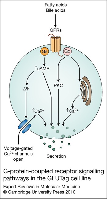
The activation of Gs-coupled receptors and consequent elevation of intracellular cyclic AMP (cAMP) has been shown to be a strong stimulus of GLP-1 release in many in vivo and in vitro studies (Refs Reference Reimer45, Reference Kieffer48, Reference Damholt, Buchan and Kofod49, Reference Simpson94, Reference Brubaker95, Reference Buchan96). cAMP acts through several downstream effectors, including protein kinase A (PKA), exchange proteins directly activated by cAMP (EPACs; RAPGEF3 and RAPGEF4) and ion channels such as cyclic-nucleotide-gated channels. In GLUTag cells, cAMP elevates proglucagon gene transcription via PKA and EPAC-dependent pathways, but also directly stimulates GLP-1 secretion (Refs Reference Drucker39, Reference Brubaker, Schloos and Drucker97) (Fig. 4). The enhancement of vesicle release not only includes a stimulatory effect on the secretory machinery, but also involves a depolarising action of cAMP on the plasma membrane, where it acts synergistically with glucose (Ref. Reference Simpson94). Although the molecular targets of cAMP have not yet been identified in primary K- and L-cells, it is clear that the combination of the adenylate cyclase activator forskolin and the phosphodiesterase inhibitor IBMX (3-isobutyl-1-methylxanthine) both elevates Ca2+ in mouse colonic L-cells and stimulates GLP-1 and GIP secretion synergistically with glucose in primary intestinal cultures (Refs Reference Reimann50, Reference Parker51).
G-protein-coupled receptor signalling pathways in the GLUTag cell line. Nutrient signalling via G-protein-coupled receptor pathways mediated by Gs and Gq stimulates glucagon-like peptide 1 (GLP-1) secretion. Gq stimulation results in activation of protein kinase C (PKC) and inositol (1,4,5)-trisphosphate-dependent release of Ca2+ from intracellular stores. Gs activation elevates intracellular cAMP levels, with consequent effects on the membrane potential (ΔΨ) and Ca2+ entry, as well as targets affecting secretion downstream of the Ca2+ signal. Inhibitory, Gi-coupled receptors have also been implicated in downregulation of incretin secretion (not shown).

Hormones and neurotransmitters known to modulate GLP-1 and/or GIP secretion via Gs-coupled receptors include pituitary adenylate cyclase activating polypeptide (PACAP/ ADCYAP1), calcitonin-gene-related peptide (CGRP; encoded by CALCA) and GIP itself (Refs Reference Hansen and Holst17, Reference Simpson94, Reference Brubaker, Schloos and Drucker97, Reference Roberge and Brubaker98). It should be noted, however, that GIP administration, although repeatedly found to stimulate GLP-1 release in rodents, does not raise GLP-1 levels in humans, indicating that species differences need to be considered. More recently, GPR119 and G-protein-coupled bile acid receptor 1 (GPBAR1/TGR5), also believed to be Gs-coupled, have come under the spotlight as potential novel drug targets to stimulate hormone secretion from L-cells. GPR119 is reportedly a receptor for the natural ligands oleoylethanolamide (OEA) and lysophosphatidylcholine (Ref. Reference Overton99), and its knockout in mice results in reduced postprandial GLP-1 levels (Ref. Reference Lan100). A small-molecule agonist of GPR119 has been shown to stimulate both GLP-1 and GIP secretion in vivo (Ref. Reference Chu101), consistent with the finding that this receptor is expressed at relatively high levels in purified mouse K- and L-cells (Refs Reference Reimann50, Reference Parker51). GPBAR1 has been identified as a receptor for bile acids (Ref. Reference Kawamata102) and is postulated to contribute to the enhanced secretion of GLP-1 observed after gastric bypass surgery (Ref. Reference le Roux103), which increases the delivery of bile components to L-cell-rich regions of the ileum and colon (Ref. Reference Patti104). In isolated perfused rat colon, the luminal infusion of bile acids increased GLP-1 levels in the portal venous effluent (Ref. Reference Plaisancie105), and in STC-1 cells GPBAR1 activation resulted in both elevated cAMP levels and GLP-1 secretion (Ref. Reference Katsuma, Hirasawa and Tsujimoto106). Consistent with this idea, GPBAR1 was also found to be highly expressed in purified mouse colonic L-cells (Ref. Reference Reimann50).
Gq-coupled receptors
Binding of agonists to Gq-coupled receptors leads to activation of PKC and inositol (1,4,5)-trisphosphate [Ins(1,4,5)P 3]-dependent release of Ca2+ from intracellular stores, and both these pathways have been demonstrated to modulate incretin secretion (Fig. 4). Bombesin, gastrin-releasing peptide and muscarinic agonists, for example, stimulate incretin release in vivo and in vitro through Gq-coupled pathways (Refs Reference Balks15, Reference Dumoulin89, Reference Brubaker95, Reference Abello107, Reference Roberge, Gronau and Brubaker108, Reference Anini, Hansotia and Brubaker109). Although the downstream molecular targets of PKC in K- and L-cells remain to be identified, the finding that bombesin triggers rapid Ca2+ transients in cell lines and primary L-cells, which at least in the former case are sensitive to the sarcoplasmic reticulum Ca2+-ATPase inhibitor thapsigargin (Ref. Reference Reimann, Ward and Gribble110), speaks for a key involvement of Ins(1,4,5)P 3-dependent Ca2+ stores in Gq-coupled incretin secretion. More-recently identified Gq-coupled receptors that are potentially involved in GLP-1 and GIP secretion are GPR40 (FFAR1) and GPR120. These are responsive to long-chain fatty acids and have been implicated in fatty acid sensing by enteroendocrine cells lines (Refs Reference Tanaka111, Reference Hirasawa112). In support of a role for GPR40 in incretin secretion in vivo, GPR40-null mice were found to exhibit diminished GIP and GLP-1 secretion when fed a high-fat diet (Ref. Reference Edfalk, Steneberg and Edlund113). Plasma GLP-1 levels were also elevated in rats after administration of the GPR120 ligand α-linolenic acid (Ref. Reference Tanaka114), although selective activation of GPR120 by this experimental approach seems unlikely. Despite the current lack of strong evidence that GPR40 and GPR120 underlie the normal incretin response to ingested fat, both L- and K-cells undoubtedly express high levels of both these receptors (Refs Reference Reimann50, Reference Parker51).
Related receptors have also been identified for medium-chain (GPR84) (Ref. Reference Wang115) and short-chain (GPR41/FFAR3 and GPR43/FFAR2) fatty acids, with expression of the latter two demonstrated in colonic L-cells by immunostaining (Refs Reference Tazoe116, Reference Karaki117). In this regard, it is interesting that luminal short-chain fatty acids, produced naturally from the bacterial fermentation of dietary fibre, have been reported to stimulate secretion of GLP-1 in perfused rat intestine (Ref. Reference Plaisancie118), and PYY (a peptide found located with GLP-1 in colonic L-cells) in rabbit colon (Ref. Reference Longo119). GPR41 is believed to be coupled to Gi/o and preferentially responsive to C3–C5-chain-length fatty acids, whereas GPR43 may couple via Gi/Gq and prefer C2/C3 chain lengths (Ref. Reference Covington120). Although there is clearly scope for the differential stimulation of alternative signalling pathways by different short-chain fatty acids, a functional link between these receptors and incretin secretion has not yet been demonstrated.
Gi-coupled receptors
In addition to the GPRs that stimulate incretin secretion, evidence also favours a role for inhibitory, Gi-coupled receptors, responding for example to somatostatin. Somatostatin is a potent inhibitor of a wide range of endocrine cell types, and has been shown to inhibit incretin secretion in both whole intestinal preparations and cell lines (Refs Reference Kieffer44, Reference Kieffer48, Reference Buchan96, Reference Hansen121). Released locally from intestinal D-cells, it is possible that somatostatin provides an additional level of control on the release of GIP and GLP-1, although the complexities of this interaction in vivo remain to be explored.
Clinical implications/applications
The range of effects of GLP-1 and GIP, not only on local gut physiology but also on glucose homeostasis, appetite and fat metabolism, have made these hormones an attractive area for drug discovery research. The action of GLP-1 that has attracted the greatest clinical interest, however, is the enhancement of glucose-stimulated insulin release from the endocrine pancreas. The finding that the insulinotropic actions of GLP-1 are preserved in subjects with type 2 diabetes (Ref. Reference Nauck122) has led to novel therapies, including GLP-1 mimetics and inhibitors of DPP4 (the ‘gliptins’), which are now in routine clinical use (Ref. Reference Drucker and Nauck123). The idea of increasing endogenous GLP-1 release from L-cells is an attractive alternative therapeutic strategy that is currently under exploration in both academia and industry.
With the increasing number of genetic variations found to contribute to the pathogenesis of diabetes and obesity, it is interesting to ask whether any might influence incretin secretion, independent of the known effect of diabetes per se on GLP-1 release. As the majority of diabetes-associated genes affect β-cell function, and L- and K-cells share common secretory pathways with pancreatic islets, this is not an unlikely scenario, although genetic association studies with GLP-1 and GIP levels are relatively few in number. It was initially suspected that the diabetes-associated TCF7L2 polymorphisms would impair GLP-1 secretion, as this transcription factor had already been shown to influence intestinal proglucagon gene expression (Ref. Reference Grant124). This idea was not, however, borne out in population studies (Ref. Reference Schafer125), and it is now believed that the principal effect of TCF7L2 is on the pancreatic islets themselves. Interestingly, polymorphisms in KCNQ1, a gene associated with diabetes particularly in the Asian population (Ref. Reference Unoki126), were found to be independently associated with plasma GLP-1 and GIP levels after an oral glucose tolerance test (Ref. Reference Mussig127). Whether the K+ channels encoded by KCNQ1 play a direct role in K- and L-cell signal transduction remains to be established.
Although GIP was the first of the incretins to be identified and is believed to be the more important physiological incretin in healthy humans, its actions are less well characterised than those of its partner GLP-1. In fact, GIP was originally named gastric inhibitory peptide due to its ability to inhibit gastric acid secretion in dogs (Ref. Reference Yip and Wolfe128), and it was only renamed glucose-dependent insulinotropic polypeptide when it was recognised that its gastric inhibitory effect was not observed at physiological concentrations in humans (Ref. Reference Baggio and Drucker6). GIP also seems to promote fat deposition in adipocytes (Refs Reference Yip129, Reference Eckel, Fujimoto and Brunzell130, Reference Kim, Nian and McIntosh131) and, intriguingly, interfering with GIP signalling by knocking out the GIP receptor, ablating the K-cells or treating with a GIP-receptor antagonist protects mice from developing severe obesity in the leptin-deficient or diet-induced obesity models (Refs Reference Althage132, Reference McClean133). Interestingly, although the loss of GIP action resulted in a concomitant impairment of the incretin effect, glucose homeostasis in these mice was improved relative to that of their obese littermates, possibly due to increased insulin sensitivity. In type 2 diabetes, the incretin effect is impaired anyway, probably because of the combination of reduced GLP-1 secretion and a decreased insulinotropic action of GIP (Refs Reference Holst7, Reference Nauck134). It therefore remains an ongoing theoretical dilemma whether treating type 2 diabetes with GIP agonists would have an overall beneficial effect by increasing the incretin effect, as has been the case with targeting the GLP-1 axis, or whether it would drive more fat deposition into adipocytes with a consequent deleterious effect on obesity. Further experiments are already under way to address these important therapeutic issues.
Acknowledgements and funding
We thank the Wellcome Trust, The Lister Institute for Preventive Medicine and MRC for funding. We also thank the peer reviewers for their helpful comments.