Case report
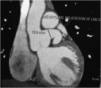
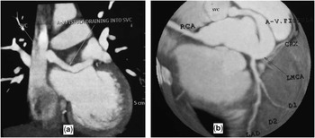
A 12-year-old boy came for the evaluation of an incidentally detected cardiac murmur on routine clinical examination. General physical examination was unremarkable. Cardiovascular examination revealed a continuous murmur associated with thrill heard best over the right second intercostal space. There was no evidence of congestive cardiac failure. Routine electrocardiogram and chest X-ray were normal. A two-dimensional echocardiography was performed that showed a dilated left main coronary artery and a 5 mm wide tract, which was seen to be originating from the left main coronary artery at its posterior aspect and having its course posterior to the aorta. Colour Doppler at the defect showed a continuous flow. Exact tract of the defect could not be established, and therefore conventional angiogram was planned. Ascending aortogram was performed with a pigtail demonstrating an aneurysmally dilated left main coronary artery, and a large fistulous communication arising from the left main coronary artery to traverse posteriorly along the aortic root to opacify the right atrium (Supplementary Video 1). A computed tomography (CT) aortogram was performed to confirm the diagnosis, which demonstrated an osteoproximal aneurysmal dilation of the left main coronary (left main coronary artery measured 11 mm) (Fig 1). The CT scan also demonstrated a long coronary arteriovenous fistula connecting the left main coronary artery and the right-sided superior vena cava, 10 cm above its junction with the right atrium (Fig 2a and b). There was no focal obstruction in the fistulous tract, but there was slight narrowing in the distal portion of the tract, with constriction seen at the superior vena cava end (Fig 2b). Other coronary arteries were normal. The anatomy of the fistula was suitable for percutaneous device closure. As the fistula was very large and to prevent its complications, we decided to close the fistula percutaneously from the venous side with a vascular plug. As the patient’s parents were not in favour of the procedure owing to social issues, we planned to keep the patient under close medical follow-up.
Computed tomography (CT) angiogram showing aneurysmal dilation of the osteoproximal left main coronary artery (11 mm in diameter).

( a ) Contrast-enhanced computed tomography (CT) scan showing the long fistulous tract draining distally into the superior vena cava, 10 cm above its junction with the right atrium. ( b ) Contrast-enhanced CT scan showing the fistulous tract from the left main coronary artery to the superior vena cava. Other coronary artery branches are normal. CRX=left circumflex artery; D=diagonal artery; LAD=left anterior descending; LMCA=left main coronary artery; RCA=right coronary artery; SVC=superior vena cava.

Discussion
Coronary arteriovenous fistulas are uncommon anomalies of the coronary artery, which was initially described by Krause in 1865. In the literature, it has been reported in about 0.2-0.25% of patients undergoing coronary arteriography.Reference Fernandes, Kadivar, Hallman, Reul, Ott and Cooley 1 It is an abnormal communication between the coronary artery and either a cardiac chamber, pulmonary artery, or venous system of the heart. Coronary arteriovenous fistulas are usually congenital in the origin but can be acquired as well. Exact aetiology of these fistulas is unknown; however, it is postulated that it may be because of failure of obliteration of the intramyocardial trabecular sinusoids, with anomalous development of the intra-trabecular spaces. Coronary arteriovenous fistulas commonly arise from the right coronary artery (55%) followed by the left anterior descending artery (35%). The involvement of the left main coronary artery is the least common and is rarely encountered. Over 90% of the fistulas drain into the venous structures of circulation. Coronary arteriovenous fistulas drain into the right ventricle in 40%, the right atrium in 26%, the pulmonary artery in 17%, the left ventricle in 3%, the coronary sinus in 7%, and the superior vena cava in 1% of the cases.Reference Dodge-Khatami, Mavroudis and Backer 2 Coronary arteriovenous fistula from the left main coronary artery to the superior vena cava is the rarest entity. Aneurysms can occur in up to 19% of coronary arteriovenous fistulae but are usually of small size. We analysed the published literature using a Medline search and identified five such cases of coronary arteriovenous fistula between the left main coronary artery and the superior vena cava. In the index-reported case, we also found an aneurysmal dilation of the left main coronary artery from its origin. Coronary arteriovenous fistulas mostly remain asymptomatic, detected either by routine general examination (as in our index case) or during routine coronary angiography. Rarely, it can present with fistula-related complications such as aneurysm formation, pressure effect on the adjacent structures, infective endocarditis, thrombosis, and significant shunt with congestive cardiac failure and rupture of the aneurysm.Reference Levin, Fellows and Abrams 3 Diagnosis of coronary arteriovenous fistula is challenging as its prevalence is low. Transoesophageal echocardiography is a good non-invasive technique for the diagnosis of these fistulas and their complications.Reference Vitarelli, De Curtis and Conde 4 A traditional gold standard diagnostic modality for coronary arteriovenous fistula is conventional coronary angiography. Although most of the coronary arteriovenous fistulas can be diagnosed by conventional coronary angiogram, the origin, draining site, and relation of the fistula with the adjacent cardiac structures may be ambiguous in some cases. To address these concerns, non-invasive modalities such as magnetic resonance imaging and contrast-enhanced computed tomography can be used as adjunct to coronary angiography not only to diagnose but also to plan the treatment stratergy.Reference Aydogan, Onursal, Cantez, Barlas, Tanman and Gurgan 5 , Reference Ropers, Moshage, Daniel, Jessl, Gottwik and Achenbach 6 In the index-reported case, the diagnosis and the treatment strategy of the fistula were better planned after CT angiogram than the conventional invasive angiogram. Natural history of the coronary arteriovenous fistulas is extremely variable. The accepted recommendation is to treat the symptomatic coronary arteriovenous fistula, but controversies exist regarding the treatment of the asymptomatic fistula.Reference Urrutia-S, Falaschi, Ott and Cooley 7 Some authors recommend closure of the fistulas even in asymptomatic patients to prevent fistula-related complications such as heart failure, endocarditis, aneurysm formation, and myocardial ischemia.Reference Urrutia-S, Falaschi, Ott and Cooley 7 Coronary arteriovenous fistula can be closed surgically or percutaneously. Surgical closure of the fistula remains the safe and effective method of treatment with good success rate.Reference Balanescu, Sangiorgi, Castelvecchio, Medda and Inglese 8 The success rate of percutaneous intervention is equally encouraging if the cases are selected carefully.Reference Perry, Rome, Keane, Baim and Lock 9 Percutaneous device closure or coil embolisation is attempted if anatomy of the fistula is favourable for intervention, for example non-tortuous vessel, fistula with distal narrowing to avoid embolism to the drainage site.Reference Perry, Rome, Keane, Baim and Lock 9 In the index case, we decided to close the arteriovenous fistula to prevent fistula-related complications (because of its size) and to relieve the patient of symptoms. We decided to close the fistula percutaneously with a vascular plug, as the anatomy was suitable for percutaneous intervention – long non-tortuous tract, device could be delivered via the distal draining site at the superior vena cava, and there was a distal constriction near the distal drainage site. As the patient’s parents did not opt for the intervention owing to social reasons, we decided to keep the patient under medical follow-up with close reference to identify fistula-related complications.
Acknowledgement
None.
Financial Support
This research received no specific grant from any funding agency, commercial or not-for-profit sectors
Conflicts of Interest
None.
Supplementary material
To view supplementary material for this article, please visit http://dx.doi.org/10.1017/S1047951114001103