Electron flow in biological systems
We begin this discussion of biofilms with a simple axiom: “Electrons must flow.” While there are a few notable exceptions to this statement, it remains a general rule of life that electron flow must occur in order for chemo-osmotic gradients to be established and adenosine triphosphate synthesized as the energy in such gradients is harvested.Reference Nealson and Rye1, Reference White2 Such is true for the mitochondria in virtually all eukaryotic cells (e.g., fungi, animal cells, and plant cells), as well as for both domains of prokaryotic cells, the bacteria, and the archaea, which lack a nucleus or organelles such as mitochondria. When electron flow stops, most eukaryotes rapidly die, while many bacteria and archaea can be more robust but eventually suffer a similar fate. This seemingly simple tenet has a major implication for microbial ecology: if redox energy is available in a natural system, and if the natural rate of electron flow is not too rapid, then it is a niche that will probably be exploited by a redox-active microbe.
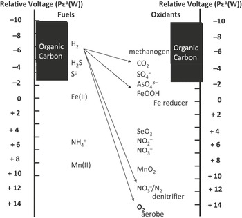
Eukaryotic cells are, as a rule, capable of utilizing only oxygen as an electron acceptor and only organic carbon as an electron donor, while prokaryotic cells (the bacteria and archaea) are often able to utilize other electron donors (e.g., inorganic molecules such as hydrogen) and/or other electron acceptors (e.g., inorganic molecules such as sulfates and nitrates). The range of electron donors available to living microbial systems is shown in Figure 1. Such a view of life’s reactionsReference Stumm and Morgan3 allows one to see the energy available for the creation of membrane gradients and relate the metabolic opportunities to environmental chemistry and geology. However, it is a misconception that bacteria and archaea are metabolically diverse as individual species. In fact, in order to accomplish what they do, they often become very highly specialized (e.g., methanotrophs often are restricted to growth on methane or methanol). Thus, it is the community of prokaryotes that is diverse, populated by individual, specialist species that are working together.
Some redox couples utilized by life. This diagram shows a selection of redox couples used by various living organisms. The relative reducing potentials of each is represented by the logarithmic scale (Pεo(W), which is the negative log of the electron potential, at a pH of 7.0 in water). A full representation of this thermodynamic approach is presented in Reference Reference Stumm and Morgan39.

For many years, it was accepted that electron acceptors had to be soluble, whether they are inorganics such as oxygen, nitrate, sulfate, or carbon dioxide, or organics such as dimethyl sulfoxide (DMSO), trimethylamine N-oxide (TMAO), or fumarate. These electron acceptors are very well documented in the literature, and reduction is commonly done on the inner membrane, where specific enzymes are found that catalyze substrate reduction.Reference Madigan and Martinko4 Of notable exception were the solid metal oxides such as Fe(III) oxides and Mn(IV) oxides, a variety of which exist in abundance in various environments. As shown in Figure 1, both Fe(III) and Mn(IV) have good potentials, with Fe(III) yielding more energy than sulfate, and Mn(IV) yielding energy at approximately the level of nitrate. Yet the biological reduction of these compounds as electron acceptors was not generally appreciated, because Mn and Fe oxides are insoluble solids, and no mechanism was available for reduction and energy conservation at the inner membrane.
This being said, there are many sites known where manganese and/or iron reduction rates are far too rapid to be explained by abiotic chemistry alone (e.g., the flux of manganese out of the sedimentsReference Aguilar and Nealson5 in Oneida Lake, NY, is on the order of 2 to 5 mM m−2 day−1). At the near-neutral pH of the sediments, the Mn(II) flux should be many orders-of-magnitude slower.Reference Dean, Moore and Nealson6 In 1987, this site was chosen as a likely habitat for microbes that could make a living via the reduction of solid manganese oxides. Using sediment samples from the zones of rapid reduction, anoxic enrichment cultures were established in which the only electron acceptor provided was solid MnO2,Reference Myers and Nealson7 and non-fermentable carbon sources, such as glycerol or lactate, were used. Under these conditions, rapid MnO2 reduction was observed, and these enrichments were used for the isolation of a microbe now named Shewanella oneidensis MR-1. It may be noted that the type species of this organism (originally named Pseudomonas putrefaciens) had been in culture collections for more than 50 years before it was tested for the ability to reduce iron. Again, the notion that solid substrates should not be used as electron acceptors kept us from asking the question.
Extracellular electron transfer and microbial fuel cells
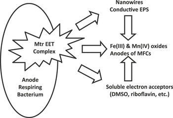
After the initial reports of Shewanella Reference Myers and Nealson7, Reference Myers and Nealson8 and another iron reducing group that would eventually be called Geobacter,Reference Lovley and Phillips9, Reference Lovley, Giovannoni, White, Champine, Phillips, Gorby and Goodwin10 a number of different metal reducers were reported, and the thought that solid substrates could be used as electron acceptors began to be tested. This marked the beginning of acceptance of the notion of extracellular electron transport (EET), which is the cornerstone of what we will discuss in the context of biofilm metabolism. The conundrum of how it might be accomplished has, over the ensuing years, revealed that a variety of approaches may be used, including: (1) outer membrane cytochromes that catalyze direct electron transfer to solid substrates,Reference Myers and Nealson7, Reference Myers and Nealson8 (2) H+ or electron shuttles that carry electrons from cells to the electron donor that is otherwise inaccessible,Reference Marsili11, Reference von Canstein, Ogawa, Shimuzu and Lloyd12 (3) conductive nanowires,Reference Gorby, Yanina, McLean, Rosso, Moyles, Dohnalkova, Beveridge, Chang, Kim, Kim, Culley, Reed, Romine, Saffarini, Hill, Shi, Elias, Kennedy, Pinchuk, Watanabe, Ishii, Logan, Nealson and Fredrickson13–Reference Reguera, Nevin, Nicoll, Covalla, Woodard and Lovley17 and, (4) other conductive matricesReference Kato, Nakamura, Kai, Watanabe and Hashimoto18, Reference Nielsen, Risgaard-Petersen, Fossing, Christensen and Sayama19 (Figure 2).
Postulated pathways for extracellular electron transport: direct transfer from outer membrane enzymes to the electron acceptor (middle arrow); the use of soluble H+ or electron shuttles to deliver electrons to the electron acceptor (bottom arrow); electron transfer via conductive nanowires or conductive matrices (top arrow). Please note: EPS, extracellular polymeric substances; Mtr EET, cytochrome-mediated extracellular electron transport; MFC, microbial fuel cell; and DMSO, dimethyl sulfoxide.

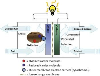
In a rather short time, Byung-Hong Kim from the Korean Institute of Technology, using Shewanella, was able to demonstrate high efficiency electron transport in microbial fuel cells,Reference Kim20 observations that have been confirmed for a number of metal reducing microbes.Reference McLean, Wange, Gorby, Wainstein, McQuaid, Ishii, Bretschger, Beyenal and Nealson21–Reference Lovley27 In such a situation, the microbiology is viewed in a slightly different mode. The microbes are contained in an anoxic chamber in which the only oxidant provided is the anode of the fuel cell. A biofilm is formed, and direct electron transfer occurs, thus obviating the need for an added electron mediator, which was previously required for microbial fuel cells (Figure 3).
Microbial fuel cell. This diagram shows one variation of a common microbial fuel cell. In the anaerobic anode chamber, organic carbon is oxidized, yielding CO2, protons, and electrons. The electrons are carried through the load to the cathode side, where they are recombined with protons that diffuse through the ion exchange membrane and molecular oxygen to yield water. In this case, the cathode reaction is catalyzed by platinum, but it can also be catalyzed by bacteria capable of aerobic respiration, as long as they have the ability to take up electrons from the cathode membrane.

Viewed in this way, the respiration of the microbial fuel cell (MFC) anode is simply an extension of the electron flow to insoluble substrates, such as iron or manganese oxides and probably requires little else. Indeed, studies utilizing a variety of mutational methods, including the deletion of relevant genes,Reference Beliaev and Saffarini28–Reference Bretschger, Obraztsova, Sturm, Chang, Gorby, Reed, Culley, Reardon, Barua, Romine, Zhou, Beliaev, Bouhenni, Saffarini, Mansfeld, Kim, Fredrickson and Nealson30 have revealed that the genes involved in the reduction of solid metal oxides are nearly identical to those needed for anode respiration.
The role of biofilms in microbial fuel cells
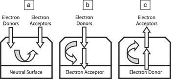
The consideration of microbial fuel cells thus brings us to the subject of biofilms in general, but with a different slant from what is usually seen. When biofilm observations and studies are done with microbes on uncharged surfaces, such as glass or plastic, we imagine it leads to a situation similar to that diagrammed in Figure 4a, in which both electron donor and electron acceptor must be obtained from the medium.Reference Hall-Stoodley, Costerton and Stoodley31 The advantages of attachment under such conditions are probably many, but metabolically, it places the biofilm community in the interesting position of competing with itself for both electron donors and acceptors. In contrast, if the surface is electrically active as an electron acceptor (Figure 4b), then bacteria will have a clear reason for attachment and maintenance of the biofilm: a non-destructible, inexhaustible supply of electron acceptors. To this end, we have had biofilms continuously producing electricity for months in our laboratories, demonstrating the robustness of these biofilm consortia.Reference Kan, Hsu, Cheung, Pirbazari and Nealson32 Indeed, it is now routine to isolate electrically active bacteria via enrichment cultures using a poised electrode as the electron acceptor.Reference McLean, Wange, Gorby, Wainstein, McQuaid, Ishii, Bretschger, Beyenal and Nealson21, Reference Thrash and Coates33, Reference Torres, Krajmalnik-Brown, Parameswaran, Marcus, Wanger, Gorby and Rittmann34
Electron flow in biofilms on differently charged surfaces. (a) Biofilm on uncharged (neutral) surface with regard to electron donors. Under these conditions, limitation for either donor or acceptor (or both) can occur. (b) Biofilm on a positively charged surface with regard to electron donors (e.g., positively charged minerals or anode of microbial fuel cell [MFC]). (c) Biofilm on negatively charged surface with regard to electron acceptor (surface of another cell; particulate organic matter; cathode of MFC).

It is also now apparent from a number of reports that microbes are also capable of using electrodes (e.g., cathodes) as sources of energy in the form of electrons (see Reference Reference Rabaey and Rozendal23 and citations therein). That is, they appear to be capable of taking up electrons, harvesting some of the energy by using this electron flow to catalyze the oxidation of oxygen. Knowing that microbes of a single species are capable of both extracellular electron donation and acceptance, however, has implications of great importance for biofilms, where cells are in intimate contact with one another, and strong redox gradients may exist.
In Figure 4, if electron flow between cells in a biofilm could be done, and if bacteria are NOT able to transfer electrons within the biofilm, then the ability for the biofilm to maintain a stable community will almost certainly be affected. As was noted previously, there are a variety of ways bacteria have evolved to move electrons to solid substrates. Whether or not a similar array can be used for the uptake of electrons is not yet known.
Mechanisms aside, if a microbe such as Shewanella spp. is capable of both electron donation and uptake, and if the resulting flow of electrons through a cell can be used to the advantage of the microbe(s), then electron flow within a biofilm could be of major importance to the community. If a biofilm is formed on an electron acceptor, then as the biofilm matures, either it must stop growing and maintain itself as a very thin layer, or it must physiologically specialize, with cells at the bottom of the biofilm maximizing their activity for electron donation, and those at the top specializing for electron uptake. Furthermore, the cells must almost certainly couple this metabolism with control of growth rate. Clearly, the cells at the bottom of the biofilm could destabilize the consortium if rapid growth continued. Cells at the top, on the other hand, might be “allowed” to continue growth, and even to detach and interact with the biofilm, under conditions of electron donor limitation.
Viewed in this way, electron flow within a biofilm becomes an integral part of the community metabolism and could occur on biofilms attached to energy yielding surfaces (other cells, dead material), as well as electron acceptor substrates. The important issue is that if microbes can solve the problem of cell-to-cell electron transport, then this type of metabolism might be viewed as an important and widespread part of the microbial metabolic world.
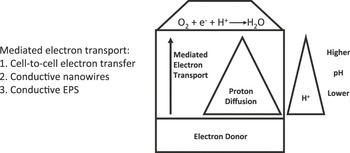
An additional consideration of this line of reasoning relates to the issue of electron flow and the molecules that are involved. We call all of the components of the electron transfer chain electron carriers, but many carry both an electron and a proton. When oxygen is used as the ultimate electron acceptor, both a proton and an electron are needed. In a microbial fuel cell, the electrons are supplied at the cathode via a wire, while the protons must diffuse across the proton diffusion membrane. Similarly, if a biofilm sitting on an electron donor was capable of EET, the flow of electrons would occur via conductivity, while the flow of protons would occur via diffusion. This would then predictReference Biffinger, Pietron, Bretschger, Nadeau, Johnson, Williams, Nealson and Ringeisen35 that an environmental pH gradient would be seen in cases of rapid electron flow (Figure 5).
Mediated electron transport. Whether electron transfer in microbial redox reactions is mediated via (1) cell-to-cell transfer, (2) conductive nanowires, and/or (3) conductive extracellular polymeric substances (EPS), electron flow will always be opposite the proton gradient.

An intriguing aspect of such a model is that such electrical contact could also act as a mode of communication in biofilm communities. If conductivity is maintained, be it via nanowires, conductive EPS, or other unknown mechanism(s), then this information should be available to the cells in other metabolic zones of the biofilm. That is, if cells at the bottom of the biofilm “know” the conditions at the top, they can respond accordingly with regard to growth, metabolism, and electron flow, effectively allowing one to view a robust biofilm community (be it a single species or a consortium) as a physiologically diverse community, sharing many traits with multicellular organisms. Another implication of this model is that within the architecture of bacterial biofilms are far more potential niches than previously described. Frequently, the activities of organisms within the community are viewed in terms of gradients of oxygen, nutrient, or waste flow. The transfer of electrons through the community creates additional gradients of flow “to” or “from” different areas of the biofilm. This inevitably creates microniches within which natural selection can act, potentially leading to the evolution of specialized cells within established biofilm communities. To date, several examples of real-time adaptive evolution within biofilms have been observed.Reference Hansen, Rainey, Haagensen and Molin36–Reference Kraigsley and Finkel38 When studying the adaptive forces at work, a consideration of the role of electron flow is likely warranted.
Concluding thoughts
We realize that this view of biofilms and microbial communities goes “against the grain” of how some may think about microbes, and our intent in this review is not to suggest that what we already know about biofilms is not correct or relevant. Rather, we hope to add another important metabolic/physiological perspective to the world of biofilms. This perspective posits that microbes on an electrically active or redox-active surface will take advantage of this position and harvest energy from it: energy that in the absence of bacterial catalysts might go untapped because of a high activation energy of reaction. An appreciation of the fact that biofilm communities may be interacting electrochemically with the surfaces they are growing on may have far-reaching implications. For example, this may warrant the redesign of bioprocess reactors, where it has generally been assumed that the surface of the reactor vessel is merely an inert platform upon which the microbes grow. It is very likely that in some industrial processes, yields could be increased when the electrochemical interactions between the microbial community and the reactor are better understood. Significant insights into the interactions between microbial biofilms and the materials from which reactors are constructed, and virtually all surfaces where biofilms may interact, will be required to begin to exploit the kinds of phenomena we describe here. We hope that this perspective will provide another angle from which the establishment, and longevity, of biofilms can be predicted and tested.
Acknowledgments
Work in the Nealson and Finkel labs is supported, in part, by a Multi-University Research Initiative (MURI) grant from the U.S. Air Force Office of Scientific Research (FA-9550-06-1-0292).