Cow milk is one of the first foods incorporated into the human diet, either replacing or supplementing maternal milk. Allergy to cow milk (CMA) concerns ∼2·5% of children below 3 years of age, the feeding of an infant, allergic to cow milk casein, may be a major problem (Anderson, Reference Anderson1996). Caseins are the main milk allergens and account for 80% of the total protein in cow milk. They include 4 fractions αS1- (32%), αS2- (10%), β- (28%) and κ-caseins (10%). Bovine αS1-casein is one of the major allergens responsible for CMA (Nakajima-Adachi et al. Reference Nakajima-Adachi, Hachimura, Ise, Honma, Nishiwaki, Hirota, Shimojo, Katsuki, Ametani, Kohno and Kaminogawa1998). It is a single-chain phosphoprotein of 199 amino acid residues, characterized by a high content of prolyl residues, which are distributed throughout the molecule. It lacks disulphide bonds, which results in its reduced structural stability (Kumosinski et al. Reference Kumosinski, Brown and Farrel1991). These factors increase the chance that the major allergenic epitopes of αS1-casein are linear rather than conformational (Cocco et al. Reference Cocco, Järvinen, Sampson and Beyer2003), thus making this casein a good subject for study of the linear epitope breakdown by proteolysis.
An increasing interest in reduction of the allergenicity of foods during processing points to the potential use of enzyme modifications to cleave the allergic proteins and to produce hypoallergenic foods (Itoh, Reference Itoh1996). Immuno-reactivity of milk proteins can be decreased by proteolysis (Peñas et al. Reference Peñas, Prestamo, Baeza, Martinez-Molero and Gomez2006). Nowadays, protein hydrolysates are also included in specific formulations, as well as hypoallergenic infant formulas to reduce their antigenicity as compared with intact protein. Proteolytic systems of lactobacilli are complex and are composed of proteinases and peptidases with different sub-cellular locations. Proteases of cells grown on milk showed similar hydrolytic activity on αS- and β-caseins (Cocco et al. Reference Cocco, Järvinen, Sampson and Beyer2003). Most of work on proteolytic activity of lactobacilli has been carried out using strains grown in culture media containing milk.
In a previous study (El-Ghaish et al. Reference El-Ghaish, Dalgalarrondo, Choiset, Sitohy, Ivanova, Haertlé and Chobert2010), we characterized the proteolytic activity of Lactobacillus fermentum IFO3956 isolated from milk products from Egypt describing the factors affecting this activity. Isolating new strains or isolates of nonstarter lactic acid bacteria (NSLAB) with technological functions potentially applicable in the food industry is scientifically or commercially highly interesting and important. These NSLAB can be used in different types of cheeses, especially those produced from raw milk due to their positive influence on cheese manufacture through lipolytic activity, citrate utilization, and production of aromatic volatile compounds as well as proteolytic and antimicrobial properties. In the current study, this strain was used in optimal conditions allowing the highest proteinase activity towards a major allergen component (αS1-casein). The obtained hydrolysates were characterized by LC/MS/MS mass spectrometry to confirm the hydrolysis of the epitopes. The binding strength of the hydrolysates with IgE was evaluated by competitive ELISA to confirm the potential impact of hydrolysis on immuno-reactivity of αS1-casein.
Material and Methods
Materials
All products were of analytical grade and obtained from different companies such as Merck, Germany (skim milk powder, calcium chloride and Triton X-100), Sigma-Aldrich, USA (glycerol, sodium citrate, Tween-20, Tween-80, sodium acetate, magnesium sulphate, manganese sulphate, cysteine, sodium dodecyl sulphate, gelatin, Tris, ammonium sulphate, trifluoroacetic acid, potassium chloride, polyvinyl alcohol and 4-methylumbelliferyl phosphate), Biokar diagnostics, France (MRS and meat extract), Fisher Bioblock Scientific, France (yeast extract and agar), Acros, USA (glucose), Panréac, France (di-potassium phosphate, potassium phosphate, glycine and sodium chloride), BD, France (peptone), Bio Basic INC., Canada (Coomassie Brilliant Blue G-250), Fisher BioReagents, USA (polyacrylamide), Applied Biosystems, Ireland (acetonitrile), Riedel-deHaën, Germany (potassium dihydrogen phosphate).
Purification of α S1-casein from total casein
Total casein was prepared from fresh milk by precipitation at pH 4·6 with 6 m-HCl followed by centrifugation (4000 g, 20 min) according to Mailliart & Ribadeau Dumas (Reference Mailliart and Ribadeau1988). αS-Caseins (αS1-+αS2-caseins) were prepared from total casein by anion exchange chromatography (FPLC; BioCad/Sprint, PerSeptive Biosystems, USA) according to Thorn et al. (Reference Thorn, Ecroyd, Sunde, Poon and Carver2008). Total casein (50 mg/ml) was re suspended in 50 mm-Tris-HCl+8 m-urea, pH 8·0+20 mm-dithiothreitol (DTT). Column source Q Sepharose High Performance (v=50 ml) was equilibrated with buffer A (25 mm-Tris-HCl +4 m-urea, pH 8·2+5 mm-DTT) and elution was performed with gradient from 0 to 30% buffer B (buffer A+1 m-NaCl) at a flow rate of 5 ml/min. αS-Caseins were collected, dialyzed and lyophilized. αS-Caseins (35 mg/ml) were re suspended in 20 mm-sodium acetate +8 m-urea, pH 6·0+20 mm-DTT. Column source Q (V=8 ml) was equilibrated with buffer C (20 mm-sodium acetate+4 m-urea, pH 6+5 mm-DTT) and elution was performed with a gradient from 0 to 30% buffer D (buffer C+1 m-NaCl) at a flow rate of 5 ml/min. αS1-Casein was collected, dialyzed and lyophilized.
Strain and growth conditions
Lb. fermentum IFO3956 was isolated from traditional Ras cheese collected from local markets of Lower Egypt (Tanta city) and identified by 16S rRNA gene amplification. The purified strain was re-grown in sterile skim milk (12·5%, w/v) supplemented with 30% (w/v) glycerol and stored at −20°C. Before use, the strain was grown twice in MRS broth.
Four different media were used to study the production of the lactobacillus proteinase: MCA (milk-citrate-agar), RSM (10% reconstituted skim milk), MRS (de Man, Rogosa and Sharpe) and CYG (casein–yeast extract–glucose). MCA is composed of 4·4% skim milk powder, 0·8% Na-citrate, 0·1% yeast extract, 0·5% glucose and 1·5% agar. RSM is 10% reconstituted skim milk and the composition of MRS (g/l) was as follows: polypeptone, 10; meat extract, 10; yeast extract, 5; glucose, 20; Tween 80, 1·08; di-potassium phosphate, 2; sodium acetate, 5; ammonium citrate, 2; magnesium sulphate, 0·2 and manganese sulphate, 0·05. CYG composition (g/l) was as follows: yeast extract, 3; peptone, 5; cysteine, 0·2; MgSO4·7H2O, 0·2; MnSO4.H2O, 0·05; sodium acetate, 5; sodium citrate, 4·4; potassium phosphate, 2; sodium caseinate, 10; glucose, 10 and Tween 80, 1 ml.
Influence of the media on the proteolytic activity
The pH of each medium was adjusted to 6·8. In case of MCA media, an aliquot (∼0·5 ml) of the activated pre-culture was applied on the top of MCA plates (Fira et al. Reference Fira, Kojic, Banina, Spasojevic, Strahinic and Topisirovic2001). The plates were incubated for 48 h at 37°C prior to cell collection. For RSM, MRS and CYG media (Hébert et al. Reference Hébert, Raya and De Giori1997), a pre-culture was prepared by inoculating these media with 5% (v/v) overnight culture and incubation at 37°C for 24 h. Fresh cells were collected from different media and re-suspended in 100 mm-Na-phosphate buffer (pH 6·8) and brought to final optical density (OD600) of 20 just before application. One volume of the cellular suspension was combined with an equivalent volume of solution of sodium caseinate (12 mg/ml) in 100 mm-Na-phosphate buffer (pH 6·8) so that the OD600 of the final combined suspension is equal to 10, and incubated for different periods (5 and 24 h) at 37°C. The control was sodium caseinate (12 mg/ml) in 100 mm-Na-phosphate buffer, pH 6·8 without addition of strain, the volume of which being replaced by the buffer in order to keep equivalent concentration of protein. All samples were analyzed by SDS-PAGE.
Extraction and characterization of cell wall-bound proteases
Cells were collected from MCA media as previously described and washed twice with 100 mm-Na-phosphate buffer (pH 6·8) containing 20 mm-CaCl2. To extract cell-wall-bound proteases, the washed cells were incubated with 1% of different detergents (SDS, Tween 80 and Triton X-100) dissolved in 100 mm-Na-phosphate buffer (pH 6·8) for 16 h at 37°C with agitation. Supernatants were collected by centrifugation (12000 g at 4°C for 20 min). The released protease was considered as an indication of intracellular enzyme (Tsakalidou et al. Reference Tsakalidou, Anastasiou, Vandenberghe, van Beeumen and Kalantzopoulos1999). The proteolytic activity of the supernatants was measured by SDS-PAGE. One volume of the supernatants was combined with an equivalent volume of solution of sodium caseinate (12 mg/ml) in 100 mm-Na-phosphate buffer (pH 6·8). The final mixtures were incubated for different periods (3 and 24 h) at 37°C. Control was prepared as described above. All samples were analyzed by gelatin Zymogram (12% polyacrylamide) and by SDS-PAGE.
SDS-PAGE
The samples were applied to a 12% polyacrylamide slab gel. The migration buffer contained 50 mm-Tris, 0·384 m-glycine and 0·1% SDS. After running at 10 mA on the stacking gel and 20 mA on the running gel, staining was performed with Coomassie Brilliant Blue G-250 followed by a convenient destaining. The gels were scanned with Image scanner III (GE Healthcare, USA) and the intensity of the bands was quantified by Image software. Analysis was performed by using Fuji Film Image Gauge V3.0 software (Fuji Photo Film Co. Ltd., Japan). Data were expressed as the ratio of the area and intensity of the band. The reduction in the intensity of band during incubation with respect to the original intensity was taken as the percentage of hydrolysis (Ong et al. Reference Ong, Henriksson and Shah2006).
Gelatin-SDS-PAGE Zymogram
The samples were applied to a 12% polyacrylamide gel containing 0·1% gelatin, where the migration buffer consisted of 50 mm-Tris, 0·384 m-glycine and 0·1% SDS. After running at 10 mA on the stacking gel and 20 mA on the running gel at 4°C, the gel was washed twice in 2·5% Triton X-100 for 30 min then twice in distilled water for 15 min. Washed gel was placed in incubation buffer (50 mm-Tris-HCl, 100 mm-NaCl, 5 mm-CaCl2, pH 7) for 16 h at 37°C before fixation in a solution of 4% ethanol/H3PO4 (1/1, v/v), twice for 60 min, then the gel was washed in 2% phosphoric acid for 60 min. Finally, the gel was transferred to a sensitization solution (17% ethanol, 15% (NH4)2SO4, 2% H3PO4) for 20 min before staining with 0·1% Coomassie Brilliant Blue G-250 and fixing in 4% ethanol/H3PO4 (1/1, v/v). The gel was rinsed twice for 10 min in distilled water and destained in 20% ethanol for 10 min before keeping in distilled water. Bands revealing activity appeared as clear zones on the blue gel. The gel was scanned with Image scanner III and the location of the clear bands was marked and compared with the standard molecular weights.
Characterization of α S1-casein hydrolysates
Preparation of hydrolysates
αS1-Casein hydrolysates produced by Lb. fermentum IFO3956 were prepared as previously described for sodium caseinate (El-Ghaish et al. Reference El-Ghaish, Dalgalarrondo, Choiset, Sitohy, Ivanova, Haertlé and Chobert2010) except the concentration of αS1-casein was 5 mg/ml. At the end of incubation periods (3, 6 and 24 h), the supernatant was analyzed by SDS-PAGE (12% polyacrylamide), liquid chromatography–mass spectrometry (LC-MS) and competitive enzyme linked immunosorbent assay (ELISA).
LC/MS/MS analyses
Mass spectrometry analyses were conducted on the platform “Biopolymers-Interaction-Structural Biology” located at the INRA centre of Nantes (INRA Research Unit 1268) (http://www.nantes.inra.fr/plateformes_et_plateaux_techniques/plateforme_bibs).
αS1-Casein peptides were separated by reversed phase-HPLC (RP-HPLC) with a Waters HPLC system (Waters 616 pump controlled by Waters 600 controller) coupled to a Finnigan LCQ ion trap spectrophotometer (Finnigan MAT, San Jose, CA). The column (2 mm×150 mm, 90 Å, 4 μm, Jupiter, PROTEO, Phenomenex) was equilibrated with solvent A (0·11% trifluoroacetic acid (TFA) in distilled water, v/v) at a flow rate of 0·15 ml/min. After injection, the column was washed for 5 min with 100% solvent A then elution was performed by applying a linear gradient to 100% of solvent B (80% acetonitrile, 20% H2O, 0·09% TFA, v/v/v) from 5 to 35 min followed by a 2 min step at 100% solvent B.
Spectra were acquired in automated MS/MS mode. The scan rate for MS mode was set between the masses of 400 to 2000 Da. Peptide identification was performed using the Mascot software (version 2.5, Matrix Science) on the MS/MS ion search mode with the following parameters: enzyme: none, peptide mass tolerance: 1·5 Da, fragment mass tolerance: 0·6 Da, fixed modification: phosphorylation. Identification was performed by comparing the obtained peptides with αS1-casein sequences.
Competitive ELISA
Wells of 96 microplates (Maxi Sorp, Nunc, Roskillde, Denmark) (plate 1) were coated with 100 μl per well of αS1-casein diluted to 5 μg/ml in 1 m-PBS pH 7·4 (0·136 m-NaCl, 2·68 mm-KCl, 1·76 mm-KH2PO4, and 10·14 mm-Na2HPO4 12 H2O) for 3 h at 30°C. Negative control (blank) was prepared by replacing αS1-casein with equal volume (100 μl) of PBS. All wells were coated with 100 μl of a mouse monoclonal anti-human IgE, clone M604199 (Ab1, Fitzgerald, Concord, USA) diluted to 1:2500 in PBS. After coating, all wells were washed three times with PBS containing 0·1% (v/v) Tween-20 (PBS/T) and saturated with 250 μl of the PBS/T solution containing 1% (w/v) polyvinyl alcohol (PBS/T/PVA) for 1 h at 37°C. Standard curve was performed with adding 100 μl of serial dilutions from 160 to 0·15 ng/ml (5000 IU) of human serum IgE (WHO international reference preparation Code 75/502), to the wells coated with the antihuman IgE antibody only (without αS1-casein).
A pool of 18 sera from patients allergic to αS1-casein was collected by the Laboratory of Immuno-Allergology of Academic Hospital, Angers, France and used after approval of the internal Ethical Committee of the hospital. The serum pool was diluted to 1:10 and mixed with equal volumes (60:60, μl:μl) of serial dilutions of αS1-casein or αS1-casein hydrolysate in 1% PBS/T/PVA (1000, 640, 160, 40, 10, 2·5, 1, 0·25, and 0·062 μg/ml) on a separate plate (plate 2) and incubated at 37°C for 1 h.
An aliquot of 100 μl was transferred from each well in the plate 2 to the wells coated with αS1-casein in plate 1 or the negative control. After an overnight incubation at 4°C, the plates were washed three times with PBS/T and incubated for 2 h at 37°C with the secondary antibody (alkaline-phosphatase-conjugated polyclonal antihuman IgE, Sigma) diluted 1:1000 on PBS/PVA. After three washings with PBS/T, the free secondary antibody was revealed by the addition of 4-methylumbelliferyl phosphate (4-MUP) substrate diluted 1:5 in Tris-HCl, pH 9·8. The fluorescence emission was measured after 90 min of incubation at 37°C with the FLx800 plate reader (Biotech Instruments, Inc., Winooski, VT) fitted with a 360 nm excitation filter and a 440 nm emission filter.
The calculation of the results was performed as described by Taheri-Kafrani et al. (Reference Taheri-Kafrani, Gaudin, Rabesona, Nioi, Agarwal, Drouet, Chobert, Bordbar and Haertlé2009). The concentration of protein (inhibitor) needed to inhibit 50% of IgE binding (IC50) was calculated from the inhibition curves by relating fluorescence intensity to inhibitor concentration. The C50% is the concentration of αS1-casein binding 50% of the IgE in the sera of the CMA patient group. The value obtained by dividing the C50% of the hydrolyzed αS1-casein by C50% of the intact αS1-casein is an indicator of the effect of the proteolysis on the IgE binding and consequently of the immuno-reactivity of the αS1-casein. The experiment was realized in triplicate.
Results and Discussion
Effect of the culture medium on proteinase activity
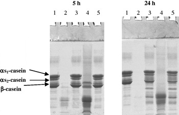
Proteinase activity of Lb. fermentum IFO3956 cells grown in different media (MCA, CYG, RSM and MRS) is shown in Fig. 1. The rate of casein hydrolysis was much higher in the milk-based media (MCA and RSM; lanes 2 and 4, respectively) than in the free-milk media (CYG and MRS; lanes 3 and 5, respectively) either after 5 or 24 h of incubation at 37°C. Highest extent of proteolysis was achieved in MCA (lane 2) followed by RSM (lane 4). However, the band intensity of casein fractions indicate that the degree of hydrolysis in MCA after 5 h is still superior to that achieved in RSM medium after 24 h incubation at 37°C. The free-milk media exhibited limited hydrolysis only, after 24 h incubation at 37°C but practically nothing after 5 h. It can also be observed that all major casein fractions were nearly hydrolysed to the same degree without any noticeable preference.
SDS-PAGE profile of Na-caseinate hydrolysate obtained by Lactobacillus fermentum IFO3956 after 5 and 24 h incubation at 37°C on different media [MCA (lane 2); CYG (lane 3); RSM (lane 4); MRS (lane 5)] as compared to control (Na-caseinate without strain, lane 1).

These results remain in agreement with those of Hébert et al. (Reference Hébert, Raya and De Giori1997), studying the influence of different media (RSM, CYG and MRS) on the proteolytic activity of Lb. helveticus CRL 581 showing that the highest protease activity was achieved in the cells growing in milk media and the lowest one in the free-milk medium (MRS). This suggests that the synthesis of proteolytic enzymes is stimulated by milk medium. Also, the micro-array analysis showed that the growth of Lb. helveticus CNRZ32 is associated with raised expression of the genes of cell-envelope associated proteinases (Pederson et al. Reference Pederson, Mileski, Weimer and Steele1999; Smeianov et al. Reference Smeianov, Wechter, Broadbent, Hughes, Rodriguez, Christensen, Ardo and Steele2007) and with raised expression of the genes of a variety of intercellular peptidases as well as with raised expression of the genes of a specific protein required for proteinase maturation in milk-based medium than MRS (Smeianov et al. Reference Smeianov, Wechter, Broadbent, Hughes, Rodriguez, Christensen, Ardo and Steele2007).
Characterization of the cell-bound proteases
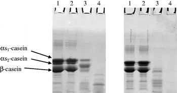
The ability of Lb. fermentum IFO3956 supernatant to hydrolyze αS1-, αS2- and β-caseins was tested after growing the bacterial cells in MCA for 48 h at 37°C, then extracting the cells with different detergents and assessing the proteolytic activity of the supernatants, after incubation with sodium caseinate for 3 and 24 h at 37°C, by SDS-PAGE (Fig. 2). Proteolysis of casein fractions could be observed after 3 h incubation at 37°C with the proteases extracted by 1% Triton X-100 (Fig. 2, lane 3) and 1% Tween-80 (Fig. 2, lane 4) while no proteolysis could be detected with the proteases extracted with 1% SDS (Fig. 2, lane 2). The proteolysis increased with the time of incubation reaching a maximum after 24 h. The protease extracted with 1% Tween-80 was more active than that extracted with 1% Triton X-100 either after 3 or 24 h incubation at 37°C with the substrate. The proteases extracted in these conditions could hydrolyze all casein fractions albeit to different degrees. Consequently, the proteases of Lb. fermentum IFO3956 can be classified to PIII type according to Kunji et al. (Reference Kunji, Mierau, Hagting, Poolman and Konings1996), being able to cleave αS1-, β-, and κ-caseins. Based on the band colour density, αS1- and αS2-caseins were completely hydrolyzed (100%) while β-casein was only hydrolyzed to 90% after incubation of sodium caseinate at 37°C for 24 h with proteases extracted with 1% Triton X-100. However, the degree of hydrolysis of these two fractions was similar (100%) in case of the proteases extracted with 1% Tween 80, independently of the time of incubation (3 or 24 h). Similar results were observed by Tzvetkova et al. (Reference Tzvetkova, Dalgalarrondo, Danova, Iliev, Ivanova, Chobert and Haertlé2007) on 21 lactobacilli isolates gathered from three types of Balkan home-made yogurts. However, the cell-wall bound proteinase of Lb. delbrueckii subsp. lactis ACA-DC178 was found to hydrolyze mainly β-casein and to lower extent αS- and κ-caseins (Tsakalidou et al. Reference Tsakalidou, Anastasiou, Vandenberghe, van Beeumen and Kalantzopoulos1999). Consequently it was classified as PI-type proteinase.
SDS-PAGE profile of Na-caseinate hydrolysate (3 and 24 h incubation at 37°C) by Lactobacillus fermentum IFO3956 supernatants obtained by treating bacterial cells with different detergents: 1% SDS (lane 2); 1% Triton X-100 (lane 3); and 1% Tween 80 (lane 4) as compared to non-hydrolyzed Na-caseinate (lane 1).

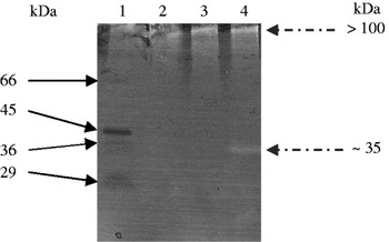
Cell-free supernatants of Lb. fermentum IFO3956 showed two clear bands (corresponding to 35 and >100 kDa) on gelatin-zymogram (Fig. 3). Similar band pattern was obtained using casein instead of gelatin as a substrate in the zymogram (data not shown). These results indicate the presence of at least two types of proteolytic enzymes of molecular masses 35 and >100 kDa. The low molecular band (35 kDa) was obtained only with 1% Tween-80 extraction (Fig. 3, lane 4). The high molecular mass band (>100 kDa) was seen in case of the extracts of the protease with either 1% Triton X-100 or 1% Tween-80 (Fig. 3, lanes 3 & 4, respectively) but not with 1% SDS extraction (Fig. 3, lane 2). The proteinase inhibition by SDS was reported by Wang et al. (Reference Wang, Huang, Wang, Liang, Yen and Sakata2008), Mohamed et al. (Reference Mohamed, Morishima, Babiker and Mori2009), and Tanskul et al. (Reference Tanskul, Hiraga, Takada, Rungratchote, Suntinanalert and Oda2009). Hence, it is possible that the band of high molecular mass may be ascribed to the mature oligomeric form of cell-wall bound proteinase while the band of low molecular mass may be its degradation product. It has been reported that the mature proteinase of Lactococcus lactis subsp. cremoris Wg2 has a molecular mass of 180–190 kDa although breakdown products of smaller sizes could be detected upon isolation (Laan & Konings, Reference Laan and Konings1989). It has been reported also that cell-envelope proteinases of LAB are synthesized as pre-proteins of approximately 2000 amino acid residues (Fernandez Espla et al. Reference Fernandez Espla, Garoult, Monnet and Rul2000).
Gelatin SDS-PAGE zymogram patterns of Lactobacillus fermentum IFO3956 supernatants obtained after cultivation for 48 h at 37°C on MCA and subsequent incubation for 24 h at 37°C with different detergents [1% SDS (lane 2); 1% Triton X-100 (lane 3) and 1% Tween 80 (lane 4)]. Lane 1 is molecular weight markers.

Characterization of α S1-casein hydrolysates
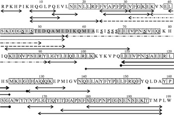
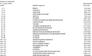
LC/MS/MS Mass Spectrometry. Figure 4 shows the sequence of bovine αS1-casein (199 amino acids). This primary structure contains as reported by Chatchatee et al. (Reference Chatchatee, Järvinen, Bardina, Beyer and Sampson2001), 8 epitopes defined as 5 major plus 3 minor IgE-binding epitopes. The major epitopes are the following peptide fragments: N17-K36, K83-K102, L109-L120, N139-F153 and Y159-T194. The minor epitopes are: E39-S48, E69-Q78 and M123-K132. These epitopes are marked on Fig. 4 by bordered boxes. Similar results were reported by Wang et al. (Reference Wang, Lin, Bardina, Goldis, Nowak-Węgrzyn, Shreffler and Sampson2010) stating that the major epitopes are the peptides F28-I44, Q78-L95, L109-S122 and A163-F179. Cerecedo et al. (Reference Cerecedo, Zamora, Shreffler, Lin, Bardina, Dieguez, Wang, Muriel, Hoz and Sampson2008) observed using peptide microarray, a single region differentially recognized by the reactive IgE group (peptide F28-E50). The reported new epitopes were already observed by Chatchatee et al. (Reference Chatchatee, Järvinen, Bardina, Beyer and Sampson2001) with slight differences so they are not indicated on the diagram. However, a new epitope reported by Lin et al. (Reference Lin, Bardina, Shreffler, Andreae, Ge, Wang, Burni, Fu, Han and Sampson2009) is different hence, it is marked on the diagram by shadowed box. Mass spectrometry analyses of hydrolysate of αS1-casein obtained after the incubation with Lb. fermentum IFO3956 for 24 h at 37°C revealed the presence of 24 peptides of different sizes, ranging from 5 to 36 amino acids (634·2662 to 1520·5089 Da) and included 9 phosphorylated peptides (F24–E55, V31–E50, K36–E55, L40–E55, K42–I71, D56–Y91, L101–S122, L109–S122 and L109–G126) (Table 1, Fig. 4). Phosphorylated peptides were shown to be much more resistant to hydrolysis by peptidases from Lb. fermentum IFO3956 than other peptides, what remains in accordance with what was observed by Deutsch et al. (Reference Deutsch, Mollé, Gagnaire, Piot, Atlan and Lortal2000) using the three LAB Lb. helveticus, Lb. delbrueckii and Streptococcus thermophilus. There was only one epitope (L109-L120) that resisted hydrolysis, which is characterized by the presence of one phosphoseryl residue located nearly in the centre and in a highly hydrophobic domain since 6 out of 12 residues are hydrophobic amino acids (2 L, 1 I, 1 V, 1 P, 1 A). Although this epitope contains 3 glutamyl residues, which are preferably hydrolyzed by this lactobacilli protease, the hydrophobic nature and the presence of phosphorylated serine hindered the enzymatic action. On the other hand, there were two phosphorylated epitopes subject to the hydrolytic action of Lb. fermentum IFO3956 (E39-K58 and E69-Q78) but their cleavage site is far away from the phosphorylated serines.
αS1-Casein sequence and peptides released by the proteolytic action of Lactobacilli fermentum IFO3956 after 24 h incubation at 37 °C, as indicated by arrows. The peptides marked by borders show the main 8 epitopes reported originally by Chatchatee et al. (Reference Chatchatee, Järvinen, Bardina, Beyer and Sampson2001) and the peptide marked by a shadow refers to a new epitope reported by Lin et al. (Reference Lin, Bardina, Shreffler, Andreae, Ge, Wang, Burni, Fu, Han and Sampson2009). The sequence uses the one-letter amino acid code and the phosphorylated serines are indicated by S.

Characterization of the 24 peptides contained in hydrolysate of αS1-casein by Lactobacillus fermentum IFO3956

It can also be observed that the proteolytic cleavage occurred preferentially at glutamic acid (E, 9 cleavages), leucine (L, 8 cleavages), proline (P, 4 cleavages). Similar results were obtained by Hébert et al. (Reference Hébert, Mamone, Picariello, Raya, Savoy, Ferranti and Addeo2008). They characterized the pattern of αS1- and β-casein breakdown and release of bioactive peptide by cell envelope proteinase from Lb. delbrueckii subsp. lactis CRL 581 and determined that this strain prefers glutamic acid (E) and glutamine (Q) from αS1-casein. The preference to cleave proline exclusively from the C-side is typical of prolyl endopeptidases, which are serine proteases widely distributed in bacteria, which can be used also to hydrolyze proline-rich prolamines (Shan et al. Reference Shan, Mart, Sollid, Gary and Khosla2004). On the other hand, it was previously reported that cell-envelope proteinases from LAB have a strong preference for hydrophobic caseins (Swaisgood, Reference Swaisgood and Fox1982). Majority of identified peptides originated from the N-terminal portion (15 peptides) and smaller number (9 peptides) from the C-terminal end of αS1-casein (Table 1 & Fig. 4).
Competitive ELISA
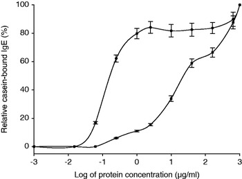
Data in Fig. 5 show the binding of intact αS1-casein or its Lb. fermentum IFO3956 hydrolysate to the IgE in sera pool of 18 CMA patients. It is observed that the binding of αS1-casein hydrolysate to IgE was considerably weaker from binding with intact αS1-casein. The C50% of intact αS1-casein was about 0·17 μg/ml while what was observed for the αS1-casein hydrolysate was as high as 24·8 μg/ml. This means that the binding of the hydrolyzed protein is about 150 times lower than that of the intact protein. This weaker binding of IgE could lead to weaker allergic reactions. Consequently, the hydrolysis of αS1-casein by Lb. fermentum IFO3956 would attenuate allergic reactions. It can be observed in Fig. 5 that for concentrations of αS1-casein equal to or higher than 1·0 μg/ml the maximum value of binding of IgE to over 80% is obtained while the binding of 1·0 μg/ml hydrolysate of αS1-casein is less than 10%. The high binding activity of αS1-casein to IgE is evidently due to the presence of numerous specific epitopes defined or still unknown. The diminution of the IgE binding of the hydrolysates of αS1-casein is due to the breakdown of some of the known epitopes of αS1-casein as seen in Fig. 5. The persistence of weak binding of the αS1-casein hydrolysates may be due to the resistance of some epitopes to the proteolysis, which still bind to IgE such as the epitope LEIVPNSVEQ (109–120). All other epitopes are cleaved by the proteolytic activity of Lb. fermentum IFO3956 at more than one site except two epitopes (E69-Q78 and N139-F153), which were cleaved at only one site. Some other unidentified epitopes may be also binding with IgE.
Binding of αS1-casein (●) and its Lactobacillus fermentum hydrolysate (▪) to IgE in the sera pool of CMA patients. Averages are indicated by horizontal black bar.

Analysis of the peptides during αS1-casein hydrolysis by Lb. fermentum IFO3956 showed the ability of the proteases of this strain to hydrolyze many allergic-epitopes (e.g. cleavage of peptide bonds F23-F24, E30-V31, E39-L40, S41-K42, E55-D56, I71-V72, Y91-L92, L98-L99, G126-I127, E141-L142, 162 and F179-S180) of αS1-casein. Consequently, the immuno-reactivity of milk protein may be considerably reduced by hydrolyzing αS1-casein with Lb. fermentum IFO3956.
Similar results were found by Tzvetkova et al. (Reference Tzvetkova, Dalgalarrondo, Danova, Iliev, Ivanova, Chobert and Haertlé2007) who studied the hydrolysis of major dairy proteins by LAB from traditional Bulgarian yogurts. It was shown that during fermentation process, milk proteins are acidified by the production of lactic acid and are hydrolyzed by proteases and peptidases from bacteria. This proteolysis is followed by a reduction of the number of epitopes and consequently by a decrease in allergenicity of hydrolyzed protein.
In conclusion, the studied strain Lb. fermentum IFO3956 displays proteolytic activity, which can be useful for the formulation of diversified novel dairy products or to improve health properties of traditional products. It can also be used for the hydrolysis of proline-rich proteins normally resisting hydrolysis like cereal prolamines. Depending on the reduced recognition and binding by IgE of αS1-casein hydrolyzed by Lb. fermentum IFO3956, this strain may be used for the production of milk and possibly milk products with reduced αS1-casein immuno-reactivity.
We would like to express our gratitude for the support of this work in the frame of project # 982164 “Study of antimicrobial and hypoallergenic products of lactic acid bacteria” to NATO ‘Science for Peace’ program, and for the doctoral fellowship of Shady El-Ghaish to the Egyptian Ministry of Foreign Affairs and Education.