Introduction
In recent years, the morphology, corresponding motion and adhesion mechanism of a wide variety of parasites have attracted interest among researchers in various fields (Taraschewski, Reference Taraschewski, Weiss and Tanowitz2000; Mostaert et al., Reference Mostaert, Crockett, Kearn, Cherny, Gazit, Serpell and Jarvis2009). In general, intestinal parasites maintain their position either by active movement within the mucosal layer (e.g. pinworm), attachment to the intestinal surface using ‘adhesion pads’, structures acting as a wedge or anchor (whipworm), rigid hooks (thorny-headed worms and many tapeworms), or clamp-like structures formed either by the stoma (several groups of nematodes), muscular pads configured as round cup-shaped suckers (tapeworms and trematodes) or elongated pads with grooves (cestodes of the family Tetraphyllidea). While tapeworms (Cestoda) can utilize a variety of attachment methods (Merchant et al., Reference Merchant, Aguilar, Avila, Robert, Flisser and Willms1998; Scholz et al., Reference Scholz, Drábek and Hanzelová1998), species in the order Cyclophyllidea use either suckers, rostellar hooks or a combination of both (Scholz et al., Reference Scholz, Drábek and Hanzelová1998; Cunningham & Olson, Reference Cunningham and Olson2010) to attach the scolex to the intestinal wall. Attachment solely by suckers allows parasites to move along the gastrointestinal (GI) tract freely, with re-attachment causing minimal or no apparent tissue trauma. The various attachment methods also provide vivid examples for researchers who are dedicated to developing actively propelled gastrointestinal medical devices for application in vivo. Medical robots, inspired by worm-like parasites, have been proposed as devices that could move successfully through the intestine (Kim et al., Reference Kim, Park, Jee and Yoon2005, Reference Kim, Laschi and Trimmer2013). While successful in vivo movement has been realized by several medical devices, there is still no perfect solution for long-term attachment of those devices (Quaglia et al., Reference Quaglia, Tognarelli, Sinibaldi, Funaro, Dario and Menciassi2013). If the long-term attachment of biosensors or biomedical devices could be achieved, physicians could continuously monitor a patient's physiological indices (such as core body temperature, pH and pressure in the GI tract) and make more accurate diagnoses. In addition, long-term attachment of these devices could enable novel approaches for slow-release and (or) time-variable drug delivery, or even the ability to manipulate nearby tissue to perform microsurgery (Valdastri et al., Reference Valdastri, Simi and Webster2012).
Artificial hooks inspired by those that occur on the rostellum of the scolex of Taenia solium have shown a remarkable ability to adhere to biological substrates (La Spina et al., Reference La Spina, Stefanini, Menciassi and Dario2005). An attachment mechanism for swallowable robotic capsules using needles to successfully fix sensors to a pig's GI tract for 6 days was developed in our previous work (Xie et al., Reference Xie, Kothari and Terry2015). Despite implementing the attachment mechanism from small-diameter needles, these hooks still penetrate into the mucosa and may cause minor tissue damage and subsequent inflammation.
Certain species of tapeworms can attach to the GI tract of vertebrates for an extended time period without creating any noticeable damage to the host, due to their ‘non-penetrative’ attachment mechanism and the relatively small geometry of the scolex. This type of attachment raises some research questions. How does the very small scolex support and sustain the whole body against forces from peristalsis and liquid flow of the GI tract? What is the exact attachment strength of the parasite? To the authors’ knowledge, these questions have not been answered in the literature. To better understand the attachment process and replicate the structure of the attachment mechanism, a method to measure the attachment strength of a parasite was designed, and the attachment strength of a series of individuals of Hymenolepis diminuta was measured.
Hymenolepis diminuta, also known as the rat tapeworm, unlike many other closely related species, including Vampirolepis nana and V. microstoma, uses only four suckers to clamp on to the mucosal villi of the intestine to maintain its position in the gut. In addition, the scolex of individuals of H. diminuta parasitizing rats regularly detaches and re-attaches to new sites in the intestine (Read & Kilejian, Reference Read and Kilejian1969). In the permissive host, while some inflammatory response has been observed (Dwinell et al., Reference Dwinell, Wise, Bass and Oaks1998), no localized tissue damage occurs.
Materials and methods
Beetles (Tenebrio molitor) infected with larvae of H. diminuta were purchased from Carolina Biological Supply (Burlington, North Carolina, USA) and the cysticercoids were fed to 12 laboratory rats (strain BN/Crl, Charles River Laboratories International, Inc., Wilmington, Massachusetts, USA). Due to the exploratory nature of the investigation and the lack of published protocols, the 12 rats were divided into three groups and infected with a varying number of cysticercoids. Individuals in group one were infected with 2–3 cysticercoids; group two with 4–6 cysticercoids and group three with 7–12 cysticercoids. Three weeks after infection, faecal pellets were checked for the presence of tapeworm eggs, and the second phase of the experiment started. Individual rats were euthanized following standard Institutional Animal Care and Use Committee (IACUC) recommendations: carbon dioxide exposure until breathing stopped, followed by cervical dislocation. The use of a general anaesthetic agent (isoflurane) was avoided deliberately, to prevent interference with tapeworm attachment. Immediately after the rats were euthanized, the small intestine was removed and placed in Hanks’ saline solution at room temperature to prevent immediate tissue degradation. Starting from the duodenum, the small intestine was opened in small segments, until an attached tapeworm was found. Intestinal tissue surrounding the attachment site (approximately 10 mm on each side) was exposed and removed. Part of the intestinal tissue along with the strobilated body of the tapeworm (usually 50–100 mm) was moved to a smaller Petri dish containing Hanks’ saline solution.
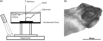
The basic idea to test the attachment strength of H. diminuta to the small intestine of the rat was to pull the tapeworm vertically when it was attached to intestinal tissue placed horizontally in a Petri dish and to measure the force required to detach the tapeworm. In this work, a tensile test machine (ADMET eXpert 5601, ADMET Inc., Norwood, Massachusetts, USA) and a digital scale (Denver Instrument XE-50, Bohemia, New York, USA) were used to measure the attachment force of H. diminuta. The accuracy of the load cell on the tensile test machine is 0.01 N and, based on the authors’ first experiment, it exceeds the range of the attachment strength of tapeworms. To solve this problem, a digital scale with a resolution of 0.0001 g (10−6 N) was used to calculate the exact attachment strength (see fig. 1a for the experimental set-up).
(a) Schematic arrangement for testing the attachment strength of Hymenolepis diminuta in the rat intestine using a tensile mechanism and digital scale. (b) Microscopy image of the scolex of H. diminuta used in the test, showing three muscular suckers with an average diameter of 86.7 μm.

The Petri dish containing the intestinal tissue and the attached tapeworm was placed on a pre-zeroed digital scale at the base of the tensile test machine. The strobilate part of the tapeworm was looped on a hook that was mounted on the tensile test machine's grip. Preliminary tests also showed that the attachment force of this worm was small compared to the weight of the tissue and the liquid surface tension, so there was no need to immobilize the intestinal tissue. The initial value of the scale was recorded as the starting point, and the tapeworm was pulled away from the tissue at a rate of 1 mm/s. A digital camera recorded the entire process and tracked all scale readings (fig. 1a).
As the tapeworm was pulled, the reading on the scale decreased and achieved a minimum value right before the tapeworm detached from the tissue, yielding the attachment strength as calculated in equation (1):
 $$\eqalign{& \hbox{Attachment strength} = \cr & \hbox{Initial scale reading} - \hbox{Minimum scale reading}.}$$
Results and discussion
Rats that were exposed to a low number of cysticercoids were inadequate subjects for the intended experiment. Two out of the four rats that received 2–3 cysticercoids did not become infected at the first attempt and needed to be re-exposed to larval tapeworms. Tapeworms in low-intensity infections (1–3 tapeworms/host) showed a different distribution and behavioural pattern than tapeworms in a more crowded (5–12 individuals/host) environment. When there were only a few tapeworms in the host, they tended to be attached to the upper part of the small intestine (approximately 5–15 mm from the pyloric sphincter), and scoleces detached at the slightest disturbance or touch during necropsy. Tapeworm scoleces in a crowded environment were more evenly distributed within the small intestine, and they were less likely to detach during the preparation phase.
Twelve tapeworms from four rats tested successfully for attachment force. Measurements varied between 0.008 g (min.) and 0.04 g (max.). The average attachment strengths of tapeworms in each rat were 0.027 g (SD = 0.009 g), 0.016 g (SD = 0.004 g), 0.014 g (SD = 0.014 g) and 0.018 g (SD = 0.001 g), respectively. The measured strengths are close among replicates and no significant differences existed among replicates from individual rats. The combined average attachment strength of H. diminuta was 0.021 g (SD = 0.011 g). The wet weight range of the worms was also measured (0.596–1.427 g), but these values only provide a minimum estimate for the full weight, since the full extraction of a single tapeworm was not feasible while focusing on the main goal of the experiment. However, in general, more competition and crowding (greater number of tapeworms per rat host) decreases tapeworm size, both in length and weight (Read, Reference Read1951). Several H. diminuta specimens extracted from the rats were relaxed in distilled water, fixed in buffered formaldehyde solution, stained and mounted on microscope slides for morphological examination. The average measurement of the suckers was 115.92 μm (±1.0782 μm) in length and 92.49 μm (±14.239 μm) in width. While the shape of suckers was slightly ovoid (fig. 1b), a circle was used for calculations by setting the diameter (D) of the circle to the average of the length and width of the ovoid. This diameter was 104.2 μm. The sucker was assumed to be hemispherical in shape, so the local force exerted by the suckers on the intestinal tissue could be calculated by the average attachment strength (F) divided by the area of the sucker.
 $$\eqalign{\hbox{Suction pressure} &= \hbox{Attachment strength}/\left({\hbox{Area of four suckers}}\right) \cr & = F{/}\left[{4 \times \pi \times \left({D/2}\right)^2}\right].}$$
The average attachment pressure of H. diminuta was calculated to be 6.145 kPa. The risk of bowel perforation with the use of suction drains with a vacuum pressure of 8–24 kPa has been reported (Graham et al., Reference Graham, Coit and Brennan1993). Since the measurements were taken between the outside margins of suckers on relaxed specimens, the estimate, 6.145 kPa, is a lower estimate of the actual pressure on the host tissue. Still, the present result indicates that the suckers’ working pressure is at or below tissue damage tolerance.
Hymenolepis diminuta (Cyclophyllidea, Hymenolepididae) is a parasitic tapeworm of rats (Rattus). On rare occasions, it can infect humans (Edelman et al., Reference Edelman, Spingarn, Nauenberg and Gregory1965), but its main life cycle involves insects as intermediate and rats as definitive hosts. This tapeworm species is often used as a model organism in scientific research focusing on tapeworm biology, due to the simplicity and safety in maintaining infected hosts. A unique feature of the genus is the lack of hooks on the scolex, and a much reduced rostellum, called an apical organ. This morphological characteristic is in contrast to those of related species, such as H. microstoma, a synonym of Vampirolepis microstoma (see Schmidt, Reference Schmidt1986), that has both a rostellum and rostellar hooks on the anterior end of the scolex. Vampirolepis microstoma, with rostellar hooks, appears to have a permanent attachment site in the host and the fully developed tapeworm does not re-locate in the intestinal tract depending on food availability (Cooreman & De Rycke, Reference Cooreman and De Rycke1972). The hooks on the rostellum are firmly embedded into the secretory ‘crypts of Lieberkühn’ at the base of the villi (Smyth, Reference Smyth1969).
The research interest of this paper focused on understanding the physical interaction between parasite attachment and host tissue. Interest in this area of investigation was driven by biomedical research and the need to place medical sensors in the intestinal tract and to investigate attachment strength in a biological system that does not use hooks. As an example of such a biological system, tapeworms in the intestine have to counteract forces created by drag as digested food moves along the intestinal tract.
The main finding is that attachment force of the tapeworm to the intestinal wall is very weak. The average attachment strength of H. diminuta was measured as 0.021 g with SD ± 0.011 g. This value is only a small portion of the potential weight of an adult tapeworm. If we consider that the tapeworm is neutrally buoyant and only fluid drag is considered as the main force, scolex attachment strength alone does not explain how this species remains fixed in the intestine. Hymenolepis diminuta has four kinds of muscles in its proglottids: longitudinal, circular, transverse and radial bundles (Lumsden & Byram, Reference Lumsden and Byram1967). These muscles create a peristaltic movement that can counteract bowel movement. In this regard, the attached scolex provides a fixed starting point. In addition, the peristaltic movement of tapeworm proglottids is independent of movement shown by the scolex, and the movement is controlled by local mechanical and chemical stimuli acting on neighbouring proglottids (Smyth, Reference Smyth1969). The mechanism that H. diminuta uses to maintain position in its host is very different from that of other intestinal parasites. The suckers, which act more like circular clamps than suction pads, grab on to the intestinal villi. Because of normal growth and digestive processes, cells in the intestinal villi are constantly being replaced and a villus can be lost and regrown rapidly following damage. For this reason, the tapeworm must have the ability to re-attach and maintain position while the scolex becomes disconnected from the intestinal wall. The ability of H. diminuta to detach, move and re-attach enables the parasite to remain in the host for a very long time. Longevity of infections is limited only by the host's lifespan (Read, Reference Read1967). Other parasites that lack the peristaltic movement, such as Moniliformis dubius (Acanthocephala), utilize hooks on the proboscis to embed and attach to the intestinal wall permanently and firmly. In contrast with the rat tapeworm, M. dubius has a shorter lifespan, measured in weeks rather than years (Crompton & Walters, Reference Crompton and Walters1972), and detached specimens are expelled from the intestinal tract.
Our estimates of the force and pressure exerted by the parasite suckers on host tissue are close to other estimates of tissue damage tolerance (Graham et al., Reference Graham, Coit and Brennan1993). A question that the experiment could not address is whether one or more suckers were clamping on the intestinal tissue. Previous studies on tapeworms fixed in situ for examination by microscopy indicate that often two suckers are clamping on the intestinal tissue (Smyth, Reference Smyth1969; Merchant et al., Reference Merchant, Aguilar, Avila, Robert, Flisser and Willms1998). If only two suckers are involved in the attachment, the calculation of the pressure on the host tissue is underestimated by a factor of two and our estimates may be slightly above the minimum tissue damage tolerance level (8 kPa).
The significance of this research is that it could be used as a guide for biomimicry researchers to design and develop an in-vivo tissue-contactable bio-robot. The strength of parasites with hooks and their attachment behaviour will be investigated in future research.
Acknowledgements
We thank S. Elizabeth Racz for helping with the study and sharing H. diminuta scolex measurements.
Financial support
This work was supported financially by Terry's University of Nebraska new faculty start-up funds, a Nebraska Research Initiative grant and the Harold W. Manter Laboratory of Parasitology. Funds were also provided partly by National Science Foundation grant DBI-1458139.
Conflict of interest
None.
Ethical standards
The authors assert that all procedures contributing to this work comply with the ethical standards of the relevant national and institutional guides on the care and use of laboratory animals. The study was approved by the University of Nebraska's (UNL's) Institutional Animal Care and Use Committee (IACUC, No. 1071).