


Statement of Research Significance
Research Question(s) or Topic(s): What are the cognitive outcomes following neurosurgical resection of frontal lobe tumors, and how can quantitative synthesis of existing evidence inform clinical management to optimize patients’ postoperative quality of life? Main Findings: Cognitive performance did not significantly change after neurosurgery. The literature shows methodological limitations, including heterogeneous assessments, variable follow-up, inconsistent reporting of baseline cognitive functioning, and small sample sizes. Study Contributions: We highlight the need for standardized and comprehensive cognitive assessments, adequately powered study cohorts, and long-term follow-up to more accurately characterize cognitive outcomes after neurosurgery, including effects involving “non-eloquent” frontal regions. We also emphasize the importance of a multidisciplinary, harmonized approach to advance understanding of post-neurosurgical cognitive effects and to enhance clinical decision-making and patient care.
Introduction
The anterior regions of the human brain play a crucial role in orchestrating complex behaviors and high-order cognitive processes. The intricate structure of the frontal lobes is reflected in a very entangled functional organization, which still remains a key area of contemporary neuroscience research (Kovach et al., Reference Kovach, Daw, Rudrauf, Tranel, O’Doherty and Adolphs2012). The structural integrity of the frontal lobes has been consistently associated with the processing of complex cognitive functions, traditionally referred to as “executive functions” (Stuss, Reference Stuss2011; Teuber, Reference Teuber1972). These functions encompass a set of higher-order processes that regulate goal-directed behavior, including planning, cognitive flexibility, inhibitory control, and monitoring of action outcomes. Executive functions are a multidimensional system of cognitive and behavioral competencies that support adaptive behavior, including both cold (i.e., cognitive) and hot (i.e., affective and social) components mediated by specific prefrontal networks (Chan et al., Reference Chan, Shum, Toulopoulou and Chen2008). Moreover, the frontal lobes are involved in attention (Nakai et al., Reference Nakai, Kato and Matsuo2005), memory (Okuda et al., Reference Okuda, Fujii, Ohtake, Tsukiura, Yamadori, Frith and Burgess2007; Ranganath et al., Reference Ranganath, Johnson and D’Esposito2003), and in some aspects of language processing (Catena Baudo et al., Reference Catena Baudo, Villamil, Paolinelli, Domenech, Cervio, Ferrara and Bendersky2023; Whitney et al., Reference Whitney, Kirk, O’Sullivan, Lambon Ralph and Jefferies2011).
Within the prefrontal cortex, several sub-regions project to and receive connections from other cortical and subcortical structures, thereby providing the neuronal substrates for integrative and goal-directed cognition. The dorsolateral prefrontal cortex is essential for working memory, problem solving, and cognitive control (Fuster, Reference Fuster1990; Goldman-Rakic, Reference Goldman-Rakic1991), as well as for error monitoring and conflict resolution (Boschin et al., Reference Boschin, Brkic, Simons and Buckley2017; Masina et al., Reference Masina, Tarantino, Vallesi and Mapelli2019). The mesial and orbitofrontal prefrontal cortex contribute to the regulation of different processes, such as emotions, decision-making, social behavior, and inhibition (Bechara, Reference Bechara2004; Jones & Graff-Radford, Reference Jones and Graff-Radford2021; Klein-Flügge et al., Reference Klein-Flügge, Bongioanni and Rushworth2022). These functional subdivisions underline the complexity of the prefrontal cortex and its relevance to our daily life functioning.
Neurosurgical resection of frontal lobe tumors presents unique challenges, particularly when lesions involve the so-called “non-eloquent” areas, which are not traditionally associated with primary neurological functions such as language processing or movement (McGirt et al., Reference McGirt, Mukherjee, Chaichana, Than, Weingart and Quinones-Hinojosa2009). Nevertheless, resections affecting the so-called “non-eloquent” frontal regions, such as the right prefrontal cortex, can markedly affect patients’ postoperative functioning, because these areas are essential for higher-order cognitive processes, such as those involved in executive functions.
This challenge becomes even more important when we consider that extensive removal of low-grade gliomas significantly increases overall survival. This has been highlighted by the results of recent studies measuring the extent of resection through T2/Fluid-Attenuated Inversion Recovery (FLAIR)-weighted MRI (Smith et al., Reference Smith, Chang, Lamborn, Chang, Prados, Cha, Tihan, VandenBerg, McDermott and Berger2008; Xia et al., Reference Xia, Fang, Chen and Sun2018). FLAIR-weighted imaging is particularly sensitive in detecting infiltrative tumor components and peritumoral abnormalities, because it suppresses cerebrospinal fluid signals and enhances the visibility of pathological hyperintensities. For this reason, FLAIR is especially useful for identifying lesions’ margins, edema, and diffuse infiltrations (Wen et al., Reference Wen, Macdonald, Reardon, Cloughesy, Sorensen, Galanis, DeGroot, Wick, Gilbert, Lassman, Tsien, Mikkelsen, Wong, Chamberlain, Stupp, Lamborn, Vogelbaum, van den Bent and Chang2010). Moreover, it plays a crucial role in differentiating tumor tissue from other lesions and providing critical information for neurosurgical planning and postoperative assessment.
In this context, standard neuroradiological protocols typically include multimodal MRI acquisitions, combining 3D T1-weighted imaging (pre- and post-contrast), T2-weighted and FLAIR sequences, and, when available, advanced techniques such as diffusion-weighted imaging, diffusion tensor imaging, and perfusion-weighted imaging (PWI; Ellingson et al., Reference Ellingson, Bendszus, Boxerman, Barboriak, Erickson, Smits, Nelson, Gerstner, Alexander, Goldmacher, Wick, Vogelbaum, Weller, Galanis, Kalpathy-Cramer, Shankar, Jacobs, Pope and Yang2015). These MRI acquisitions allow a precise characterization of tumor extent, infiltration patterns, and relations with white matter tracts, thereby supporting a more effective and safe neurosurgical approach.
Other recent findings have suggested that intraoperative mapping should be extended beyond language and motor functions to include a broader range of cognitive domains, with the goal of supporting patients in resuming their normal familial, social, and professional activities (Duffau, Reference Duffau2018).
Despite the growing interest in the field, there is still a lack of systematic and quantitative investigations specifically addressing the effects of resection of frontal lobe tumors (Duffau, Reference Duffau2009). Although, in previous reviews and meta-analyses, cognitive outcomes in brain tumor patients have been explored (De Roeck et al., Reference De Roeck, Gillebert, Van Aert, Vanmeenen, Klein, Taphoorn, Gehring, Lambrecht and Sleurs2023; Ng et al., Reference Ng, See, Ang, Tan, Ang and King2019; Schei et al., Reference Schei, Solheim, Salvesen, Hansen and Sagberg2022), the picture that emerges is far from uniform. Some authors have reported significant postoperative cognitive decline, while others found only transient or no impairments (Hendi et al., Reference Hendi, Rahmani, Larijani, Ajam Zibadi, Raminfard, Shariat Moharari, Gerganov and Alimohamadi2022; Zangrossi et al., Reference Zangrossi, Silvestri, Bisio, Bertoldo, De Pellegrin, Vallesi, Della Puppa, D’Avella, Denaro, Scienza, Mondini, Semenza and Corbetta2022). Adding to this variability, in many investigations heterogeneous populations, tumors of different histological types (e.g., extra-axial tumors with intra-axial lesions), and heterogeneous tumor locations across the brain have been included (Kirkman et al., Reference Kirkman, Hunn, Thomas and Tolmie2022).
In the present systematic review and meta-analysis, we aimed to advance our current knowledge by synthesizing available evidence on cognitive outcomes following neurosurgical resection of frontal lobe tumors. Specifically, our objective was to provide a quantitative assessment of the cognitive effects of neurosurgery, thereby contributing to the understanding of the impact of neurosurgical intervention. Finally, we aimed to provide insights to improve clinical management, for maximizing both neuro-oncological efficacy and postoperative quality of life.
Method
Ethical approval was not requested because this systematic review and meta-analysis involves the collection and synthesis of archival data from previously published clinical trials in which informed consent had already been obtained by the original investigators, and all ethical aspects are considered adequately addressed.
Literature search
The present study was conducted according to the most recently updated guidelines of the Preferred Reporting Items for Systematic Reviews and Meta-Analyses - PRISMA (Page et al., Reference Page, McKenzie, Bossuyt, Boutron, Hoffmann, Mulrow, Shamseer, Tetzlaff, Akl, Brennan, Chou, Glanville, Grimshaw, Hróbjartsson, Lalu, Li, Loder, Mayo-Wilson, McDonald and Moher2020). A systematic literature search was conducted on three databases: PubMed, PsycINFO, and Embase. Search terms indicating “neoplasm” were combined with the terms “frontal” or “prefrontal.” For a comprehensive search strategy for all databases, please refer to the supplementary material. The supplementary material for this article can be found at [https://doi.org/10.17605/OSF.IO/X7V39]. The search was carried out on February 29th, 2024 and no restriction on the publication date range was applied.
The included studies met the following inclusion criteria: (i) the presence of a brain neoplasm; (ii) tumor localization within the frontal cortex; (iii) availability of neuropsychological assessments; (iv) availability of both pre- and post-neurosurgery neuropsychological assessments; (v) publication in a peer-reviewed English-language journal; and (vi) ethical approval by a medical ethics committee or institutional review board.
Studies were excluded if they met any of the following criteria: (i) the article was a review, case report, letter to the editor, or conference paper; (ii) neuropsychological assessment was conducted at only one time point (either pre- or post-neurosurgery); (iii) the full text was unavailable; (iv) the necessary statistical data were not provided (e.g., mean scores, standard deviations, or z-scores), for both pre- and post-neurosurgery neuropsychological tests.
Studies with sufficient information to calculate effect sizes directly, or for which data were available from the corresponding authors upon request, were also included in the meta-analysis.
The PRISMA flow chart for study selection is displayed in Figure 1. Articles were screened by three independent reviewers (EP, FM, KP), who used Rayyan (Ouzzani et al., Reference Ouzzani, Hammady, Fedorowicz and Elmagarmid2016). Any disagreements were resolved through discussion among the three reviewers, with each point being thoroughly debated until a mutual agreement was reached.
PRISMA flowchart illustrating the stages of the systematic review process.

Initially, 1,316 articles were identified through databases and registers (PubMed, PsycInfo, and Embase). After removing duplicates (n = 646), 670 records were screened. After title and abstract screening, 114 articles remained. Of these, 64 were excluded from the systematic review and 99 from the meta-analysis. Consequently, the final numbers of included articles were 50 for the systematic review and 15 for the meta-analysis. Further details on the reasons for exclusion are provided in Figure 1.
Quality assessment
To evaluate the methodological quality of the included studies, we employed tools developed by the Joanna Briggs Institute (JBI), selected according to the design of each study. For studies included in the meta-analysis, we used the JBI Checklist for Cohort Studies (Aromataris et al., Reference Aromataris, Lockwood, Porritt, Pilla and Jordan2024; Moola et al., Reference Moola, Munn, Tufunaru, Aromataris, Sears, Sfetc, Currie, Lisy, Qureshi, Mattis, Mu, Aromataris, Lockwood, Porritt, Pilla and Jordan2024) and the JBI Checklist for Case Series (Munn et al., Reference Munn, Barker, Moola, Tufanaru, Stern, McArthur, Stephenson and Aromataris2019). For studies included in the systematic review, we applied the same tools, and we additionally used the JBI Checklist for Case-Control Studies (Aromataris et al., Reference Aromataris, Lockwood, Porritt, Pilla and Jordan2024; Moola et al., Reference Moola, Munn, Tufunaru, Aromataris, Sears, Sfetc, Currie, Lisy, Qureshi, Mattis, Mu, Aromataris, Lockwood, Porritt, Pilla and Jordan2024).
Given the presence of single-group cohort studies within our dataset, we implemented slight adaptations to the original JBI Checklist for Cohort Studies to enable its application to these designs. All changes to the checklist items are transparently documented in each table at the following OSF link [https://doi.org/10.17605/OSF.IO/X7V39]. Quality assessments were performed independently by two reviewers (CS, AS). Any discrepancies in scoring or interpretation were discussed and resolved through consensus. No third-party arbitration was necessary. Inter-rater agreement prior to consensus was recorded but not used to calculate formal statistics, because our aim was to reach a unified judgement. The independent scores assigned by each reviewer and the final consensus scores are openly available on OSF [https://doi.org/10.17605/OSF.IO/X7V39].
Data extraction
We carried out data extraction by using a standardized template. The extracted data encompassed study characteristics (i.e., authors, year of publication, and country), patients’ characteristics (i.e., sample size, type of tumor and location, biological sex, age, tumor grade, type of neurosurgery), and neuropsychological assessment details (i.e., mean scores on each test, standard deviations, or z-scores at various time points).
Neuropsychological tests were categorized into four broad domains based on the DSM-5 definition of cognitive functions: Executive functions (which also included attention), Language, Learning and memory, and Visuo-spatial functions. Furthermore, screening tests such as the Mini Mental State Examination (Folstein et al., Reference Folstein, Folstein and McHugh2014) and the Montreal Cognitive Assessment (Nasreddine et al., Reference Nasreddine, Phillips, Bédirian, Charbonneau, Whitehead, Collin, Cummings and Chertkow2005) were classified under the domain “General cognition,” whereas the Wechsler Adult Intelligence Scale (WAIS; Wechsler, Reference Wechsler2012) was included under the domain “Intelligence.”
Statistical analysis
All quantitative analyses were conducted through R (R Development Core Team, version 4.1.2, R Core Team, 2023) with the metafor package (Viechtbauer, Reference Viechtbauer2010).
Effect size
The effect sizes were estimated from studies in which a single experimental design was used, where the same group of participants performed the same cognitive tasks/tests both before and after the neurosurgical intervention. This repeated-measures design (pre- and post-neurosurgery neuropsychological assessments) allowed us to compare cognitive changes within participants and to examine performance across different cognitive domains. The effect size was calculated as the standardized mean difference between pre- and post-neurosurgery neuropsychological assessments, using a paired-sample Cohen’s d (Cohen, Reference Cohen1969). For each comparison, the raw score standardization method (Morris & DeShon, Reference Morris and DeShon2002) was applied. This method involves dividing the mean difference by the pooled standard deviation of raw scores, rather than the standard deviation of the difference, to improve comparability of effect sizes metrics.
Sampling variances were computed through appropriate formulas based on the paired-sample designs. Because the correlations between pre- and post-neurosurgery assessments were not reported in the included studies, they could not be directly extracted. Nonetheless, in repeated-measures effect size calculations, the assumed pre-post correlation affects the estimated sampling variance. Therefore, following recommendations for meta-analyses involving repeated-measures designs (Borenstein et al., Reference Borenstein, Hedges, Higgins and Rothstein2009; Morris & DeShon, Reference Morris and DeShon2002), we conducted sensitivity analyses, recalculating the sampling variances by using three plausible pre-post correlation values: .5, .7, and .9. These values were selected to represent moderate, moderately high, and high correlations between repeated neuropsychological assessments. The main analyses reported in the manuscript were based on r = .7. Sampling variances computed with the other correlations (i.e., r = .5 and r = .9) were used to evaluate the robustness of our results. The sensitivity analyses showed that the choice of correlation value did not substantially affect the findings. The full sensitivity analyses are reported in the supplementary material (Table S2). The supplementary material for this article can be found at [https://doi.org/10.17605/OSF.IO/X7V39].
When multiple outcomes were reported, we combined the effect sizes by accounting for the correlation between the outcomes (Borenstein et al., Reference Borenstein, Hedges, Higgins and Rothstein2009). A range of plausible correlations was used to ensure robustness (i.e., .5, .7, .9), and the sensitivity analysis confirmed that these values had minimal impact on the overall results (Table S2, supplementary material). The supplementary material for this article can be found at [https://doi.org/10.17605/OSF.IO/X7V39]. As before, the reported results are based on a correlation of 0.7.
All effect sizes were adjusted for small sample bias using Hedges’ g correction factor, as implemented in the metafor package (Hedges, Reference Hedges1981).
Meta-analyses models
To evaluate the impact of neurosurgical interventions on cognitive performance, we conducted a meta-analysis of neuropsychological outcomes reported across studies that included both pre- and post-neurosurgery assessments. The analysis was performed in two stages.
In the first stage, a main multilevel random-effects model was conducted, pooling data from all studies and combining all cognitive domains into a single model. This allowed for an overall estimate of the cognitive effects of neurosurgery, thereby accounting for between-studies variability. Because, in several studies, multiple cognitive domains were considered as outcomes, a correlation of .5 was assumed between repeated cognitive measures. This assumption allowed different cognitive domains to be treated as correlated variables.
To account for the correlation between sampling errors across cognitive domains, a covariance matrix (V) was constructed based on the underlying data structure, by means of the vcalc function. This matrix (Figure S1, the supplementary material) was incorporated into the model, which was estimated through the rma.mv function. The supplementary material for this article can be found at [https://doi.org/10.17605/OSF.IO/X7V39]. In general, all models were fitted through restricted maximum likelihood estimation (REML). Beyond this model, which was based on the entire pool of studies, two meta-regressions were conducted to examine the influence of potential moderators. Specifically, whether the timing of post-intervention neuropsychological assessments and the mean age of patients affected the results.
In the second stage, subgroup meta-analyses were conducted for each cognitive domain, according to the classification reported in Table S1 of the supplementary material. The supplementary material for this article can be found at [https://doi.org/10.17605/OSF.IO/X7V39]. The cognitive domains included: (1) General cognition, evaluated using broad screening tools such as the Montreal Cognitive Assessment (Nasreddine et al., Reference Nasreddine, Phillips, Bédirian, Charbonneau, Whitehead, Collin, Cummings and Chertkow2005) and the Mini-Mental State Examination (Folstein et al., Reference Folstein, Folstein and McHugh2014); (2) Executive functions; (3) Language; (4) Learning and memory; and (5) Visuo-spatial functions. The domains of Praxis and Intelligence were excluded from the analysis because they were investigated in only two and one study, respectively. Overall, this domain-specific approach enabled us to examine potential differences in the impact of neurosurgery on distinct cognitive functions. To assess the robustness of the findings, and because some neuropsychological tests could plausibly be assigned to more than one cognitive domain, we repeated the subgroup analyses using a slightly modified cognitive-domain classification. The results based on the original classification are reported in Table S3 (supplementary material), whereas those based on the alternative classification are reported in Table S4 (supplementary material). The two sets of results were substantially consistent, supporting the robustness of the findings. The supplementary material for this article can be found at [https://doi.org/10.17605/OSF.IO/X7V39].
In addition, given that the brain tumors were located in frontal regions, we conducted an analysis focused specifically on executive-function components. Because executive functions represent a multidimensional construct (Stuss, Reference Stuss2011; Teuber, Reference Teuber1972), we selected only those cognitive measures that could be mapped onto the three-factor model proposed by Miyake et al. (Reference Miyake, Friedman, Emerson, Witzki, Howerter and Wager2000): inhibition, set shifting/flexibility, and updating/working memory. Measures that were executive-related but did not clearly isolate one of these components, such as processing speed or verbal fluency tasks, were excluded from this component-level analysis. Further details on the classification used for the executive-function component analysis are reported in Table S5 of the supplementary material. The supplementary material for this article can be found at [https://doi.org/10.17605/OSF.IO/X7V39].
We then fitted a multilevel random-effects model using the same nesting structure adopted in the main multilevel random-effects model. As described above, a covariance matrix was also incorporated into the model (Figure S2, supplementary material). The supplementary material for this article can be found at [https://doi.org/10.17605/OSF.IO/X7V39]. In this case, the covariance matrix was constructed to account for the correlation between sampling errors across executive-function components extracted from the same study contrast (i.e., inhibition, set shifting/flexibility, and updating/working memory). The model was estimated using REML. The executive component was also entered in a further model as a categorical moderator to test whether postoperative change differed across inhibition, set shifting/flexibility, and updating/working memory.
Heterogeneity was assessed through multiple methods, including the Q-test and the I 2 statistic. Through the Q-test, we evaluated whether the variability observed across studies was greater than what would have been expected by chance. In contrast, through the I 2 statistic, we quantified the proportion of the total variation, including both within-study and between-studies variability, that can be attributed to true heterogeneity rather than to sampling error. Note that a significant Q-test result combined with an I 2 value exceeding 75% is generally considered indicative of substantial heterogeneity (Higgins & Thompson, Reference Higgins and Thompson2002). Potential publication bias was explored through visual inspection of funnel plots (Sterne & Egger, Reference Sterne and Egger2005). All relevant data and R scripts are available on OSF [https://doi.org/10.17605/OSF.IO/X7V39].
Results
Literature search
A detailed overview of all the 50 studies that we included in the systematic review is provided in Table 1. For each study, the sample sizes, the tumor type and location (when specified), the timing of follow-up assessments, and the administered neuropsychological tests were reported. Of the 50 studies, 15 were included in the meta-analysis, whereas the remaining 35 were excluded because of insufficient data, such as missing outcome measures or incomplete reporting of necessary statistics. The contents of Table 1 allow for a clear comparison of study characteristics and facilitate the understanding of the methodological variability across the included studies.
Sample sizes, tumor location and type (when specified), time from neurosurgery to cognitive assessment, and cognitive tests used in the studies. For studies included in the meta-analysis that comprised more than one contrast (i.e., independent samples), the sample size and tumor location were specified for each contrast

Table 1. Long description
The table provides a detailed overview of 50 studies, focusing on sample sizes, tumor types, locations, time from neurosurgery to cognitive assessment, and cognitive tests used. The table has 50 rows and 10 columns. Column headers include Study ID, Sample size and tumor location, Tumor type, Time from neurosurgery to cognitive assessment, Cognitive tests, Meta-analysis assessment, and Notes. Row labels are study identifiers. Each row provides specific details for each study. For example, Row 1: Study ID 1, Sample size and tumor location 30 Right frontal lobe, Tumor type Diffuse astrocytoma, Time from neurosurgery to cognitive assessment 6 months, Cognitive tests RCF, TMT, WMS, Meta-analysis assessment No, Notes None. Row 2: Study ID 2, Sample size and tumor location 20 Right frontal lobe, Tumor type Oligodendroglioma, Time from neurosurgery to cognitive assessment 3 months, Cognitive tests RCF, TMT, WMS, Meta-analysis assessment No, Notes None. Row 3: Study ID 3, Sample size and tumor location 15 Left frontal lobe, Tumor type Glioblastoma, Time from neurosurgery to cognitive assessment 6 months, Cognitive tests RCF, TMT, WMS, Meta-analysis assessment Yes, Notes None. The table includes studies with various tumor types and locations, different times from neurosurgery to cognitive assessment, and a range of cognitive tests. The meta-analysis assessment column indicates whether each study was included in the meta-analysis. The notes column provides additional information if available.
Tests – SLTA: Standard Language Test of Aphasia; WAIS: Wechsler Adult Intelligence Scale; WMS: Wechsler Memory Scale; FAB: Frontal Assessment Battery; DO80: DO 80 Picture Naming; TMT-A: Trail Making Test – Part A; TMT-B: Trail Making Test – Part B; OCS: Oxford Cognitive Screen (Demeyere et al., Reference Demeyere, Riddoch, Slavkova, Bickerton and Humphreys2015); ENPA: Neuropsychological Exam of Aphasia (Capasso & Miceli, Reference Capasso and Miceli2001); RAVLT: Rey Auditory Verbal Learning Test; MFTC: Multiple Features Targets Cancellation; BADA: Batteria per l’Analisi dei Disturbi Afasici (Miceli et al., Reference Miceli, Laudanna, Burani and Capasso1994); BDI: Beck Depression Inventory; BAI: Beck Anxiety Inventory; WASI: Wechsler Abbreviated Scale of Intelligence; TOPF: Test of Premorbid Functioning (Wechsler, Reference Wechsler2011); NAB = Neuropsychological Assessment Battery; WCST = Wisconsin card sorting Test; BADS = Behavioral Assessment of the Dysexecutive Syndrome; AAT = Aachen aphasia test (Huber et al., Reference Huber, Poeck and Willmes1984); BNT = Benton naming test; CDT = Clock Drawing Test; STAI = State–Trait Anxiety Inventory; BDAE: Boston Diagnostic Aphasia Examination; BORB: Birmingham Object Recognition Battery; TAS: Toronto Alexithymia Scale; VOSP: Visual Object and Space Perception Battery; MoCA: Montreal Cognitive Assessment; MMSE: Mini-Mental State Examination (for all tests not otherwise specified, a complete description can be found in Lezak et al., Reference Lezak, Howieson, Bigler and Tranel2012).
Other abbreviations – LGG = low-grade glioma; HGG = high-grade glioma; RH = right hemisphere; LH = left hemisphere; BH = both hemispheres.
Quality assessment
Meta-analysis
The methodological quality of the 15 studies included in the meta-analysis was assessed through the JBI Checklist for Cohort Studies (n = 13) and the JBI Checklist for Case Series (n = 2). Among the cohort studies, 5 were rated as having good overall quality and 8 as fair. Of the 2 case series, one was rated as fair and the other one as poor. Albeit overall fair quality, the most common methodological limitations among cohort studies included insufficient reporting on eligibility criteria or low comparability of participants at baseline, inadequate control for potential confounders, and a lack of clearly described strategies for handling incomplete follow-up or missing data.
Case series were limited by poorly defined inclusion and exclusion criteria, non-consecutive or incomplete participant recruitment, and sparse reporting of clinical and/or demographic characteristics. Although some of these issues reflect inherent limitations of the specific design (Munn et al., Reference Munn, Barker, Moola, Tufanaru, Stern, McArthur, Stephenson and Aromataris2019), they nonetheless reduce the reliability and generalizability of the reported findings. The individual ratings provided by each reviewer, along with the inter-rater agreement table, are available at the following OSF link [https://doi.org/10.17605/OSF.IO/X7V39].
Systematic review
The methodological quality of the 35 studies included in the systematic review was assessed through the JBI critical appraisal tools, selected according to each study’s design: the JBI Checklist for Cohort Studies (n = 28), the JBI Checklist for Case Series (n = 6), and the JBI Checklist for Case-Control Studies (n = 1).
Among the cohort studies, 10 were rated as having good overall quality, whereas 18 were rated as having fair quality. Despite generally acceptable methodological standards, some recurring limitations were observed. These comprised incomplete or unclear reporting of inclusion criteria, insufficient control for confounding variables, incomplete follow-up and/or inadequate strategies to manage missing data.
Of the 6 case series, 4 were rated as fair and 2 as poor. Methodological concerns in this group involved poorly defined inclusion and exclusion criteria, non-consecutive or incomplete participant recruitment, and limited reporting of demographic and clinical characteristics. All these limitations hindered the interpretability and reproducibility of the findings. By contrast, the single case–control study (Mattavelli et al., Reference Mattavelli, Casarotti, Forgiarini, Riva, Bello and Papagno2012) received a fair rating, meeting five out of eight applicable items.
All studies were independently evaluated by two reviewers (AS, CS). Discrepancies in ratings were resolved through discussion and consensus. A detailed summary of individual and consensus ratings can be accessed at the following OSF link [https://doi.org/10.17605/OSF.IO/X7V39].
Overall neuropsychological effects of neurosurgery
In this model, results from all included studies (n = 15) and all cognitive domains were combined. When in a study was included more than one independent sample, each sample was treated as a separate contrast within that study. For example, in one of the included studies (Wang et al., Reference Wang, Guo, Wang, Liu, Chen, Zhai, Wang, Jiao, Zhao, Fan, Xue, Gao, Ji and Wang2023), two independent patient groups with different tumor locations were assessed. Consequently, this study contributed two distinct pre- versus post-neurosurgery contrasts to the analysis, resulting in one study with two contrasts in total. Overall, this model included 19 contrasts derived from the 15 included studies. In each of the 15 studies, multiple cognitive measurements were provided, resulting in a total of 59 observations across the entire pool of studies.
Without accounting for moderators, the overall mean weighted effect size was not significant [estimate: 0.0819, SE = 0.1249, z = 0.6558, p = .512, 95% CI: −0.1629 to 0.3267]. The Q-test indicated significant between-study heterogeneity [Q(df = 58) = 210.5424, p < .0001]. The I 2 statistic suggested moderate between-studies (59.61%) and low within-study (20.94%) variance. The asymmetry in the funnel plot suggested a potential publication bias in the literature (Figure 2).
Funnel plot showing an asymmetrical distribution of the effect sizes. The traced line in the middle of the funnel represents the overall effect size, the traced lines forming the sides of the funnel are the 95% confidence intervals.

Timing of post-intervention neuropsychological assessments as moderator
To examine whether the timing of post-intervention neuropsychological assessments influenced the outcomes, this moderator was included in the meta-regression. The test for moderators showed a nonsignificant result [QM(df = 1) = 0.2054, p = .6504]. No significant effect of this moderator was found [estimate: −0.0002, SE = 0.0005, z = −0.4533, p = .6504, 95% CI: −0.0011 to 0.0007], indicating that the timing of neuropsychological assessment did not significantly impact the overall effect size.
Despite the inclusion of this moderator, the test for residual heterogeneity remained significant [QE(df = 57) = 209.6828, p < .0001], indicating that a substantial amount of variability remained unexplained. The I 2 statistic revealed moderate heterogeneity because of between-studies variance (60.67%) and low heterogeneity because of within-study variance (20.96%). These results suggested that the timing of neuropsychological assessments did not meaningfully account for the heterogeneity observed across studies.
Mean age of patients as moderator
To assess whether the mean age of patients influenced the outcomes, this variable was included as a moderator in the meta-regression analysis. The moderator test yielded a nonsignificant result [QM(df = 1) = 0.2942, p = .5875], indicating that mean age did not have a statistically significant impact on the overall effect size. The overall mean weighted effect size was also nonsignificant [estimate: −0.0054, SE = 0.0100, z = −0.5424, p = .5875, 95% CI: −0.025–0.0141], suggesting that differences in the average age of patients, across studies, were not meaningfully associated with neuropsychological outcomes.
Even with the inclusion of age, residual heterogeneity remained significant [QE(df = 47) = 116.5078, p < .0001], indicating that a considerable amount of variability remained unexplained. The I2 statistic showed that approximately 35.62% of the heterogeneity could be attributed to between-studies variance, whereas within-cluster variance was negligible. Overall, these findings suggested that mean age did not substantially account for the heterogeneity in neuropsychological outcomes observed across studies.
Domain-specific cognitive effects of neurosurgery
General cognition
A multivariate meta-analysis model was fitted as a subgroup analysis including only studies where the domain of General cognition was investigated. The model was based on 8 contrasts nested within 6 studies. The overall mean weighted effect size was not statistically significant [estimate: 0.6274, SE = 0.4622, z = 1.3574, p = .1746, 95% CI: −0.2785–1.5333], indicating no clear evidence of a consistent difference in cognitive performance between pre- and post-intervention assessments, within the domain of General cognition (Figure 3).
Forest plots of change in the domain of General cognition after neurosurgical resection.

Executive functions
As a subgroup analysis, a multivariate meta-analytic model was conducted including only studies where the domain of Executive functions was examined. This model included 16 contrasts nested within 12 independent studies. The overall mean weighted effect size was small and it was not statistically significant [estimate: 0.0652, SE = 0.1488, z = 0.4378, p = .6615, 95% CI: −0.2265–0.3568], thereby suggesting no consistent evidence of a change in cognitive performance from pre- to post-intervention within the domain of Executive functions (Figure 4).
Forest plots of change in the domain of Executive functions after neurosurgical resection.

To further characterize the executive-function domain, we repeated the analysis by including only measures that could be classified as inhibition, set shifting/flexibility, or updating/working memory, in line with the three-factor model (Miyake et al., Reference Miyake, Friedman, Emerson, Witzki, Howerter and Wager2000). Across these component-specific executive measures, the overall weighted effect size was not statistically significant [estimate: −0.0708, SE = 0.0840, z = −0.8429, p = .3993, 95% CI: −0.2354–0.0938]. Heterogeneity remained significant [Q(df = 22) = 125.0164, p < .0001].
The executive component was also entered in a further model as a categorical moderator to test whether postoperative change differed across inhibition, set shifting/flexibility, and updating/working memory. The test of the moderator did not reach statistical significance [QM(df = 3) = 7.1222, p = .0681], indicating no evidence that postoperative change differed across executive-function components. Residual heterogeneity remained significant [QE(df = 20) = 119.6105, p < .0001].
Language
A multivariate meta-analysis model was fitted as a subgroup analysis including only studies in which the domain of Language was investigated. The model was based on 15 contrasts nested within 12 studies. The overall mean weighted effect size was not statistically significant [estimate: 0.0801, SE = 0.2403, z = 0.3335, p = .7388, 95% CI: −0.3909–0.5512], indicating no clear evidence of a consistent difference in cognitive performance between pre- and post-intervention assessments within the domain of Language (Figure 5).
Forest plots of change in the domain of Language after neurosurgical resection.

Learning and memory
A multivariate meta-analysis model was fitted as a subgroup analysis for studies in which the domain of Learning and memory was investigated (Figure 6). The model was based on 13 contrasts nested within 9 studies. The overall mean weighted effect size was not statistically significant [estimate: 0.1903, SE = 0.2504, z = 0.7599, p = .4473, 95% CI: −0.3005–0.6811], thereby indicating no clear evidence of a consistent difference in cognitive performance between pre- and post-intervention assessments within the domain of Learning and memory.
Forest plots of change in the domain of Learning and memory after neurosurgical resection.

Visuo-spatial functions
A multivariate meta-analysis model was applied as a subgroup analysis for studies, in which the focus was on the domain of Visuo-spatial functions. The model included 6 contrasts nested within 5 studies. The overall mean weighted effect size was not statistically significant [estimate: 0.5460, SE = 0.4026, z = 1.3563, p = .1750, 95% CI: −0.2430–1.3350], thereby suggesting no substantial evidence of a consistent difference in cognitive performance between pre- and post-intervention assessments within the domain of Visuo-spatial functions (Figure 7).
Forest plots of change in the domain of Visuo-spatial functions after neurosurgical resection.

Discussion
The complexity of tumor resection procedures depends on a wide range of factors, including the location of the lesions involved in neurosurgery. The most sensitive areas, known as “eloquent” regions, are located near the cortical and subcortical networks that allow language and movement. In contrast, tumor resection in the frontal lobes, in particular the right prefrontal cortex, is deemed relatively straightforward, as these regions are generally considered as more “expendable” (i.e., less functionally critical) compared to eloquent areas. As a result, the frontal lobes are frequently used as neurosurgical corridors to access deeper brain structures. This approach might lead neurosurgeons to adopt a less conservative approach, potentially involving the removal of tissue that contributes to important cognitive processes.
Nevertheless, the frontal lobes are deeply implicated in complex cognitive processes, which are less overt than speech or motor processes, but they are equally fundamental for daily functioning (e.g., the ability to work and interact normally with relatives/friends). Indeed, it is widely known that alterations in these domains may lead to difficulties in organizing goal-directed actions, disability to adapt to environmental changes, impaired inhibition of inappropriate responses, and deficit in maintaining and manipulating information. In addition, damage to orbitofrontal and medial prefrontal regions may result in changes in social cognition, emotional regulation and decision-making. In this regard, it has been recently reported that certain regions within the right lateral orbitofrontal region are predictive of neuropsychological outcomes following tumor resection. These results highlight the complexity of frontally-mediated brain function and reinforce the importance of considering “non-eloquent” regions when planning neurosurgical interventions (Chohan et al., Reference Chohan, Flores, Wertz and Jung2024).
Because of recent methodological innovations, a shift has been proposed from purely neuroanatomical to connectivity-based models of neurosurgical planning. For instance, a study has introduced the concept of “connectotomy,” in which resection impact is not represented in voxel-wise anatomical space, but in terms of structural disconnection within brain networks (Herbet et al., Reference Herbet, Duffau and Mandonnet2024). Therefore, it is essential to investigate the short- and long-term consequences of neurosurgical resection of the frontal lobes to assess their impact on patients’ quality of life.
In the present meta-analysis, we synthesized results from studies examining the neuropsychological outcomes in patients who underwent resection of tumors located in the frontal lobes. From an initial pool of 50 studies that included both pre- and post-intervention neuropsychological assessments, 15 studies met the methodological criteria for the inclusion in the meta-analysis. The analysis of the included cohort studies and randomized controlled trials reveals a general lack of homogeneous and standard assessment protocols. A substantial variability across studies, in terms of cognitive domains assessed, the assessment tools employed, and the timing of follow-ups, was also found.
Through our quantitative analysis, we did not detect statistically significant cognitive changes following neurosurgery. This suggests that, in the available literature, no consistent statistically significant pre-post cognitive change emerged at the level of broad cognitive domains. However, the review of the available evidence did not allow us to determine whether patients’ preoperative cognitive functioning was generally within the normal range. Several studies reported deficits in specific cognitive domains, whereas others provided insufficient information or lacked comparable normative references to interpret baseline performance. Consequently, the nonsignificant effects observed cannot be interpreted as reflecting either preserved baseline cognition or a lack of space for further decline (i.e., a floor effect). Clarifying this issue will require more standardized reporting of baseline cognitive performance, as well as adequately powered studies.
The component-level analysis of executive functions qualifies this overall pattern. When only tests that could be mapped onto inhibition, set shifting/flexibility, or updating/working memory were considered, the overall postoperative change was not statistically significant. The executive component did not significantly moderate the effect, indicating no reliable evidence that postoperative change differed across inhibition, set shifting/flexibility, and updating/working memory.
Our findings could have relevant clinical implications. For instance, with reference to neuro-oncological teams, our results might provide evidence to guide counseling and help patients and their families. Indeed, our findings could enable patients to set realistic expectations and to reassure them about the impact of neurosurgery on cognitive functions. Although the extent of resection is a key factor for improving survival, we showed that it does not compromise cognitive integrity in a significant way. Nonetheless, these finding should be interpreted with caution, as even subtle executive or behavioral changes may have a meaningful impact on patients’ functioning.
At the same time, several important limitations must be acknowledged. First of all, the quality of studies and the variability among patients might introduce a range of confounding factors. Indeed, in many studies, there were important methodological limitations, such as the presence of inadequate control groups and small sample sizes.
From a neuropsychological perspective, additional limitations should be considered. The variability in neuropsychological assessment protocols might also play a crucial role in the lack of significant effects and, also, suggests the need to establish shared consensus guidelines for evaluating cognitive changes in the context of frontal lobe tumor removal. At a more specific level, there is a lack of standardization in the selection of cognitive tests, with different batteries assessing partially overlapping domains, limiting comparability of results. In addition, many studies have used screening tools rather than comprehensive batteries, reducing sensitivity to subtle deficits. This limitation is especially relevant to executive functions, which are challenging to measure because they often overlap with other cognitive domains and are not fully captured by standard instruments. Finally, the ecological validity of commonly used neuropsychological tests is often limited, as these measures may not fully capture real-life cognitive demands.
A further potential limitation concerns post-neurosurgical cognitive assessments, which might be affected by practice effects. To partially address this issue, parallel forms of the tests should be used. Nevertheless, it was not always specified whether these were adopted in the included studies. Moreover, for some measures, parallel versions are not available. In this regard, the lack of systematic control for practice effects across longitudinal assessment may have led to an underestimation of postoperative cognitive decline (Calamia et al., Reference Calamia, Markon and Tranel2013).
Another important limitation concerns the lack of detailed information about the intraoperative monitoring protocols. On the basis of the studies included in the present systematic review and meta-analysis, all neurosurgical procedures were performed through awake approaches. Nevertheless, in most studies, clear descriptions of the specific intraoperative strategies, for cognitive monitoring, were not reported. It remains unclear whether only traditional eloquent functions were assessed, or whether more complex domains (e.g., executive functions) were also taken into account. This gap, in the available data, limits our understanding of the actual degree of cognitive protection implemented during neurosurgery. Importantly, the choice of awake neurosurgery itself might have contributed to the generally preserved cognitive outcomes observed in our analysis. Indeed, awake neurosurgery enables real-time functional monitoring and potentially allows neurosurgeons to better spare critical areas involved in cognition (Duffau, Reference Duffau2018).
Several patient-related and neurosurgical variables might influence cognitive trajectories, including age, tumor type/size/location, duration of preoperative symptoms, the neurosurgeon’s expertise, and the neurosurgical technique employed. Moreover, adjuvant treatments, such as chemotherapy and/or radiotherapy, commonly administered depending on tumor subtype and residual tumor burden, could have both positive and negative effects on cognition, particularly on executive functions (Hilverda et al., Reference Hilverda, Bosma, Heimans, Postma, Peter Vandertop, Slotman, Buter, Reijneveld and Klein2010; Koutsarnakis et al., Reference Koutsarnakis, Neromyliotis, Komaitis, Mazarakis, O’Hara, Stranjalis and Chumas2021; Laaker et al., Reference Laaker, Cantelon, Davis, Lloyd, Agyeman, Hiltz, Smith, Konsman and Reyes2023; Tanzilli et al., Reference Tanzilli, Pace, Prosperini, Oppido, Telera, Rasile, Farneti, Marucci and Villani2022). Although we originally aimed to include these variables in our analyses as moderators, the lack of published data precluded this possibility. We highlight the need for more comprehensive and transparent reporting of results, in future studies.
The duration of post-intervention follow-ups is another critical factor. Indeed, in many studies, cognition was only assessed shortly after neurosurgery. This approach might not be sufficient to capture delayed effects of neurosurgery or to test for cognitive reorganization. In future studies, longitudinal follow-ups should be included, with multiple assessment points over extended periods.
A considerable number of relevant studies had to be excluded from our meta-analysis because of missing data. Despite our attempt to contact the authors, only a few responded, often citing data-sharing restrictions related to ethical approval. This significantly restricts the comprehensiveness of our meta-analysis, and, therefore, its results should be interpreted with caution. Eventually, the selection of studies might have introduced biases. For example, studies with nonsignificant findings might have been less likely to be published, which could affect the representativeness of the available evidence.
Taken together, our findings point to potential directions for future research. First, the development of standardized protocols for neuropsychological assessment, tailored to specific tumor locations, would harmonize pre- and post-intervention cognitive evaluations and enhance data comparability. Indeed, different regions of the brain are associated with specific cognitive functions. Thus, neurosurgical interventions, in these areas, are likely to affect different cognitive domains. For instance, tumor resection in the temporo-parietal region could affect language functions, spatial reasoning or sensory integration, whereas neurosurgery in the frontal regions could impact executive functioning.
Standardizing the timing of assessments, particularly in the post-intervention follow-up phases, is crucial. Preferably, neuropsychological assessments should be repeated at multiple time points, thereby allowing to track both immediate effects and long-term recovery patterns. In parallel, we believe that it is essential to systematically document all variables that might influence neuropsychological outcomes, including patient age, tumor type/size, the extent of tumor and healthy tissue resection, the neurosurgical approach, the pharmacological treatments, and any additional therapies, such as chemotherapy or radiotherapy. To summarize, standardizing data collection and reporting, together with strategies to overcome current ethical limitations (e.g., privacy legislation), would allow the development of larger and more comprehensive databases, thereby enhancing the robustness and generalizability of future finding.
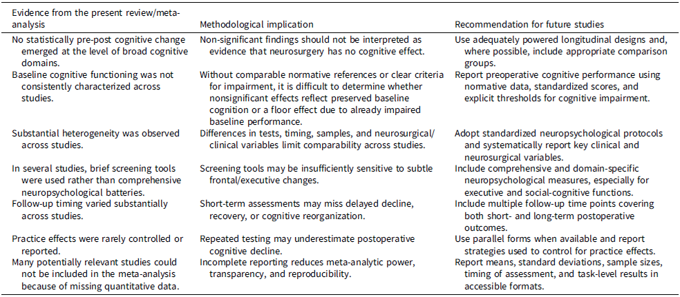
Based on the limitations identified in the available literature and on the results of the present meta-analysis, we summarized key methodological implications and recommendations for future studies in Table 2. This table is intended to synthesize the methodological implications of the present review and meta-analysis and to guide future research on cognitive outcomes after frontal lobe tumor neurosurgery.
Summary of current evidence and methodological recommendations

Table 2. Long description
A table summarizing evidence from a review and meta-analysis on cognitive outcomes after frontal lobe tumor neurosurgery, focusing on methodological implications and recommendations for future studies. The table has three columns: Evidence from the present review/meta-analysis, Methodological implication, and Recommendation for future studies. It contains six rows with the following data: Row 1: No statistically prepost cognitive change emerged at the level of broad cognitive domains, Non-significant findings should not be interpreted as evidence that neurosurgery has no cognitive effect, Use adequately powered longitudinal designs and, where possible, include appropriate comparison groups. Row 2: Baseline cognitive functioning was not consistently characterized across studies, Without comparable normative references or clear criteria for impairment, it is difficult to determine whether nonsignificant effects reflect preserved baseline cognition or a floor effect due to already impaired baseline performance, Report preoperative cognitive performance using normative data, standardized scores, and explicit thresholds for cognitive impairment. Row 3: Substantial heterogeneity was observed across studies, Differences in tests, timing, samples, and neurosurgical/clinical variables limit comparability across studies, Adopt standardized neuropsychological protocols and systematically report key clinical and neurosurgical variables. Row 4: In several studies, brief screening tools were used rather than comprehensive neuropsychological batteries, Screening tools may be insufficiently sensitive to subtle frontal/executive changes, Include comprehensive and domain-specific neuropsychological measures, especially for executive and social-cognitive functions. Row 5: Follow-up timing varied substantially across studies, Short-term assessments may miss delayed decline, recovery, or cognitive reorganization, Include multiple follow-up time points covering both short- and long-term postoperative outcomes. Row 6: Practice effects were rarely controlled or reported, Repeated testing may underestimate postoperative cognitive decline, Use parallel forms when available and report strategies used to control for practice effects. Row 7: Many potentially relevant studies could not be included in the meta-analysis because of missing quantitative data, Incomplete reporting reduces meta-analytic power, transparency, and reproducibility, Report means, standard deviations, sample sizes, timing of assessment, and task-level results in accessible formats.
Conclusions
The results of our meta-analysis did not reveal any significant evidence of cognitive changes following neurosurgical resection of frontal lobe tumors. Although the removal of tumors in the frontal lobes is a critical therapeutic intervention, its cognitive implications are more complex than commonly assumed. Our findings call for a thorough reconsideration of post-intervention expectations and highlight the importance of a multidisciplinary approach to patient management. Only through well-designed studies and collaborations among neurosurgeons, neuropsychologists, and other specialists, our understanding of the cognitive impact of frontal lobe tumor neurosurgery could be widened, thereby improving patients’ care and everyday life outcomes.
Supplementary material
The supplementary material for this article can be found at https://doi.org/10.1017/S135561772610215X.
Acknowledgements
None.
Author contributions
Elisa Pellencin: Conceptualization, Methodology, Writing - original draft preparation, Writing - review and editing; Fabio Masina: Conceptualization, Methodology, Formal analysis and investigation, Writing - review and editing; Nicolò Castelli: Writing - review and editing; Giovanni Carone: Writing - review and editing; Alexa Schincariol: Methodology, Writing - review and editing; Cristina Scarpazza: Methodology, Writing - review and editing; Alessandro Perin: Writing - review and editing, Supervision; Konstantinos Priftis: Conceptualization, Methodology, Writing - original draft preparation, Writing - review and editing, Supervision.
Funding statement
No financial support was received for this study.
Competing interests
The authors declare no conflicts of interest with respect to the contents of the current study.




 Open access

