Introduction
Field vegetables are subject to infestations by a complex of root maggot species in the genus Delia (Diptera: Anthomyiidae) (Finch Reference Finch1989). Two Delia species, Delia antiqua (Meigen) and Delia radicum (Linnaeus), are known to be principle pest species of Canadian allium (Amaryllidaceae) and cruciferous (Brassicaceae) vegetables, respectively (Finch Reference Finch1989; Howard et al. Reference Howard, Garland, Seaman and Grafius1996), but other Delia species, such as Delia platura (Meigen) and Delia florilega (Zetterstedt) (Mlynarek et al. Reference Mlynarek, MacDonald, Sim, Hiltz, McDonald and Blatt2020), are commonly found in those same fields and have been observed to feed on decomposing or fresh crucifers, Allium, legumes, and even certain cereals (Griffiths Reference Griffiths1993; Savage et al. Reference Savage, Fortier, Fournier and Bellavance2016). Without confirming or performing species identification, the presence of multiple Delia species where some might be secondary invaders can make it difficult to determine what control measures are necessary and to conduct research into new control measures (Mlynarek et al. Reference Mlynarek, MacDonald, Sim, Hiltz, McDonald and Blatt2020).
Accurate identification of Delia species can be challenging due to delicate or shared morphological characters among species (Griffiths Reference Griffiths1991, Reference Griffiths1993; Savage et al. Reference Savage, Fortier, Fournier and Bellavance2016). At the larval stage, D. radicum can easily be distinguished from other Delia species by the absence of tubercle X, but identification of D. antiqua, D. platura, and D. florilega relies on the number of papillae on the paired anterior spiracles (Savage et al. Reference Savage, Fortier, Fournier and Bellavance2016): D. antiqua has nine to 12 papillae per spiracle, a number that overlaps with D. florilega and D. platura, both of which can have six to nine papillae (Savage et al. Reference Savage, Fortier, Fournier and Bellavance2016). In addition, the papillae can vary in number between the two spiracles on a single larva and are often difficult to count, especially in early larval stages. The adults are only slightly easier to identify, with accurate identification relying on the presence of hairs on the ventral surface of the wing’s costal vein, the number and placement of tibial and prealar bristles on the legs and thorax, respectively, and the size of the parafacial. However, overlap in adult characters also prevents differentiation between D. platura and D. florilega females (Darvas and Szappanos Reference Darvas and Szappanos2003; Savage et al. Reference Savage, Fortier, Fournier and Bellavance2016). Additionally, the bodies of adult flies are often damaged during trapping, resulting in missing wings and bristles, which increases the risk of misidentification among all four species. The difficulty of morphological identification makes finding alternative methods to accurately identify these species necessary.
Identifying species using molecular techniques is increasingly popular for morphologically difficult groups (Hebert et al. Reference Hebert, Ratnasingham, Zakharov, Telfer, Levesque-Beaudin and Milton2016; Van der Heyden et al. Reference Van der Heyden, Fortier and Savage2020). Closely related insect species have been effectively identified in the past using polymerase chain reaction, followed by restriction fragment length polymorphism analysis (Hoy Reference Hoy2013). This method amplifies a particular gene, then uses carefully selected restriction enzymes to cut at target sites, resulting in species-specific bands when the digested polymerase chain reaction product is viewed on an agarose gel (Hoy Reference Hoy2013). Comparative band lengths created with restriction enzymes have successfully been used as a rapid diagnostic technique in agricultural studies (Brunner et al. Reference Brunner, Fleming and Frey2002; Lewter and Szalanski Reference Lewter and Szalanski2007; Arneodo et al. Reference Arneodo, Balbi, Flores and Sciocco-Cap2015; Hua et al. Reference Hua, Zhao, Zhang, Hu, Zhang and Li2018), including those identifying closely related Diptera species such as the members of the Calliphoridae (Ratcliffe et al. Reference Ratcliffe, Webb, Weinzievl and Robertson2003), Tephritidae (Armstrong et al. Reference Armstrong, Cameron and Frampton1997; Muraji and Nakahara Reference Muraji and Nakahara2002), and Drosophilidae families (Kim et al. Reference Kim, Tripodi, Johnson and Szalanski2014).
The objective of the present study was to identify and verify cut sites for restriction enzymes in the insect mitochondrial DNA barcode region, the cytochrome c oxidase subunit 1 (CO1) gene, that will allow for the rapid identification of economically important and frequently encountered Anthomyiidae species, especially in vegetable-growing regions in Canada – for example, Nova Scotia, southern Québec, and southern Ontario – and elsewhere in the world. Four Delia species commonly found in field vegetable crops were chosen: two molecularly distinct lines of D. platura (H-Line and N-line, as presented in Savage et al. Reference Savage, Fortier, Fournier and Bellavance2016), D. florilega, D. radicum, and D. antiqua. Botanophila fugax (Meigen) (Diptera: Anthomyiidae), a species that is morphologically similar to Delia but is known not to damage crops (Savage et al. Reference Savage, Fortier, Fournier and Bellavance2016), was also included in the study. Restriction enzymes were selected and tested for their ability to differentiate among the Anthomyiidae species. Accurate identification of Anthomyiidae maggots and field-collected flies via a simple polymerase chain reaction–restriction fragment length polymorphism technique can improve research into the development of management strategies.
Methods
Sampling and identification
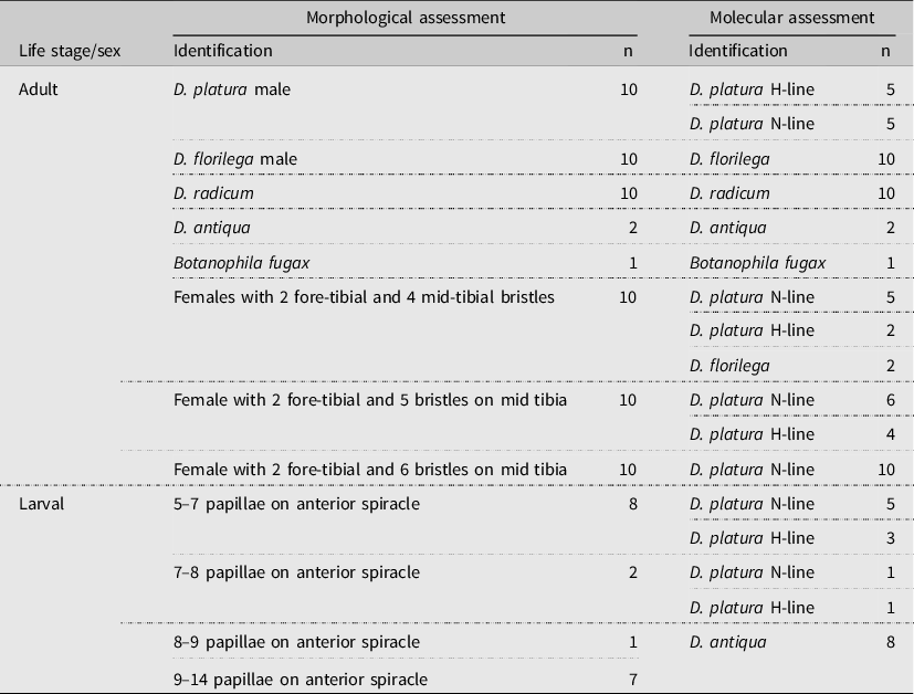
Adult flies were collected using blue sticky traps (Distributions Solida, Saint-Ferréol-les-Neiges, Québec, Canada) from onion and brassica fields in the vicinity of the Agriculture and Agri-Food Canada Harrow Research and Development Centre, Harrow, Ontario, Canada (42.0° N, –82.89° W). The collected flies remained on the sticky cards at room temperature for a maximum of six months until processing. Morphological identification of the Delia species caught in fields in southern Ontario was performed only on specimens in good condition using the Savage et al. (Reference Savage, Fortier, Fournier and Bellavance2016) key. Adult Delia females with two fore-tibial bristles were assumed to be either Delia florilega or D. platura but were not identified to species because D. florilega females, which have four mid-tibial bristles, cannot be distinguished from females of D. platura, which may have four to six mid-tibial bristles (Savage et al. Reference Savage, Fortier, Fournier and Bellavance2016). The D. platura or D. florilega females were grouped based on mid-tibial bristle number (Table 1), and only the adult males of these species were used to analyse the efficiency of restriction enzymes. A leg from a B. fugax specimen was received from the Centre for Biodiversity Genomics, University of Guelph (Guelph, Ontario, Canada) because this species was not collected in our fields (Supplemental material).
Morphological and molecular identification of Delia and Botanophila species from vegetable fields in eastern Canada with the sample size (n) for each species.

Larval flies were hand collected from the Harrow Research and Development Centre, from the Muck Crops Research Station (Kettleby, Ontario, Canada) of the University of Guelph (44° N, –76° W), and from a D. platura lab colony at the Agriculture and Agri-Food Canada Kentville Research and Development Centre, Kentville, Nova Scotia (45.07° N, –64.48° W). The larval flies were preserved in 70% ethanol at room temperature and were grouped by number of papillae on the anterior spiracle.
DNA extraction
Ten adult specimens were used to extract the DNA and to test the restriction enzymes for each of the focal anthomyiid species when available (Table 1), and larval specimens were used to test the efficacy of the restriction-enzyme method on immature specimens. A leg or the thoracic muscles were taken from the adult flies, whereas the entire larva, except for the anterior section containing the papillae, was used for DNA extraction. Total genomic DNA was extracted using the DNeasy blood and tissue kit (cat. no. 20051; QIAGEN, Toronto, Ontario, Canada) following the manufacturer’s protocol, with the exception that tissue was incubated at 56 °C overnight.
Amplification and purification of CO1
The standard 658-bp CO1 mitochondrial DNA barcode region was amplified using Folmer et al. (Reference Folmer, Black, Hoeh, Lutz and Vrijenhoek1994) primers LepF1 (5′-ATTCAACCAATCATAAAGATATTGG-3′) and LepR1 (5′-TAAACTTCTGGATGTCCAAAAAATCA-3′; Integrated DNA Technologies, Coralville, Iowa, United States of America). Polymerase chain reaction products were amplified using 2 μL of genomic DNA, 5 μL of 10× polymerase chain reaction buffer containing Mg, 2 μL of 25 mM MgCl2 (FroggaBio, Concord, Ontario, Canada), 1 μL of 10 mM dNTP Mix (FroggaBio), 0.4 μL each of 50 μM LepF1 forward and LepR1 reverse primers, 1.25 U Taq DNA polymerase (FroggaBio), and DNA-free H2O for a total reaction volume of 50 μL (all reagents were acquired from FroggaBio). Thermocycling conditions consisted of an initial denaturation step at 94 °C for three minutes, followed by 30 cycles of 94 °C for 30 seconds, 51 °C for 30 seconds, and 72 °C for one minute, and a final extension at 72 °C for 10 minutes. An additional primer set, LCO1490 (5′-GGTCAACAAATCATAAAGATATTGG-3′) and HCO2198 (5′-TAAACTTCAGGGTGACCAAAAAATCA-3′), was tested with D. radicum and D. florilega specimens and run using the same thermocycling conditions as the LepF1/LepR1, except for a 52 °C annealing temperature and 35 amplification cycles employed because a Wolbachia (Anaplasmataceae) infection caused unexpected noise in the first set of primers (see the section, Wolbachia screening, below).
Purification methods were tested on all the polymerase chain reaction products to determine which method – a spin column kit (Epoch Life Science, Sugar Land, Texas, United States of America) or ExoSap-IT (Thermo Fisher Scientific, Waltham, Massachusetts, United States of America) – was suited for restriction fragment length polymorphism analysis on Delia specimens. The polymerase chain reaction products were purified following the manufacturer’s protocol for both methods before digestion with the restriction enzymes.
Restriction-enzyme analysis
The CO1 mitochondrial DNA barcodes for each anthomyiid species were downloaded from sequence data provided by Savage et al. (Reference Savage, Fortier, Fournier and Bellavance2016) and Barcode of Life database (BOLD) BINS (Delia radicum BOLD: AAG2465, Delia antiqua BOLD: AAL1158, Delia florilega BOLD: ACR4394, Delia platura H-line BOLD: AAG2511, Delia platura N-line BOLD: AAA3453, and Botanophila fugax BOLD: AAG2462). Ten sequences from each BOLD BIN were used in the VIRS programme, a visual tool for identifying restriction sites (available from http://bis.zju.edu.cn/virs/; accessed 6 February 2019) to find all the restriction enzymes that cut the mitochondrial DNA barcodes of the genetically distinct lines and species. Based on the species and restriction-enzyme sequences, only four restriction-enzyme candidates were flagged and tested.
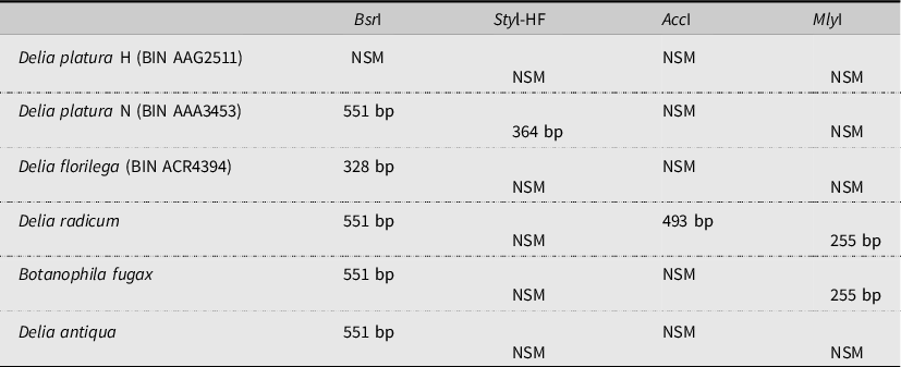
Restriction enzymes for each of these Delia species were then compared to determine which enzymes would make species-specific cuts that could be visualised on an agarose gel. Four candidate restriction enzymes (acquired from Integrated DNA Technologies) were identified (Table 2): AccI (5′-GT↓MKAC-3′), BsrI (5′-ACTGGN↓-3′ and 5′-NC↓CAGT-3′), StyI (5′-C↓CWWGG-3′), and MlyI (5′-GAGTC(N)5 ↓-3′, and 5′-↓(N)5GACTC-3′).
Estimated cleavage site (in base pairs) of four restriction-enzyme sites for the trimmed cytochrome CO1 barcode fragment (658 bp) in Delia platura H-line, D. platura N-line, D. florilega, D. radicum, Botanophila fugax, and D. antiqua. NSM, no sequence match.

The restriction enzymes were initially run separately to confirm where the cuts occurred, then they were combined into double digests, where buffer and band pattern would allow, increasing efficiency of the polymerase chain reaction–restriction fragment length polymorphism method. A restriction digest using the enzymes BsrI and StyI was performed in a single 15-μL reaction containing 6 μL of purified polymerase chain reaction product, 2 U BsrI, 5 U of StyI, 1.5 μL of Buffer 3.1, and DNA-free H2O. The BsrI–StyI reaction was incubated at 37 °C for one hour, then at 65 °C for one hour, followed by enzyme inactivation at 80 °C for 20 minutes. The enzymes MlyI and AccI were run together in a second 15-μL restriction digest reaction, containing 6 μL of purified polymerase chain reaction product, 3 U MlyI, 3 U AccI, 1.5 μL CutSmart buffer, and DNA-free H2O. The MlyI–AccI reaction was incubated at 37 °C for two hours, followed by an enzyme inactivation step at 80 °C for 20 minutes.
Visualisation and sequencing
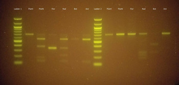
Polymerase chain reaction and restriction digest products were visualised using 1.5% agarose gels stained with SYBR safe (Thermo Fisher Scientific). Visualisation of the gels was performed using a blue-light DNA transilluminator (blueBox S, Minipcr, Cambridge, Massachusetts, United States of America). Running gels at 95 V for 25 minutes was sufficient to confirm amplification of the targeted regions and species identification based on expected restriction-enzyme cuts (Table 2; Fig. 1); however, full separation of all bands can be seen using 90 V for one hour.
Digest of the restriction-enzyme sites with BsrI–StyI (Ladder 1) and AccI–MlyI (Ladder 2) for the CO1 barcode fragment amplified with LepF/LepR in Delia platura H-line (PlatH), D. platura N-line (PlatN), D. florilega (Flor), D. radicum (Rad), Botanophila fugax (Bot), and D. antiqua (Ant). Band lengths given are relative to cut-site positions on CO1 sequences trimmed of primers. Gels were run at 90 V for one hour to fully separate bands using a 1-Kb ladder.

The uncut polymerase chain reaction product of all the Delia specimens examined was sent for sequencing at the Genome Québec Innovation Centre (McGill University, Montréal, Québec, Canada). These sequences (Supplemental material; Genbank: MW303324–MW303401) were used to verify the accuracy of the restriction-enzyme cuts. We visualised the sequences individually for quality using FinchTV, version 1.4.0 (Geospiza, Inc.; Seattle, Washington, United States of America, http://www.geospiza.com). For B. fugax, the barcode region of these specimens was previously sequenced by Guelph University.
Wolbachia screening
Due to indications of contamination in the D. radicum and D. florilega sequences, all Delia samples were screened for the presence of a Wolbachia sp. by using a primer set for the wsp gene, wsp_F1 (5′-GTCCAATARSTGATGARGAAAC-3′) and wsp_R1 (5′-CYGCACCAAYAGYRCTRTAAA-3′). The protocol for amplifying the wsp gene followed those set out by PUBMED (https://pubmlst.org/wolbachia/wsp/info/protocol.shtml). Seven samples that showed the presence of Wolbachia were sequenced (Genbank: MW320349–MW320355), of which four were from D. florilega and three were from D. radicum.
Results
The polymerase chain reaction products digested with enzymes BsrI, StyI, AccI, and MlyI revealed the presence of products with the expected band sizes, herein given as lengths relative to sequences trimmed of primers, that create distinct patterns for each of the anthomyiid species analysed in this study (Fig.1; Table 2; Supplemental material). We found that BsrI consistently distinguished sequences of the D. platura H-line, D. florilega, and males from the other Delia species in this study – all of which were cut by BsrI at 551 bp (Table 2). The D. platura H-line (n = 10) males were unique in that no digestion occurred with BsrI, whereas a restriction site was present only at 328 bp in D. florilega (n = 10) males (Fig. 1; Table 2). The D. radicum specimens (n = 10) were cut at 551 bp; however, the majority displayed an additional, unexpected restriction cut by BsrI that resulted in an extra two bands at around 350 bp and just under 200 bp. The StyI restriction site was present only in D. platura N-line males, whereas the AccI restriction site was unique to D. radicum specimens (Table 2). Both D. radicum and B. fugax are cut at 255 bp by the MlyI enzyme (Table 2).
The restriction enzymes can be combined into two double digests that allow identification of all species included in this study (Fig. 1). The combination of BsrI and StyI allows the two D. platura genetic lines and D. florilega to be identified in a single reaction; however, careful sizing of the bands is required because the extra bands in most D. radicum individuals created a pattern similar in appearance to that of the D. platura N-line (Fig. 1). The appearance of the extra bands in D. radicum specimens was significantly reduced so as to be negligible when the CO1 region was amplified with LCO1490/HCO2198 primers rather than with LepF/LepR before the BsrI and StyI restriction digest. The second restriction digest with AccI and MlyI distinguished D. radicum from all other Delia spp., including the D. platura N-line, no matter which primer pair was used (Fig. 1). The AccI–MlyI enzyme pair, run in CutSmart buffer (New England Biolabs (NEB), Ipswich, Massachusetts, United States of America), which contains no salt, performs poorly unless the polymerase chain reaction products are purified through spin columns rather than by ExoSAP-IT.
The Delia females with a combination of two fore-tibial bristles and six mid-tibial bristles all had band patterns that matched those of the D. platura N-line, whereas females with five mid-tibial bristles proved to be a mix of both genetic lines of D. platura, and females with four mid-tibial bristles proved to be a mix of the D. platura genetic lines and of D. florilega (Table 1). The adult D. antiqua had band patterns that matched those of larvae that had nine or more papillae on at least one spiracle (Table 1). Larvae with seven or fewer papillae on at least one spiracle had band patterns matching those of the male D. platura lines (Table 1).
Sequencing of the CO1 gene regions confirmed the accuracy of the restriction-enzyme band patterns in identifying species; however, the forward and reverse sequences from multiple D. radicum specimens amplified with the LepF1/LepR1 primers were overlaid with another sequence. Sequences from three specimens that lacked the overlay were subsequently used to infer the accurate D. radicum sequence of other specimens when the overlaid sequence was isolated. The isolated sequence was put through the National Center for Biotechnology Information’s nBLAST and aligned with Wolbachia sequences with a 98.2–99.5% identity. The potential Wolbachia CO1 sequence that was isolated from the D. radicum sequences was found to have a restriction site for BsrI at 197 bp, a cut position that matches the unexpected band sizes seen in the majority of D. radicum specimens. Screening with the wsp Wolbachia primers confirmed the presence of Wolbachia in all D. radicum specimens, and sequencing confirmed a 100% match with sequences in nBLAST for Wolbachia in D. radicum. Eight D. florilega specimens also had Wolbachia wsp sequences that matched 100% to Wolbachia from calyptratea species, whereas weak signals were observed in two D. platura N-line specimens and one D. platura H-line specimen. Alignments of the wsp sequences showed variation among the Delia species.
Discussion
The economically important Delia species can be rapidly and accurately identified through the use of polymerase chain reaction–restriction fragment length polymorphism analysis of the insect mitochondrial DNA barcode region, the CO1 gene. A single restriction enzyme, BsrI, is able to distinguish between the most morphologically challenging species, D. platura and D. florilega, as well as between the two genetic lines of D. platura. A single restriction enzyme, MlyI, is able to identify B. fugax from the Delia species. Combining BsrI with the other three restriction enzymes – StyI, AccI, and MlyI – into double digestion reactions allows for rapid diagnostics among the anthomyiid species tested. Studies or routine pest monitoring that requires identification of Anthomyiidae flies in vegetable field crops can use any number of or all four of these enzymes, depending on the focus of the monitoring.
This is the first polymerase chain reaction–restriction fragment length polymorphism method to differentiate Delia species using restriction enzymes and, as such, there were areas in need of exploration and optimisation. We found no interference during DNA extraction by the presence of a special “dry-touch” adhesive found on sticky cards (Distributions Solida), allowing field collection methods (i.e., sticky cards) to remain unaltered. Although the restriction enzymes used with a buffer containing salt appeared unaffected by the purification process, using a spin column purification kit improved performance and band clarity for the restriction enzymes run in CutSmart buffer, which contains no salt. Salt interference, due to residues in the final polymerase chain reaction product that ExoSAP-IT does not remove, is likely responsible for the poor performance of restriction enzymes optimised to run with little to no salt in the reaction.
The appearance of unexpected bands caused by BsrI in D. radicum specimens was significantly reduced when the LCO1490/HCO2197 primer set was used for amplification, although the results remained unchanged for the other Delia species. The unexpected bands observed in D. radicum specimens were likely the result of unintentional amplification of Wolbachia sp. by the LepF1/LepR1 primers that are calculated to be responsible for 86.77% of accidental Wolbachia sequences on nBLAST (Smith et al. Reference Smith, Bertrand, Crosby, Eveleigh, Fernandez-Triana and Fisher2012). The confirmation of Wolbachia sp. in our D. radicum specimens and the presence of a BsrI cut site at the appropriate location causing the unplanned bands seen in D. radicum specimens support the interference of Wolbachia when LepF1/LepR1 primers are used. Although the Wolbachia restriction site would give bands of similar size to those expected by BsrI in D. florilega, of which 67% of our specimens were infected, we observed the correct band patterns in uninfected D. florilega specimens. In addition, sequencing of the CO1 regions in all Delia spp. other than D. radicum did not reveal any contamination, with sequences coming back visually clean and successfully aligning to their respective species on nBLAST. The polymerase chain reaction–restriction fragment length polymorphism method developed for Delia is, therefore, a practical and effective identification method compared to traditional morphological approaches in research.
Research was previously hindered by the challenges of Delia morphological identification due to shared characters among species, but the addition of the analysis examined in the present study to the “toolbox”, as well as being a cheaper option than sequencing, allows for species-level identification and offers the sensitivity to distinguish even among genetic lines. The use of the restriction enzyme BsrI showed that females of the two D. platura genetic lines overlap morphologically when four and five mid-tibial bristles are present, but six mid-tibial bristles, previously unreported in Savage et al. (Reference Savage, Fortier, Fournier and Bellavance2016), are likely a unique trait to the N-line (Table 1). Combining morphological and behavioural analyses with identification via polymerase chain reaction–restriction fragment length polymorphism may illuminate additional differences between the two genetic lines and among other Delia species. Other anthomyiid species of economic importance, such as D. floralis (Fallé) and D. planipalpis (Stein), have the potential to be identified by polymerase chain reaction–restriction fragment length polymorphism when additional restriction enzymes are included. Unfortunately, specimens were not collected from eastern Canada nor were they available to this study for confirmation. Research into if, how, and when each root maggot species attacks field vegetables crops is no longer limited by uncertainty over species identification.
The ability to accurately identify all stages of a pest’s life cycle and to distinguish among closely related species is critical for ecological and integrated pest management research. Bioassays performed by Mlynarek et al. (Reference Mlynarek, MacDonald, Sim, Hiltz, McDonald and Blatt2020) found that D. antiqua is the only species collected from onions in eastern Canada that actively causes damage at all growth stages of the onions. The ability to accurately identify field-collected Delia spp. in onion fields without having to rear or morphologically identify specimens will allow management practices to be applied only when D. antiqua is present.
Recently, a high-resolution melting assay method was introduced to differentiate between the seedcorn maggot species complex (Van der Heyden et al. Reference Van der Heyden, Fortier and Savage2020), which will provide more options to those looking to identify Delia species. Both the high-resolution melting assay method and the method investigated in the present study are based on amplifying the barcode gene. The main difference between the two methods is that high-resolution melting uses real-time polymerase chain reaction, whereas our method does not. Additionally, the high-resolution melting method can efficiently differentiate between D. florilega and the two lines of D. platura, whereas our method can identify five species and the two genetically distinct lines of D. platura.
Using polymerase chain reaction–restriction fragment length polymorphism is an efficient identification tool that is cost effective and requires only basic molecular equipment (e.g., regular thermocycler). Advantages of this method over traditional morphological identification include the ability to identify damaged specimens and the flexibility to distinguish among species with morphologically shared characters (e.g., D. florilega and D. platura larvae and female adults) and among distinct genetic lines. The use of restriction fragment length polymorphism in Delia identification will allow fast identification of species commonly found in field crops and will inform the development of targeted and effective management strategies.
Supplementary material
To view supplementary material for this article, please visit https://doi.org/10.4039/tce.2021.22.
Acknowledgements
We thank Dr. Suzanne Blatt from the Kentville Research and Development Centre, Agriculture and Agri-Food Canada, Dr. Mary Ruth MacDonald from the Muck Crop Research Station, University of Guelph, Valerie Levesque-Beaudin from the University of Guelph for sending specimens, and Dr. Roselyne Labbé from Harrow Research and Development Centre for comments on a draft of the manuscript. The project was funded by the PMC-AAFC.