Introduction
Chronic fatigue syndrome (CFS) is most probably a multifactorial condition in which psychological and social factors are implicated alongside biological changes (Wessely et al. Reference Wessely, Hotopf and Sharpe1998). Of the biological factors identified to date, one of the most replicated is of neuroendocrine perturbation, especially to the hypothalamic–pituitary–adrenal (HPA) axis. A comprehensive review concluded that there is evidence of reduced basal cortisol output in CFS, most consistently shown by 24-h urinary free cortisol (UFC) measurement, but also seen in sequential salivary free cortisol and blood sampling (Cleare, Reference Cleare2003). Subsequent studies have tended to confirm these conclusions (Cevik et al. Reference Cevik, Gur, Acar, Nas and Sarac2004; Jerjes et al. Reference Jerjes, Cleare, Wessely, Wood and Taylor2005, Reference Jerjes, Peters, Taylor, Wood, Wessely and Cleare2006). The reason CFS patients have lowered cortisol output is not clear; one possibility is that it is a primary factor in the development of the illness, although it has also been hypothesized that lowered cortisol itself is of multifactorial aetiology in CFS and occurs, in part, secondary to aspects of CFS, such as inactivity, sleep disturbance or stress (Cleare, Reference Cleare2004). A recent study suggests that impaired cortisol responses in CFS are restricted to those with a history of childhood abuse, which is itself sixfold higher in CFS than in controls (Heim et al. Reference Heim, Nater, Maloney, Boneva, Jones and Reeves2009).
Regardless of cause, there are suggestions that lowered cortisol may be of clinical relevance. Two randomized controlled trials have shown that low-dose cortisol replacement therapy can lead to significant short-term reductions in fatigue and other features of CFS (McKenzie et al. Reference McKenzie, O'Fallon, Dale, Demitrack, Sharma, Deloria, Garica-Borreguero, Blackwelder and Straus1998; Cleare et al. Reference Cleare, Heap, Malhi, Wessely, O'Keane and Miell1999). This suggests that low cortisol may be one of several maintaining factors in the illness. Whether a primary or secondary factor, once hypocortisolism has developed it may itself lead to symptoms and represent a maintaining factor in illness chronicity.
Cognitive behavioural therapy (CBT) is one of the evidence-based therapies that is recommended for treating CFS (Whiting et al. Reference Whiting, Bagnall, Sowden, Cornell, Mulrow and Ramirez2001; Reid et al. Reference Reid, Chalder, Cleare, Hotopf and Wessely2005) and is recommended in the guidelines from the UK National Institute of Health and Clinical Excellence. CBT is individually tailored, but important components include changing unhelpful patterns of rest and activity (which can include profound inactivity or intermittent bursts of overactivity interspersed with inactivity), improving sleep patterns, increasing exercise capacity, identifying unhelpful cognitions about the illness or the coping strategies used, using problem-solving techniques to reduce stress, and treating anxiety and depression if present (Wessely et al. Reference Wessely, Hotopf and Sharpe1998). Not all patients respond to CBT, and several factors are associated with poor response to therapy, including physical illness attributions, treatment-resistant depression (Butler et al. Reference Butler, Chalder, Ron and Wessely1991), certain illness cognitions (Deale et al. Reference Deale, Chalder and Wessely2000), a passive activity pattern, and focusing on bodily symptoms (Prins et al. Reference Prins, Bleijenberg, Bazelmans, Elving, de Boo, Severens, van der Wilt, Spinhoven and van der Meer2001).
No studies have yet looked at whether there might be any biological factors that predict preferential response to CBT in CFS. We hypothesized that if hypocortisolism is indeed a biological maintaining factor in CFS, then patients with lower cortisol levels would show a lesser response to CBT. A parallel to this comes from research in major depression, which is characterized by hypercortisolism in contrast to the hypocortisolism of CFS, where those with the highest degree of HPA axis overactivity have the lowest response rates to CBT (Thase et al. Reference Thase, Dube, Bowler, Howland, Myers, Friedman and Jarrett1996).
Method
Subjects
Subject selection and diagnosis
Patients aged 18–65 years were recruited into the study from consecutive referrals to the CFS clinic at King's College Hospital, London. All patients had undergone medical screening to exclude detectable organic illness, including a minimum of physical examination, urinalysis, full blood count, urea and electrolytes, thyroid function tests, liver function tests, 0900 h cortisol (to screen for Addison's disease) and erythrocyte sedimentation rate (ESR). Patients were interviewed using a semi-structured interview for CFS (Sharpe et al. Reference Sharpe, Chalder, Palmer and Wessely1997) and were included if they met both international consensus criteria for CFS (Sharpe et al. Reference Sharpe, Archard, Banatvala, Borysiewicz, Clare, David, Edwards, Hawton, Lambert, Lane, McDonald, Mowbray, Pearson, Peto, Preedy, Smith, Smith, Taylor, Tyrrell, Wessely and White1991; Fukuda et al. Reference Fukuda, Straus, Hickie, Sharpe, Dobbins and Komaroff1994), did not have fibromyalgia according to American College of Rheumatology (ACR) criteria (Wolfe et al. Reference Wolfe, Smythe, Yunus, Bennett, Bombardier, Goldenberg, Tugwell, Campbell, Abeles, Clark, Fam, Farber, Feichtner, Franklin, Gatter, Hamaty, Lessard, Lichtbroun, Masi, McGain, Reynolds, Romano, Russell and Sheon1990) and were judged suitable to receive CBT by the assessing clinician. Psychiatric assessment was undertaken using the Schedules for Clinical Assessment for Neuropsychiatry (SCAN), adapted for DSM-IV (APA, 1994). Female patients were tested during days 1–7 of their menstrual cycle, pregnancy having been excluded prior to testing.
Medication
The majority of patients (64/84) were free from psychotropic medication, steroids or medication known to affect the HPA axis for a minimum of 2 months prior to endocrine testing, including all of the subgroup who gave saliva samples. For the UFC measurement, a minority of subjects were taking such medication. Therefore, for the UFC results, we performed statistical analyses twice, once with all patients included and once with only drug-free patients.
Medication defined as liable to affect the HPA axis was: any psychotropic medication (i.e. antidepressant, antipsychotic, anxiolytic/hypnotic or mood stabilizer); any corticosteroid derivative; any drug designed to act on the HPA axis; anti-epileptic medication; or other mediation that, after checking the British National Formulary, was judged likely to alter the HPA axis. Of the patients taking such medication, all were taking an antidepressant (one of whom also took sodium valproate and zopiclone) except one taking propranolol and one pizotifen. Of the patients taking medication that was not considered liable to affect the HPA axis, two were taking an antihypertensive, two non-steroidal analgesia, one an antibiotic, one a statin, and two an H2-antagonist (one of whom also took domperidone).
Sample size and characteristics
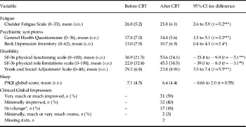
Eighty-four patients (59 female) entered the study, with a mean age of 40.4 (s.d.=10.5) years. The mean length of illness at entry to the study was 4.8 (s.d.=3.2) years. Twenty-three of 84 patients had a co-morbid diagnosis of a current major depressive episode according to DSM-IV criteria. Other clinical descriptors are shown in Table 1. A prior power calculation had estimated a sample size requirement of 60 subjects on the basis of previous data; we extended recruitment beyond this as the response rate to CBT was lower than initially predicted and so as to allow a sufficiently powered analysis of the subgroup not taking medication liable to affect the HPA axis.
Clinical measures before and after CBT

CBT, Cognitive behavioural therapy; SF-36, 36-item short-form health survey; PSQI, Pittsburgh Sleep Quality Index; CI, confidence interval; s.d., standard deviation.
Asterisks represent values significantly improved after CBT by the paired t test
* p<0.05, ** p<0.01, *** p<0.001.
a Three drop-outs rated as unchanged.
Clinical assessment
All patients filled out the following questionnaires at baseline and after completion of CBT:
Fatigue: Chalder Fatigue Scale (Chalder et al. Reference Chalder, Berelowitz, Hirsch, Pawlikowska, Wallace, Wessely and Wright1993). Fatigue problem rating scale, incorporating 0–8 Likert scales for severity and interference with life (Deale et al. Reference Deale, Chalder, Marks and Wessely1997).
Psychiatric symptoms: General Health Questionnaire-12 (GHQ-12; Goldberg & Blackwell, Reference Goldberg and Blackwell1970); Beck Depression Inventory (Beck et al. Reference Beck, Ward and Mendelson1961).
Functional capacity: Medical Outcomes Survey (MOS) Short-Form 36-item (SF-36) physical function subscales (Ware & Sherbourne, Reference Ware and Sherbourne1992); Work and Social Adjustment Scale (WSAS; Mundt et al. Reference Mundt, Marks, Shear and Griest2002).
Sleep disturbance: Pittsburgh Sleep Quality Index (PSQI; Buysse et al. Reference Buysse, Reynolds, Monk, Berman and Kupfer1989).
All patients were rated by their therapists after completion of CBT on a clinical global impression improvement scale (Guy, Reference Guy1976) blind to the endocrine results; this was chosen prospectively as the primary measure of therapeutic response, with a value of 1 or 2 (very much or much improved) taken to indicate a response to therapy.
The institutional ethics committee approved all procedures. After complete description of the study to the subjects, written informed consent was obtained.
CBT
CBT for CFS has been described in detail elsewhere (Wessely et al. Reference Wessely, Hotopf and Sharpe1998). We used experienced therapists and adhered to set clinical protocols (Deale et al. Reference Deale, Chalder, Marks and Wessely1997). The standard course of therapy lasts for 12–15 sessions. To standardize procedures, we retested after 6 months or 15 sessions of therapy, whichever was sooner. Treatment was received as part of usual clinical care and therefore there was no untreated control group.
24-h UFC excretion
Subjects were given a plastic bottle containing 0.1 g boric acid as a preservative and standard instructions for a 24-h collection starting at 0900 hours (Cleare et al. Reference Cleare, Blair, Chambers and Wessely2001a). Upon receipt, the volume was measured, the container shaken, and 20 ml frozen until assay.
Salivary cortisol
Samples were taken using untreated salivettes. A detailed protocol for the salivary cortisol collections is described elsewhere, including precautions taken to avoid false high values (Roberts et al. Reference Roberts, Wessely, Chalder, Papadopoulos and Cleare2004). Testing was undertaken at home on any normal weekday except Mondays. Patients provided a sample of saliva at 0800, 1200, 1600 and 2000 hours. Samples were kept in the refrigerator overnight and sent back in the post in the morning. On arrival at the laboratory, they were frozen at −20°C until assay.
Hormone assays
UFC
UFC was assayed using the Technicon Immuno-1 assay (Bayer plc, UK) on extracted and reconstituted samples. The lower limit of the assay was 20 nmol/l; values that were reported as <20 nmol/l were assigned a value half way between the lower limit of detection and zero.
Saliva cortisol
After defrosting and centrifuging, cortisol was measured in duplicate using a time-resolved fluoroimmunoassay as described elsewhere (Pariante et al. Reference Pariante, Papadopoulos, Poon, Checkley, English, Kerwin and Lightman2002), except that the rabbit cortisol antibody (product no. 2330-5105, batch 21051565; Biogenesis, UK) and the europium-labelled cortisol were diluted 1/4500 and 1/65 respectively in assay buffer before use. All samples of one subject were analysed in the same run.
Statistical analysis
All data were checked to confirm that they were normally distributed, and parametric statistics were used.
For the salivary cortisol day curve, we assessed the total output from 0800 to 2000 hours by calculating the area under the curve (AUC) using the trapezoidal method. In addition, we took two secondary measures: the mean value throughout the day and the diurnal change (difference between first and last samples), given previous suggestions that this latter variable might be altered in CFS (Cleare, Reference Cleare2003; Nater et al. Reference Nater, Youngblood, Jones, Unger, Miller, Reeves and Heim2008).
Response to CBT was analysed by comparing clinical data before and after CBT using a paired t test. Data were analysed using an intention-to-treat analysis with those dropping out of therapy rated as non-responders. The effect of endocrine data on the response to CBT was calculated by comparing UFC and salivary cortisol variables between responders and non-responders to CBT using an independent samples t test. Clinical Global Impression (CGI) outcome data were missing in two cases, and these analyses were performed on 82 subjects only. We also performed a correlational analysis, using Pearson's product-moment coefficients, looking at the relationship between endocrine values and clinical measures. Finally, we looked at the outcome of treatment based upon predefining a ‘low UFC’ group, using 59 nmol/day based on our previous results using the same laboratory and test (Cleare et al. Reference Cleare, Blair, Chambers and Wessely2001a).
Means are given plus standard deviations (s.d.) or with 95% confidence intervals (CIs) in parentheses. Effect sizes are calculated where relevant using Cohen's d.
Results
Three patients dropped out while having CBT and were rated as treatment non-responders. CGI data were missing in two patients.
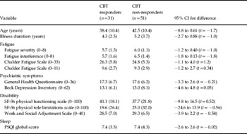
Clinically, CBT showed moderate efficacy in this group, with significant overall reductions in fatigue, disability and psychiatric symptoms (Table 1). Of the 82 patients with usable data, there were 31 responders (39%) and 51 non-responders (61%) on the CGI (Table 1). Analysis of baseline clinical features did not distinguish any clinical variables that were significantly associated with response to CBT (Table 2).
Baseline clinical features in subsequent responders and non-responders to CBT

CBT, Cognitive behavioural therapy; PSQI, Pittsburgh Sleep Quality Index; SF-36, 36-item short-form health survey; CI, confidence interval.
Values given as mean (standard deviation).
Comparison of baseline clinical variables by the independent t test revealed that no differences were statistically significant.
We examined the pretreatment data to see if there was any confounding effect of co-morbid depression on the endocrine variables. There was no significant difference between those with CFS alone and those with CFS and co-morbid depression on either 24-h UFC (92.8±59.0 and 82.1±50.4 nmol/day respectively, 95% CI −17 to 38.3) or on the salivary cortisol output (AUC values 73.2±23.0 and 67.3±20.7 nmol/l h respectively, 95% CI −6 to 17.7). This is similar to other research findings (Cleare et al. Reference Cleare, Miell, Heap, Sookdeo, Young, Malhi and O'Keane2001b; Roberts et al. Reference Roberts, Wessely, Chalder, Papadopoulos and Cleare2004) and we did not stratify for depression in the subsequent analyses.
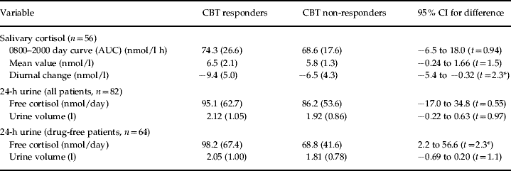
Looking at all patients, the pretreatment UFC was higher, but non-significantly so, in subsequent responders than non-responders. However, after excluding those taking medication liable to affect the HPA axis, there was a significant difference between those who went on to respond to CBT, in that responders had higher pretreatment UFC values (Table 3). The effect size calculation for this (using Cohen's d and a pooled s.d.) was 0.57, suggesting a medium size effect of UFC on CBT response.
Baseline endocrine measures in subsequent responders and non-responders to CBT

CBT, Cognitive behavioural therapy; AUC, area under the curve; CI, confidence interval.
Values are given as mean (standard deviation).
Asterisks show those variables that differed between responders and non-responders by an independent t test
(* p<0.05).
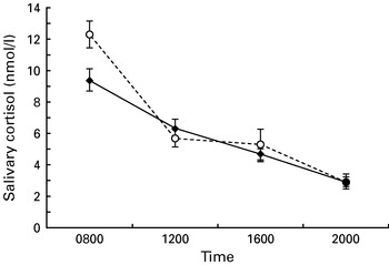
Total salivary cortisol output (AUC) was non-significantly higher in subsequent CBT responders than in non-responders (Table 3). However, the diurnal change in salivary cortisol was significantly different, being higher in subsequent responders than non-responders, indicating a flatter slope before treatment in the subsequent CBT non-responders. The effect size calculation for this (Cohen's d) was 0.62, a size of medium effect. Inspection of the diurnal profiles in salivary cortisol (Fig. 1) suggests that the main contributor to the flatter slope was the lower 0800 h value in the subsequent CBT non-responders.
Salivary cortisol levels across the day in patients with chronic fatigue syndrome (CFS) divided into those who subsequently responded to cognitive behavioural therapy (CBT responders; - -○- -) and those who subsequently did not respond to CBT (CBT non-responders; —◆—). Diurnal change is significantly less in CBT non-responders than in CBT responders (p<0.05, see Table 3).

We found that 29/82 subjects had a UFC value pretreatment of 59 nmol/day or less, and were categorized as having ‘low cortisol’ according to our predefined cut-off. The response rate in this group was 28%, compared to 43% in the remaining 53/82 subjects, a lower rate, but not of statistical significance (χ=1.99, p=0.16). There were no significant differences in any of the clinical variables between the ‘low cortisol’ subgroup and the rest of the patients.
To minimize the number of correlations, we used only the main endocrine outcomes (24-h UFC and the salivary cortisol AUC) and correlated them cross-sectionally with the questionnaire measures of fatigue (Chalder Fatigue Scale), psychiatric symptoms (GHQ), disability (WSAS) and sleep disturbance (PSQI) at baseline. We found a significant negative correlation between 24-h UFC and WSAS scores (r=−0.23, p=0.038), suggesting that lower cortisol was associated with higher levels of disability.
Discussion
As outlined in the introduction, one of the most often reported biological changes in CFS is HPA axis dysfunction, with reduced circulating cortisol likely to be present in at least a subgroup of patients with CFS (Cleare, Reference Cleare2003). What has been uncertain is the clinical relevance of this mild hypocortisolism. This study highlights intriguing implications about the link between endocrine dysfunction, CFS and CBT.
First, we were able to detect a clinical correlation between low basal cortisol levels (UFC) and higher disability scores in untreated CFS patients. The direction of causality in the correlation is not discernable from this study. It is certainly plausible that low cortisol levels lead to worsened functional capacity, or the perception of worsened functional capacity, given the association of low cortisol with fatigability in other states such as Addison's disease. However, equally plausible is that patients with worsened disability are less physically active, and hence have lower cortisol levels secondary to their level of activity, although we have no measure of actual levels of physical activity. The magnitude of the correlation was relatively low at −0.23, reinforcing that a relatively small amount of the variance in disability is explained by cortisol levels, and that many other factors are also likely to be relevant in determining disability in CFS.
Second, we found that those with a more dysregulated HPA axis, that is a lower basal cortisol (UFC) and a flatter diurnal cortisol slope (saliva), respond less well to a standard 12–15 sessions of CBT. No other studies have investigated the prognostic implications of HPA axis changes in CFS. Our findings add to the previous research noting that cortisol supplementation can lessen fatigue and disability, and provide further evidence that low cortisol levels could be acting as an additional, biological maintaining factor in some patients. We suggest that the additional effects of lowered cortisol make CBT either less effective or more difficult to implement in these patients. This might imply that such patients require a longer duration of therapy, or perhaps a modified version of therapy. Others have reported that those who respond less well to CBT are more persistently physically inactive (Prins et al. Reference Prins, Bleijenberg, Bazelmans, Elving, de Boo, Severens, van der Wilt, Spinhoven and van der Meer2001). If low cortisol is at least partly influenced by inactivity in CFS, then this may represent a similar group of CBT non-responsive patients.
Another possible implication of this finding derives from our previous demonstration that some patients benefit from hydrocortisone replacement therapy (Cleare et al. Reference Cleare, Heap, Malhi, Wessely, O'Keane and Miell1999). It may be that those with low levels of pretreatment cortisol could benefit from an initial dual treatment approach with low-dose hydrocortisone, which might facilitate subsequent CBT. However, if inactivity and sleep disturbance have indeed contributed to lowered cortisol levels, then CBT would seem to be the optimal way to reverse those factors and raise cortisol, in the long term, and without potential adverse effects of pharmacological steroid replacement.
An alternative explanation for the findings might be that the lower CBT responses in low-cortisol subjects is not attributable to the fact that they had low cortisol levels, but instead occurred because CBT was not flexible enough to cater to the particular needs of those patients, or was not the right approach. However, we could not see any differences on initial clinical assessment in those defined with ‘low cortisol’ compared to the rest of the group, or between the responders and non-responders, and all subjects had been judged initially suitable for CBT. Thus, although there may be unmeasured differences that characterized the non-responders, of those we collected cortisol was the one that most differentiated between subsequent non-responders and responders.
It is interesting that the main finding with regard to 24-h UFC predicting poor CBT response was only detectable in those who were free of medication likely to affect the HPA axis. This underscores the importance of studying medication-free patients wherever possible when attempting to understand the influence of biological factors in CFS.
It is also important to note that this was not a trial of CBT. We found an overall response rate of 39% in this group when assessed immediately after 6 months of therapy, but some studies have suggested that further improvement can occur in the 6 months following the end of the main therapy sessions (Deale et al. Reference Deale, Chalder, Marks and Wessely1997). Finally, it is notable that our findings on the influence of HPA axis changes on the response to CBT mirror those seen in depression. Thus, in major depression, which is characterized by HPA axis overactivity, CBT is less effective in those with a more overactive HPA axis, whereas we now show that in CFS, characterized by HPA axis underactivity, CBT is less effective in those with a more underactive HPA axis. This further attests to significant biological differences between major depression and CFS.
There are several limitations to the study that need discussion. This study was designed primarily to measure adrenal output of cortisol because this is the most reproduced finding in the CFS literature to date (Cleare, Reference Cleare2003). However, this means that we have not assessed other aspects of the HPA axis that may be of clinical relevance. As described, we assessed patients at 6 months to standardize the amount of CBT received. We cannot from these data look at the role of the HPA axis on the longer-term effects of CBT or on relapse. As noted earlier, one possibility is that it is the speed of response to CBT that is affected rather than the absolute response. Our sample showed a relatively low response rate to CBT, although response rates are somewhat lower outside of clinical trials than the rates seen within clinical trials (Quarmby et al. Reference Quarmby, Rimes, Deale, Wessely and Chalder2007) and our sample was selected to be compliant with endocrine research. Finally, our sample size may not have been sufficient to detect a priori who will have a poorer response to CBT using a measure such as the 24-h UFC, and larger samples will be needed to assess this possibility. At this stage we would not recommend that any patients be excluded from the opportunity to benefit from CBT, given the poor long-term prognosis of established CFS in the absence of treatment (Wessely et al. Reference Wessely, Hotopf and Sharpe1998).
In conclusion, this study suggests that HPA axis changes (reduced cortisol levels and a flattened diurnal release of cortisol) are of clinical relevance in CFS because they are associated with a poorer response to CBT. Regardless of whether it is one of the primary causes of the illness or an effect of the illness secondary to disrupted sleep, physical inactivity and deconditioning, and chronic stress, low cortisol levels could be an important maintaining factor contributing to symptom chronicity in at least some patients with CFS. This might imply that such patients require a longer duration of therapy, or perhaps a modified version of therapy, or alternative treatments alongside CBT to obtain maximum benefit.
Acknowledgements
We are grateful to D. Blair for her help in subject recruitment and to L. Poon in the National Affective Disorders Unit at The Bethlem Royal Hospital for help in specimen collection and sample processing. The study was funded by a project grant from the Linbury Trust. Dr Cleare, Professor Wessely and Professor Chalder are is supported by the NIHR Biomedical Research Centre at the South London and Maudsley NHS Foundation Trust and the Institute of Psychiatry (King's College London). Additional support for the study was provided by the Psychiatry Research Trust and the King's College Hospital Joint Research Committee. The funding sources had no further role in study design; in the collection, analysis and interpretation of data; in the writing of the report; and in the decision to submit the paper for publication.
Declaration of Interest
None.