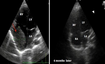
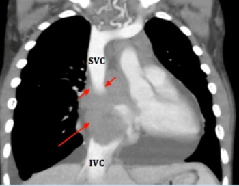
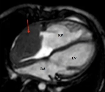
A nine-year-old male with a background of B-cell acute lymphoblastic leukaemia diagnosed at 2 years of age was referred for paediatric cardiology evaluation due to bradycardia, palpebral oedema, respiratory distress, increased neck volume, and odynophagia. He had a systolic murmur in the tricuspid area. The chest X-ray showed increased cardiothoracic ratio (70%). An electrocardiogram showed alternate sinus and junctional rhythm and periods of complete atrioventricular blockade. Transthoracic echocardiogram (ETT) showed a large mass inside the right atrium measuring 65 × 50 mm (Fig 1), that obstructed the superior caval vein drainage. There was moderate tricuspid regurgitation, dilated right-sided chambers, and pericardial and left pleural effusion. Cardiac function was globally preserved. Cardiac CT angiography (Fig 2) showed superior caval vein and sinus node artery compression by the mass. Cardiac MRI confirmed normal cardiac function and excluded extracardiac extension of the mass (Fig 3). Myelogram was performed and confirmed late relapse of acute lymphoblastic leukaemia. He was started on enoxaparin and initiated chemotherapy according to IntReALL 2010 sr protocol. Stem cell transplantation was indicated after minimal residual disease results above the positive cut-off at the end of induction. Six months after the start of chemotherapy, the ETT demonstrated a significant reduction of the cardiac mass, with a persisting residual mass (Fig 1, right), and decompression of the superior caval vein. In order to confirm absence of lymphoblasts in the residual mass that would be an indication for local radiotherapy (resulting in additional cardiac toxicity), cardiac biopsy under fluoroscopic and transesophageal echocardiogram guidance was performed. The sheath and the bioptome were inserted into the inferior caval vein, so that the tip of the sheath and the bioptome were directed to the mass inside the right atrium; after the sheath was in position, the bioptome was gently advanced to the end of the sheath, the jaws were opened against the mass and closed to obtain a piece; keeping the jaws closed, the bioptome was removed, and the specimen was placed in a container; multiple specimens were obtained. The procedure occurred without complications such as pneumothorax, arrhythmias, tricuspid valve injury, air and clot embolism, atrial perforation or pericardial tamponade.
2D transthoracic echocardiogram, apical four-chamber view, demonstrating giant mass into the right atrium (arrow), left atrium (LA), right atrium (RA), right ventricle (RV), left ventricle (LV).

Cardiac angio TC coronal view, demonstrating superior caval vein (SVC) compression (short arrow), mass inside the right auricle (long arrow).

Cardiac MRI scan, four-chamber view, demonstrating giant mass into the right atrium (arrow), left atrium (LA), right ventricle (RV), left ventricle (LV).

The biopsy did not reveal blast cells so allogeneic stem cell transplantation was performed and patient remained in remission.
He is currently asymptomatic. Chemotherapy induced complete disease response, with a remaining soft tissue mass with no active disease and a favourable clinical outcome.
Most Acute lymphoblastic leukaemia (ALL) relapses occur during treatment or within the first 2 years after treatment completion and can occur in an isolated form or combined with the involvement of another site, less frequently involving other extramedullary site. Reference Locatelli, Schrappe, Bernardo and Rutella1 Primary cardiac tumours are rare, and metastasis is more common. Reference Kumar, Tigadi and Azrin2 The investigation of atrial mass usually requires multimodality imaging and sometimes a biopsy to affirm histological diagnosis. The echocardiography is usually the initial modality of imaging. Reference Kumar, Tigadi and Azrin2,Reference Obeid, al Mudamgha and Smulyan3 The MRI or contrast-enhanced CT scan is sensitive for detecting tumour infiltration into the myocardium or mediastinal structures. The biopsy of a cardiac mass remains a difficult and risky procedure, but the guidance of fluoroscopy or transesophageal echocardiography decreases the risk of heart lesion. With intensive combination chemotherapy and allogeneic haematopoietic stem cell transplantation, 60% of all children with late combined relapsed B-cell ALL can be cured. Reference Locatelli, Schrappe, Bernardo and Rutella1
Acknowledgements
We thank, Graça Sousa and Ana Teixeira for their contribution.
Financial support
This research received no specific grant from any funding agency, commercial, or not-for-profit sectors.
Conflicts of interest
None.