Introduction
Nectonema Verrill, 1879 is the sole marine genus in the phylum Nematomorpha (horsehair worms). Nectonema differs morphologically from other nematomorphs in having natatory bristles (present only in adults) and dorsal and ventral nerve cords, among other characters (Schmidt-Rhaesa Reference Schmidt-Rhaesa and Schmidt-Rhaesa2013). Since the first discovery of Nectonema around 1870 (Ward Reference Ward1892a), knowledge of this genus has accumulated through reports on detailed external and internal morphology (e.g., Ward Reference Ward1892a), the parasitic nature of its juveniles (Ward Reference Ward1892b), egg structure (Ward Reference Ward1892a; Huus Reference Huus1932), mating behaviour (Huus Reference Huus1932), development in the host (Huus Reference Huus1932; Schmidt-Rhaesa Reference Schmidt-Rhaesa1996), species diversity (Verrill Reference Verrill1879; Nierstrasz Reference Nierstrasz1907; Bock Reference Bock1913; Brinkmann Reference Brinkmann1930; Poinar and Brockerhoff Reference Poinar and Brockerhoff2001), host diversity (e.g., Schmidt-Rhaesa et al. Reference Schmidt-Rhaesa, Pohle and Gaudette2013, Kakui et al. Reference Kakui, Fukuchi and Shimada2021; Kakui Reference Kakui2024), and the chromosome-level genome (Cunha et al. Reference Cunha, de Medeiros, Lord, Sørensen and Giribet2023). Nectonema nonetheless remains enigmatic, with much of its biology – such as early development, true species and host diversity, and habitat use – remaining poorly known.
Nectonema nematomorphs have been reported as parasites in various decapod and isopod hosts (Supplementary Table S1). Although most of these records involved juvenile individuals lacking natatory bristles, some adults have been collected from inside host crustaceans (e.g., Brinkmann Reference Brinkmann1930; Schmidt-Rhaesa Reference Schmidt-Rhaesa1996) and likely represented individuals just before emergence. Some studies have suggested that Nectonema species exhibit high host specificity (Brinkmann Reference Brinkmann1930; Born Reference Born1967), although there are contradictory reports of several species occurring in a wide range of hosts (Supplementary Table S1). It remains unclear whether the latter species with apparently broad host utilisation include cryptic species with narrow specificity, implying higher true species diversity than is presently known.
Nectonema is taxonomically challenging. Morphological differences among species are subtle, and the developmental stages and sexes of specimens used for species descriptions have not been consistent. There are currently five named species. Poinar and Brockerhoff (Reference Poinar and Brockerhoff2001) distinguished them based on the colouration of the anterior body region (dark or translucent), presence or absence of an anterior chamber, arrangement of rows of natatory bristles in adults, and stoma shape in juveniles, among other characters. Males, females, and juveniles have been described only for Nectonema agile Verrill, Reference Verrill1879 (type locality: Massachusetts, USA, northwestern Atlantic) and Nectonema munidae Brinkmann, Reference Brinkmann1930 (type locality: near Bergen, Norway, northeastern Atlantic). For the other species, information is available only for males in Nectonema melanocephalum Nierstrasz, Reference Nierstrasz1907 (type locality: Pulau Kawassang, Indonesia, eastern Indian Ocean), females in Nectonema svensksundi Bock, Reference Bock1913 (type locality: Spitzbergen Island, Norway, Arctic), and juveniles in Nectonema zealandica Poinar and Brockerhoff, Reference Poinar and Brockerhoff2001 (type locality: South Island, New Zealand, southern Pacific). The latter three species have not been reported since their original descriptions.
During a faunal survey around Kikaijima Island, Japan, the second author collected one shrimp parasitised by a Nectonema nematomorph, which we here describe as a new species. This is the fifth published record of Nectonema from Japanese waters (Oku et al. Reference Oku, Fukumoto, Ohbayashi and Koike1983; Yoshida Reference Yoshida2016; Kakui et al. Reference Kakui, Fukuchi and Shimada2021; Kakui Reference Kakui2024; this study). For phylogenetic inference, future DNA barcoding efforts, and accurate host–parasite pairing, we determined a partial sequence for the 18S rRNA (18S) gene from the nematomorph and a partial sequence for the cytochrome c oxidase subunit I (COI) gene from the host. To assess our species’ taxonomic position, we conducted a phylogenetic analysis based on 18S from the new species and other Nectonema taxa for which 18S data were available.
Material and methods
The infected shrimp was collected by snorkeling from coral rubble at 4 m depth at Keraji, Kikaijima Island, Ryukyu Islands, Japan (28.288667°N, 129.975722°E) on 27 May 2025. It was photographed alive and then dissected to extract the nematomorph. The latter was photographed and video-recorded alive. The host and nematomorph were then fixed and preserved in 99% ethanol.
The fixed nematomorph was cut into several pieces. The anterior and posterior parts were mounted on a glass slide in glycerin and observed with an Olympus BX53 compound microscope. One of two subanterior fragments was used for DNA extraction, and another fragment was treated with hexamethyldisilazane (HMDS; Nation Reference Nation1983), sputter-coated with gold, and observed at 15 kV accelerating voltage with a scanning electron microscope (SEM; S-3000N, Hitachi, Japan). The remaining part was preserved in 99% ethanol. The host was dissected with forceps, observed with a Nikon SMZ1500 stereoscopic microscope, and identified to genus based on Banner and Banner (Reference Banner and Banner1982), Poore and Ahyong (Reference Poore and Ahyong2023), Anker (Reference Anker2023), and Ashrafi et al. (Reference Ashrafi, Ďuriš and Anker2024). Illustrations were prepared with Adobe Illustrator CS6 from draft line drawings made with a drawing tube. The body length of the fresh nematomorph was measured from the anterior to the posterior end, and the body width at the widest portion. The carapace length of the host was measured from the orbital margin to the midpoint of the posterodorsal margin. All measurements were made from digital images by using ImageJ (Schneider et al. Reference Schneider, Rasband and Eliceiri2012). The studied material was deposited in the Invertebrate Collection of the Hokkaido University Museum (ICHUM), Sapporo, Japan, under catalog number ICHUM9108.
Total DNA was extracted from part of the body of the nematomorph and from muscle tissue of the host by using a NucleoSpin Tissue XS Kit (Macherey-Nagel, Germany). For COI, primers LCO1490 and HCO2198 (Folmer et al. Reference Folmer, Black, Hoeh, Lutz and Vrijenhoek1994; Supplementary Table S2) were used for PCR amplification and cycle sequencing (CS); COI amplification was unsuccessful for the nematomorph. For 18S, primers SR1 and SR12 (Nakayama et al. Reference Nakayama, Watanabe, Mitsui, Uchida and Inouye1996) were used for PCR amplification (18S amplification was unsuccessful for the host), and 18S-b3F, 18S-b3R, 18S-b4F, 18S-a4R, 18S-b5F, 18S-b6F, 18S-a6R, and 18S-b8F (Kakui et al. Reference Kakui, Katoh, Hiruta, Kobayashi and Kajihara2011, Reference Kakui, Fukuchi and Shimada2021; Kakui and Shimada Reference Kakui and Shimada2017, Reference Kakui and Shimada2022; Kakui and Hiruta Reference Kakui and Hiruta2022; Supplementary Table S2) for CS. PCR amplification conditions for COI and 18S with KOD One PCR Master Mix (Toyobo, Japan) were 45 cycles of 98°C for 10 s, 56°C (COI) or 60°C (18S) for 5 s, and 68°C for 1 s (COI) or 10 s (18S). All nucleotide sequences were determined with a QuantumDye Terminator v3.1 Cycle Sequencing Kit (Quintara Biosciences, USA) and a 3130 or 3730 DNA Analyzer (Life Technologies, USA). Fragments were assembled by using MEGA7 (Kumar et al. Reference Kumar, Stecher and Tamura2016). The sequences were deposited in the International Nucleotide Sequence Database (INSD) through the DNA Data Bank of Japan (DDBJ). BLAST searches (Altschul et al. Reference Altschul, Gish, Miller, Myers and Lipman1990) were conducted to determine the nucleotide sequences in the INSD most similar to those we determined.
The 18S dataset for phylogenetic analysis comprised the dataset from Kakui (Reference Kakui2024) and the one nematomorph sequence we determined. The sequences were aligned (1710 positions in the aligned dataset; see Supplementary Files S1 and S2) as described by Kakui (Reference Kakui2024). Methods for selecting the optimal Q mixture model (MIX{TIM3+FO,JC+FO}+R2), the maximum likelihood (ML) analysis, estimation of branch support (1000 pseudoreplicates for both Shimodaira–Hasegawa-like approximate likelihood ratio tests [SH-aLRT] and standard bootstraps [BS]), and tree drawing were as described by Katsuragawa et al. (Reference Katsuragawa, Shiraki, Shimomura and Kakui2025). Kimura (Reference Kimura1980) 2-parameter (K2P) distances among Nectonema species were calculated with MEGA7.
This article is registered in ZooBank under: https://zoobank.org/References/A291194E-1583-4A0D-A3BD-2C57C8FD518A.
Results and discussion
Host animal
The host shrimp, a female lacking both chelipeds, was identified morphologically as Alpheus sp. A BLAST search showed the 658-bp partial COI sequence we determined from the host was most similar to the COI sequence from Alpheus edwardsii H. Milne Edwards, 1837 (PP651964; query cover 100%, identity score 83.41%; McIlroy et al. Reference McIlroy, Guibert, Archana, Chung, Emmett Duffy, Gotama, Hui, Knowlton, Leray, Meyer, Panagiotou, Paulay, Russell, Thompson and Baker2024), which supported our generic identification based on morphology.
Taxonomy
Nectonema Verrill, 1879
[New Japanese name: Oyogi-hariganemushi-zoku]
Nectonema shimadai sp. nov.
[New Japanese name: Asahi-oyogi-hariganemushi]
Figs. 1–3, Supplementary Movies S1, S2
https://zoobank.org/NomenclaturalActs/E4F98AAF-7AB2-43F0-B77C-FABE40E541C0
Type host
Alpheus sp. (Decapoda: Caridea: Alpheidae).
Occurrence in host
Occupying host body cavity from anterior end of cephalothorax to posterior end of pleonite 5 (Fig. 1a, b).
Nectonema shimadai sp. nov. parasitic in Alpheus sp. (a, b) Alpheus sp. containing the holotype of Nectonema shimadai sp. nov., dorsal (a) and left (b) views, fresh specimen. (c) Nectonema shimadai sp. nov., holotype, partly damaged fresh specimen. (c’, c”) Same, enlarged views of the anterior and posterior ends, respectively. Arrowhead, giant cell; arrow, female gonopore.

Type locality
Keraji, Kikaijima Island, Ryukyu Islands, Japan (28.288667°N, 129.975722°E), 4 m depth.
Type material
Holotype. Adult female, ICHUM9108, one slide, one SEM stub, and one vial with nematomorph (one vial with host included under same catalog number); body length 49.0 mm, body width 340 μm, ex Alpheus sp. (female, carapace length 3.7 mm), collected at the type locality on 27 May 2025 by Shoki Shiraki.
Etymology
The specific name is a noun in the genitive case and honours Daisuke Shimada, a nematode taxonomist and long-standing collaborator with the first author in research on Nematoida.
Representative sequences
The 18S sequence (LC909668; 1894 bp) was determined from the holotype. The COI sequence (LC909669; 658 bp, encoding 219 amino acids) was determined from the host shrimp.
Description of a female
Body (Fig. 1c; Supplementary Movie S1) elongate, nearly cylindrical but gradually tapering towards both ends, 144 times as long as wide. Anterior region translucent; remaining part salmon pink when alive. Fine transverse striations present from subanterior to subposterior region (Figs. 2, 3). Dorsal and ventral surfaces each with two rows of natatory bristles; many bristles lost, but observed from subanterior to subposterior regions (Fig. 2a, c, d). Anterior end (Figs. 1c’, 2a, b; Supplementary Movie S2) covered with short bristles. Anterior margin bilobed in dorsal view, with shallow median furrow. Cephalic papillae not observed. Distinct anterior chamber (ca. 0.3 mm long) present, with four giant cells. Mouth opening anteroventrally. Oral cavity reduced. Sclerotised proboscis absent. Pharynx extending beyond septum, forming loop posteriorly. Intestine not observed. Developing gonads (?) in the body cavity. Posterior end (Figs. 1c”, 2c, d) rounded; gonopore opening posteriorly.
Nectonema shimadai sp. nov., holotype, ethanol-fixed, deformed specimen in glycerin. (a, b) Anterior portion, right dorsal view, drawing (a) and light micrograph (b); ventral rows of natatory bristles not shown in (a). (c, d) Posterior portion, lateral view, drawing (c), and light micrograph (d). In (a), internal structures (pharynx and giant cells) are drawn in gray lines. Abbreviations: ac, anterior chamber; gc, giant cell; gp, gonopore; mo, mouth; nb, natatory bristle; ph, pharynx.

Nectonema shimadai sp. nov., holotype, fragment of laterally flattened portion, lateral view, SEM image showing fine transverse striations on body.

Males and juveniles unknown.
Phylogenetic analysis and genetic divergence
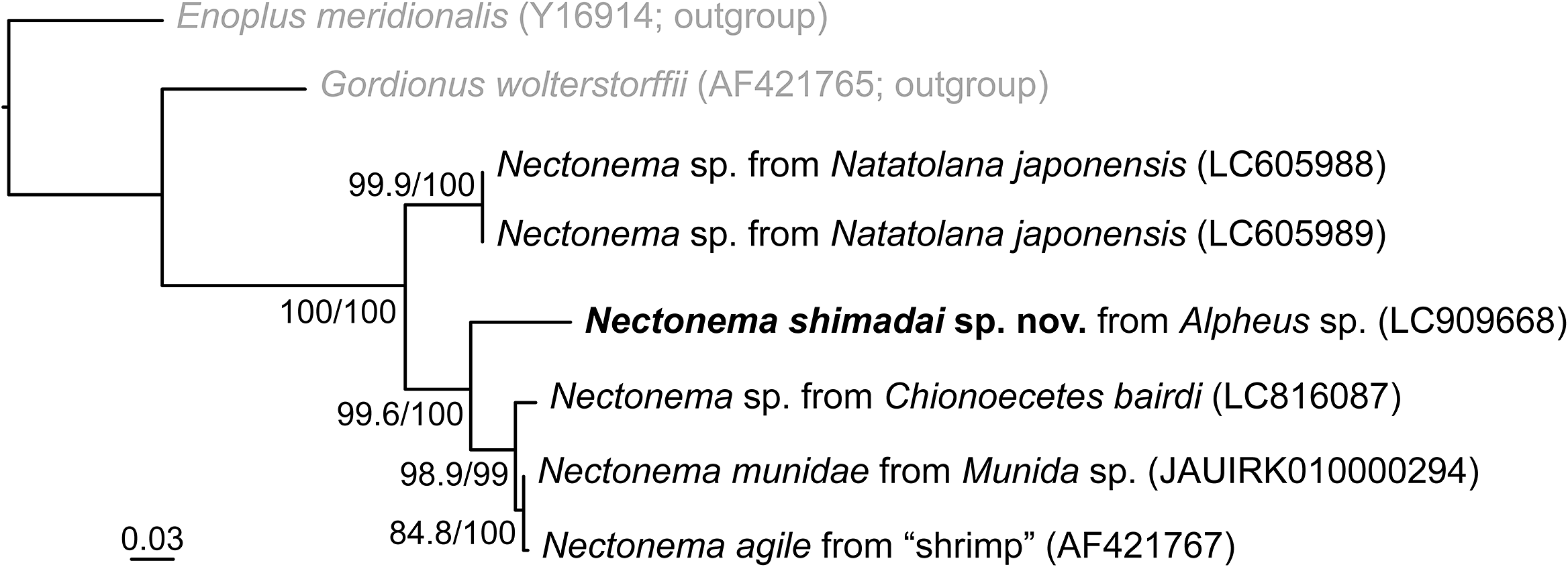
The 18S sequence of N. shimadai sp. nov. was 12.8%, 9.8%, 9.6%, and 9.3% divergent (K2P distance) from homologous sequences from Nectonema sp. parasitic in the isopod Natatolana japonensis (LC605988 and LC605989; Kakui et al. Reference Kakui, Fukuchi and Shimada2021), Nectonema sp. parasitic in the crab Chionoecetes bairdi (LC816087; Kakui Reference Kakui2024), N. munidae (JAUIRH010000294; Cunha et al. Reference Cunha, de Medeiros, Lord, Sørensen and Giribet2023), and N. agile (AF421767; Bleidorn et al. Reference Bleidorn, Schmidt-Rhaesa and Garey2002), respectively. In the 18S-based ML tree (Fig. 4), the four decapod-parasitic species form a highly supported clade (SH-aLRT/BS = 99.6%/100%), with N. shimadai sp. nov. the sister taxon to a highly supported (98.9%/99%) clade comprising the others. Pairwise K2P distances between N. shimadai sp. nov. and members of the latter clade (9.3–12.8%) were greater than those among the three species within the clade (0.3–2.3%).
Maximum-likelihood tree for Nectonema reconstructed from 18S sequences (1710 positions); numbers near nodes are Shimodaira–Hasegawa-like approximate likelihood ratio test (SH-aLRT)/standard bootstrap (BS) values as percentages; scale at bottom indicates branch length in substitutions per site.

Remarks
Nectonema shimadai sp. nov. has a translucent anterior body region and a distinct anterior chamber, character states that distinguish it from adults of the congeners N. melanocephalum and N. svensksundi, which have a dark anterior body region and lack a distinct anterior chamber. Like N. shimadai sp. nov., adult N. agile and N. munidae have a translucent anterior body region and a distinct anterior chamber, but N. shimadai sp. nov. differs from them in having a salmon-pink body colour when alive (grayish or yellowish white in N. agile; whitish-yellow [females] or semi-transparent [males] in N. munidae; Verrill Reference Verrill1879; Brinkmann Reference Brinkmann1930). The 18S data showed that N. shimadai sp. nov. is highly divergent from these two species. In addition, the three species may parasitise different hosts, although N. agile and N. munidae have been reported from various decapod species (Supplementary Table S1; see also discussion below). The currently known host for N. shimadai sp. nov. is Alpheus sp. (Caridea: Alpheidae). The regular hosts of N. agile and N. munidae are considered to be Palaemon vulgaris Say, 1818 (Caridea: Palaemonidae) and Munida tenuimana G.O. Sars, 1872 (Anomura: Munididae), respectively (Brinkmann Reference Brinkmann1930; Born Reference Born1967).
Our female of Nectonema shimadai sp. nov. is 340 μm wide, which is narrower than juveniles (including “pre-adult” sensu Poinar and Brockerhoff [Reference Poinar and Brockerhoff2001]) of N. zealandica, even when compared with juveniles that are shorter than our specimen. Poinar and Brockerhoff (Reference Poinar and Brockerhoff2001, 151) gave the length of N. zealandica as 13 mm (parasitic juvenile) to 300 mm (pre-adult), and the maximum width as 472 μm (parasitic juvenile) to 737 μm (pre-adult). In addition, although it may be different between juveniles and adults, the colour of fresh N. zealandica juveniles was white to cream rather than salmon pink as in N. shimadai sp. nov. Nectonema zealandica uses a host decapod in a different infraorder from that of N. shimadai sp. nov. and was reported from far from Japan: it was parasitic in Hemigrapsus sexdentatus (H. Milne Edwards, 1837) (Brachyura: Varunidae) from South Island, New Zealand, southern Pacific. Given the various differences, we concluded that N. shimadai sp. nov. is not conspecific with N. zealandica.
Alpheidae, the family to which the host bearing N. shimadai sp. nov. belongs, is one of the most species-rich decapod families (De Grave et al. Reference De Grave, Decock, Dekeyzer, Davie, Fransen, Boyko, Poore, Macpherson, Ahyong, Crandall, de Mazancourt, Osawa, Chan, Ng, Lemaitre, van der Meij and Santos2023), yet published reports of Nectonema utilising species in this family are scarce. Prior to our study, Yamashita et al. (Reference Yamashita, Komai and Kon2023: fig. 1B) presented an image of an alpheid likely parasitised by a Nectonema nematomorph. Host surveys targeting alpheids will likely reveal additional cases of Nectonema infection.
Species sharing the same host group or ocean were not monophyletic in our 18S tree. Nectonema shimadai sp. nov., a caridean parasite from the northwestern Pacific, was the sister group to a clade comprising Nectonema sp. (a brachyuran parasite from the northwestern Pacific), N. munidae (an anomuran parasite from the northeastern Atlantic), and N. agile (a caridean parasite from the northwestern Atlantic). In addition, N. shimadai sp. nov. showed a markedly larger genetic distance from members of the clade than the latter did among themselves. Nectonema shimadai sp. nov. was collected from warm, shallow water, whereas the other three were from cold, shallow to deep waters (cf. Spalding et al. Reference Spalding, Fox, Allen, Davidson, Ferdaña, Finlayson, Halpern, Jorge, Lombana, Lourie, Martin, McManus, Molnar, Recchia and Robertson2007). The inferred tree might thus reflect genetically distant warm- and cold-water lineages within decapod-parasitic nematomorphs.
The true species diversity of Nectonema may be much higher than currently recognised. Information is lacking for the early stages in the life cycle of Nectonema, i.e., from hatching to the stage preceding infection of the definitive host. We thus cannot rule out the possibility that this group uses a broadly distributed planktonic paratenic host, which would enable passive long-distance dispersal. However, given that Nectonema appears to have a short adult lifespan (Brinkmann Reference Brinkmann1930) and high host specificity (Brinkmann Reference Brinkmann1930; Born Reference Born1967), it likely contains many locally endemic species. The broad trans-Atlantic distribution and high host diversity reported for N. agile suggest that its species boundaries merit closer examination, especially in regard to whether it represents a single species or a multiple-species complex. Nectonema remains taxonomically challenging, but continued accumulation of molecular data from both juvenile and adult stages, together with the formal description of clearly distinguishable adults, represents a steady and important approach to elucidating the taxonomy.
Supplementary material
The supplementary material for this article can be found at http://doi.org/10.1017/S0022149X26101229.
Acknowledgements
We thank Yoshinobu Matsushima and Rintaro Ono for help in sampling; the staff of the KIKAI Institute for Coral Reef Sciences for providing laboratory facilities; Yuki Oya for help in sample processing; Shogo Sekiguchi and Yoshinobu Matsushima for help in molecular work; and Matthew H. Dick for reviewing the manuscript and editing the English.
Financial support
This study was supported in part by a grant (25B002) from the Kurita Water and Environment Foundation and a Grant-in-Aid for JSPS Fellows (JP23KJ0063) from the Japan Society for the Promotion of Science (JSPS).
Competing interests
The authors declare none.