Introduction
Vymazalováite, Pd3Bi2S2, was found in vein-disseminated pyrite-chalcopyrite-galena ores in the Talnakh deposit, Noril'sk, Russian Federation at 69°30'20”N and 88°27'17”E. The host rocks are diopside- hydrogrossular-serpentine metasomatites developed in diopside-monticellite skarns where monticellite is replaced by hydrogrossular and serpentine. These rocks lie ~30 m below the lower contact of the Talnakh intrusion (eastern part of the Komsomolsky mine). Mineralization represents the outermost part of the disseminated ores and is characterized by a lack of Ni minerals, also with no Ni content in the platinum-group minerals. The ores are known for their large galena content in an association of pyrite and chalcopyrite with abundant Pd-, Pt-, Ag- and Au-bearing minerals. Silver and gold not only form alloys but also are commonly incorporated in sulfides, selenides, sulfo-selenides and telluro-sulfoselenides (Sluzhenikin and Mohkov, Reference Sluzhenikin and Mokhov2015). These ores were formed during hydrothermal replacement of skarns.
Vymazalováite forms small (from a few μm to 35 µm) inclusions or euhedral grains, generally rectangular in polished section, in intergrowths of polarite, sobolevskite, acanthite and unnamed (Pd,Ag)5BiS2 in galena, rarely in chalcopyrite. Less commonly, the mineral occurs in aggregates (from a few μm to 35 µm) composed of intergrowths of telargpalite, braggite, vysotskite, sopcheite, stibiopalladinite, sobolevskite, moncheite, kotulskite, malyshevite (PdCuBiS3), insizwaite, acanthite, Au-bearing silver and the newly described mineral kravtsovite (PdAg2S) (Vymazalová et al. Reference Vymazalová, Laufek, Sluzhenikin, Stanley, Kozlov, Chareev and Lukashova2017) in association with pyrite, chalcopyrite and galena (Fig. 1).
(a) reflected light image of vymazalováite enclosed in galena and chalcopyrite; (b) back-scattered electron (BSE) images of an enlargement (marked as square) showing vymazalováite intergrown with acanthite in chalcopyrite; (c) reflected-light image of vymazalováite grain rimmed by polarite and sobolovskite, intergrown with acanthite and chalcopyrite in galena; (d) similar area to (c) in BSE; (e) reflected-light image illustrating euhedral grain of vymazalováite enclosed in galena; and (f) similar area to (e) in BSE. Key: vym – vymazalováite; gn – galena, ccp – chalcopyrite, sob – sobolevskite; pol – polarite; ac – acanthite; di - diopside.

Both the mineral and name were approved by the Commission on New Minerals, Nomenclature and Classification of the International Mineralogical Association (IMA 2016-105). The mineral name vymazalováite is chosen to honour Dr Anna Vymazalová (b. 1974) research scientist at the Czech Geological Survey for her contributions to the ore and experimental mineralogy of the platinum-group minerals and for her participation in the description of several new minerals. The holotype (polished section) is deposited at the Department of Earth Sciences of the Natural History Museum, London, UK, catalogue No BM 2016, 150. According to the chemical composition the mineral belongs to the 02.BC group (sulfides with Pd) of the Strunz & Nickel classification scheme (Strunz and Nickel, Reference Strunz and Nickel2001).
Appearance, physical and optical properties
In plane-polarized reflected light, vymazalováite is creamy grey in the assemblage with galena and chalcopyrite (Fig. 1). It is isotropic and displays no internal reflections.
Vymazalováite is opaque with a metallic lustre. The powder of synthetic vymazalováite is grey in colour and has a grey streak. Synthetic vymazalováite is brittle. The density calculated on the basis of the empirical formula and cell dimensions of synthetic vymazalováite is 9.25 g/cm3.
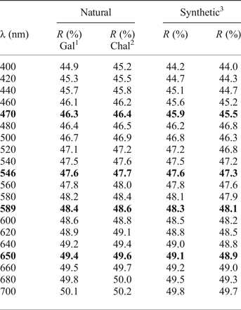
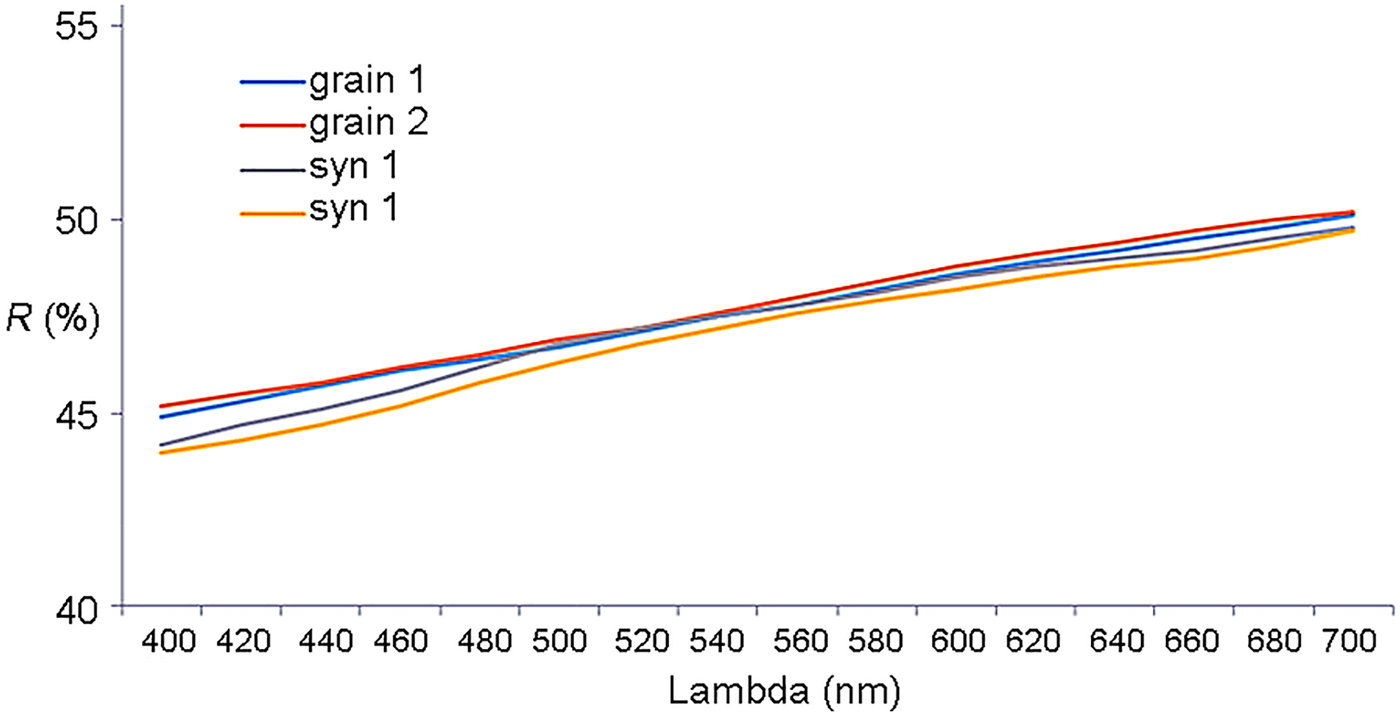
Reflectance measurements were made in air relative to a WTiC standard on both natural and synthetic vymazalováite using a J & M TIDAS diode array spectrometer attached to a Zeiss Axiotron microscope. The results are tabulated (Table 1) and illustrated in Fig. 2. The reflectance spectra for natural and synthetic vymazalováite are in good agreement bearing in mind the effect of the minor element substitutions in the natural grains and the fine grain size of the inclusions in galena and chalcopyrite.
Reflectance data for vymazalováite and synthetic grains of Pd3Bi2S2. Grain 1 is of an inclusion in galena. Grain 2 an inclusion in chalcopyrite.
Reflectance data for natural and synthetic vymazalováite.*

*Wavelengths required by the Commission on Ore Mineralogy are given in bold. R is the reflectance at a given wavelength for a cubic mineral.
1Gal is for an inclusion in galena;
2Chal is for an inclusion in chalcopyrite.
3Two grains of the synthetic vymazalováite were measured.
Chemical composition
Electron probe micro-analysis (EPMA) on grains of natural vymazalováite were obtained using an Inca Wave 500 (WDS, Oxford Instruments) installed on an SEM Lyra 3GM (TESCAN), at the TESCAN demonstration laboratory in Moscow, Russia, with analytical conditions 20 kV, 10 nA and counting times 30 s (on peak positions)/2×15 s (background on left and right positions). The spectra were collected on PbMα, BiMα, PdLα, AgLα, SKα and SeLα lines with standards of pure Se, Pd, Ag, Bi, synthetic PbTe and natural FeS2 (pyrite).
Synthetic vymazalováite was investigated using a CAMECA SX-100 electron probe microanalyser in wavelength-dispersive mode with an electron beam focused to 1–2 µm at the Institute of Geology of the Czech Academy of Sciences, Prague. Pure elements and FeS2 were used as standards and the radiations measured were BiMα PdLα, SKα, with an accelerating voltage of 15 kV and a beam current of 10 nA measured on the Faraday cup.
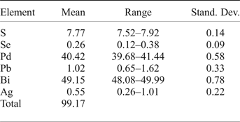
Results of the EPMA are given in Table 2. The empirical formula calculated on the basis of 7 atoms per formula unit is Pd3.05(Bi1.89Ag0.04Pb0.04)Σ1.97(S1.95Se0.03)Σ1.98 for natural vymazalováite and Pd3.04Bi1.97S1.99 for synthetic vymazalováite giving a simplified formula of Pd3Bi2S2.
Electron probe micro-analyses (wt.%) for vymazalováite (mean of 7 analyses).

Stand. Dev: standard deviation.
Synthetic analogue
The small size and nature of the intergrowths of vymazalováite with polarite, galena and chalcopyrite prevented its extraction and isolation in an amount sufficient for the relevant crystallographic and structural investigations. Therefore, these investigations were performed on the synthetic phase Pd3Bi2S2, being equivalent to natural vymazalováite.
The synthetic Pd3Bi2S2 phase was prepared using an evacuated silica glass tube method in the Laboratory of Experimental Mineralogy of the Czech Geological Survey in Prague. Palladium (99.95%), bismuth (99.999%) and sulfur (99.999%) were used as starting materials for synthesis. The evacuated tube with its charge was sealed and annealed. After cooling using a cold-water bath, the charge was ground into powder in acetone using an agate mortar and mixed thoroughly to homogenize. The pulverized charge was sealed in an evacuated silica-glass tube again and heated at 400°C for 70 days. The experimental product was quenched rapidly in cold water.
Crystallography
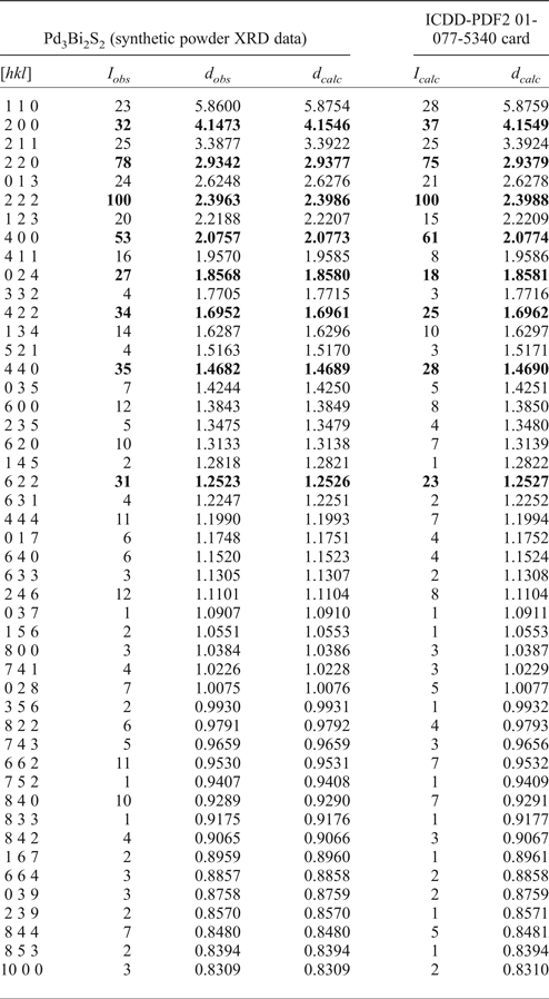
The crystal structure of synthetic Pd3Bi2S2 was solved using single-crystal work by Weihrich et al. (Reference Weihrich, Matar, Eyert, Rau, Zabel, Andratschke, Anusca and Bernert2007) and Weihrich and Anusca (Reference Weihrich and Anusca2006) (ICDS database card No. 417634) and also confirmed by experimental study and the powder X-ray diffraction (XRD) data obtained from the synthetic analogue (provided by A. Vymazalová). The data are as follows: the mineral is cubic, space group I213, with a = 8.3097(9) Å, V = 573.79(1) Å3 and Z = 4. The strongest lines in the powder XRD pattern of synthetic vymazalováite [d in Å (I) (hkl)] are: 4.15(32)(200), 2.93(78)(220), 2.40(100)(220), 2.08(53)(400), 1.695(34)(422), 1.468(35)(440) and 1.252(31)(622). Complete X-ray data are listed in the International Centre for Diffraction Data database (ICDD-PDF2 01-077-5340 card). X-ray data for synthetic Pd3Bi2S2 are given in Table 3.
Powder XRD data (d in Å) for synthetic vymazalováite compared to data taken from ICDD-PDF2 01-077-5340 card (I ≥ 1).

The eight strongest lines are given in bold.
The structural identity of the natural vymazalováite with the structural model proposed by Weihrich et al. (Reference Weihrich, Matar, Eyert, Rau, Zabel, Andratschke, Anusca and Bernert2007) for the synthetic Pd3Bi2S2 phase was confirmed by electron back-scatter diffraction (EBSD) measurements on the natural sample.
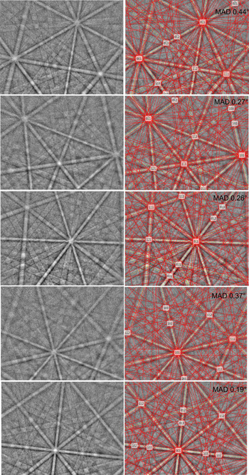
TESCAN Lyra 3 GMH and Mira 3 scanning electron microscopes combined with Oxford Instruments X-Max80 (EDS), NordlysMax3 (EBSD) and Inca Wave500 (WDS) detectors were used for the measurements. The surface of the natural sample was prepared for investigation by polishing with colloidal silica and/or ion milling with a focused ion gun (FIB). EBSD patterns (EBSPs) were collected and processed using a proprietary computer program AztecHKL (Oxford Instruments). The centres of twelve Kikuchi bands were automatically detected using the Hough transform routine with a resolution of 60 (internal Hough space resolution parameter in the AztecHKL software). The solid angles calculated from the patterns were compared with a synthetic Pd3Bi2S2 match [using Weihrich et al. (Reference Weihrich, Matar, Eyert, Rau, Zabel, Andratschke, Anusca and Bernert2007) crystal-structure data converted with Twist software (Oxford Instruments)] to index the patterns. The EBSD patterns (also known as Kikuchi patterns) obtained from the natural material (twenty-two measurements on different spots on natural vymazalováite) were found to match the patterns generated from the structural model described by Weihrich et al. (Reference Weihrich, Matar, Eyert, Rau, Zabel, Andratschke, Anusca and Bernert2007) for synthetic Pd3Bi2S2. The values of the mean angular deviation (MAD, i.e. goodness of fit of the solution) between the calculated and measured Kikuchi bands range between 0.16° and 0.49°. These values reveal a very good match; as long as values of mean angular deviation are <1°, they are considered as indicators of an acceptable fit (see Fig. 3).
EBSD images of vymazalováite obtained from grains with different orientations; in the right pane the Kikuchi bands are solved (indexed) using 92 reflector planes. Acquisition conditions: accelerating voltage = 30 kV, beam current = 0.3 nA, no binning (full EBSP resolution is 1344 x 1024). Indexing conditions: refined accuracy mode, 12 bands, 92 reflectors.

The crystal structure of vymazalováite is a structure type of corderoite (Hg3S2Cl2). It belongs to the antiperovskite superstructure, and is structurally related to pašavaite (Pd3Pb2Te2), shandite and parkerite and can be called a ‘cubic parkerite’. It is related closely to the monoclinic parkerite, differing in the occupation of one Pd position only. The three structure types of shandites, monoclinic and cubic parkerites could be derived from a modelling scheme that uses a CsCl or perovskite superstructure (Weihrich et al. Reference Weihrich, Matar, Eyert, Rau, Zabel, Andratschke, Anusca and Bernert2007). On the basis of its chemical composition the mineral is the Bi analogue of laflammeite (Pd3Pb2S2).
Acknowledgements
This work was supported by the Russian Scientific Foundation (grant No 14-07-00693) at the IGEM RAS. The authors thank the two referees, Louis Cabri and David Vaughan, and the Editor Peter Williams whose comments enhanced the manuscript.