Introduction
Among the numerous species considered to be invasive in coastal environments around the world, many decapod crustaceans have been recorded, such as Carcinus maenas (Linnaeus, 1758) (Geller et al., Reference Geller, Walton, Grosholz and Ruiz1997); Callinectes sapidus Rathbun, 1896 (Stasolla & Innocenti, Reference Stasolla and Innocenti2014; Garcia et al., Reference Garcia, Pinya, Colomar, París, Puig, Rebassa and Mayol2018); Charybdis hellerii (A. Milne-Edwards, 1867) (Campos & Türkay, Reference Campos and Türkay1989; Mantelatto & Luiz-Diaz, Reference Mantelatto and Luiz-Diaz1999; Dineen et al., Reference Dineen, Clark, Hines, Reed and Heather2001; Evans et al., Reference Evans, Zill and Paulay2018); Lysmata lipkei Okuno & Fiedler, 2010 (Alves et al., Reference Alves, Lima, Hirose, Martinez, Dolabella and Barros-Alves2018); Lysmata vittata (Stimpson, 1860) (Abdelsalam, Reference Abdelsalam2018; Alves et al., Reference Alves, Lima, Hirose, Martinez, Dolabella and Barros-Alves2018, Reference Alves, López-Greco, Barros-Alves and Hirose2019); and Nikoides sibogae De Man, 1918 (Levitt et al., Reference Levitt, De Grave and Shenkar2014). However, the invasive potential of a species depends on morphological, reproductive, metabolic and life-history aspects, which are related to the phenotypic plasticity of the species (Lardies & Bozinovic, Reference Lardies and Bozinovic2008; Lenz et al., Reference Lenz, Ahmed, Canning-Clode, Díaz, Eichhorn, Fabritzek, Gama, Garcia, von Juterzenka, Kraufvelin, Machura, Oberschelp, Paiva, Penna, Ribeiro, Thiel, Wohlgemuth, Zamani and Wahl2018; Negri et al., Reference Negri, Schubart and Mantelatto2018). Although little is known about the biological characteristics that potentiate the invasive success of these decapods, some characteristics, mainly related to reproduction, are important and can contribute to the success of invader species, including high genetic variability, short generation time, rapid growth, early sexual maturity, high reproductive capacity, and natural mechanisms of rapid dispersal (Ricciardi & Rasmussen, Reference Ricciardi and Rasmussen1998).
Furthermore, the availability of food during the planktonic development of these crustaceans is another relevant factor for the invasion of new territories, since the dissemination patterns of marine organism populations are directly related to their dispersal ability (Hiller & Lessios, Reference Hiller and Lessios2017). Therefore, larvae that are independent of exogenous food would have great invasive success, since the nutrients transferred by the parental female to the egg are sufficient to sustain the full larval development (or part of it). Thus, these larvae present a high resistance to starvation in the first days after hatching (Anger, Reference Anger2001).
Considering this, the literature describes at least two indices to assess the effects of food deprivation. Anger & Dawirs (Reference Anger and Dawirs1981) first demonstrated the existence of a ‘point of no return’ (PNR) in decapod larvae that corresponds to the limit from which the larvae can no longer recover from nutritional stress caused by previous starvation, even though they are subsequently fed, whereas the ‘point of reserve saturation’ (PRS) is defined by the non-food intake required for the subsequent moult to the next larval stage. PRS represents the earliest time of food access, until which the accumulated food resources are enough to successfully complete the current larval phase (Anger, Reference Anger2001; Gebauer et al., Reference Gebauer, Paschke and Anger2010). Thus, PNR and PRS are used to determine the nutritional vulnerability index (NVI), which can fluctuate from values slightly higher than zero (low nutritional vulnerability) to infinity (high nutritional vulnerability) (Gebauer et al., Reference Gebauer, Paschke and Anger2010). Therefore, these parameters are useful for comparisons of nutritional vulnerability among different species and developmental life stages (Anger, Reference Anger1995; Figueiredo et al., Reference Figueiredo, Penha-Lopes, Narciso and Lin2008; Stumpf et al., Reference Stumpf, Calvo, Pietrokovsky and López Greco2010; Calvo et al., Reference Calvo, Tropea, Anger and López-Greco2012; Guerao et al., Reference Guerao, Simeó, Anger, Urzúa and Rotllant2012; Pantaleão et al., Reference Pantaleão, Barros-Alves, Tropea, Alves, Negreiros-Fransozo and López-Greco2015; Barros-Alves et al., Reference Barros-Alves, Alves, Antunes, López-Greco and Negreiros-Fransozo2018). Knowledge of these critical points in early larval stages is key to evaluate the physiological state of the larvae in their natural environment (Paschke et al., Reference Paschke, Gebauer, Buchholz and Anger2004; Rotllant et al., Reference Rotllant, Moyano, Andrés, Estévez, Díaz and Gisbert2010).
Thus, in this study, we tested the hypothesis that larvae of invasive decapod crustaceans are able to develop through their initial stages, even when exposed to different periods of starvation, which would facilitate the invasive process of the species. For this, we used the shrimp Lysmata vittata as a model system. This shrimp is widely distributed in the Indian and Pacific Oceans (Marin et al., Reference Marin, Korn and Kornienko2012a, Reference Marin, Korn and Kornienko2012b; Wang & Sha, Reference Wang and Sha2018), and is considered to be an invader in the Atlantic Ocean and has already been registered in New Zealand (Ahyong, Reference Ahyong2010) and off the coast of Brazil (Soledade et al., Reference Soledade, Baeza, Boehs, Simões, Santos, Costa and Almeida2013; Almeida et al., Reference Almeida, Barros-Alves, Hirose and Alves2018; Alves et al., Reference Alves, Lima, Hirose, Martinez, Dolabella and Barros-Alves2018, Reference Alves, López-Greco, Barros-Alves and Hirose2019). In general, L. vittata shows dense populations within invaded areas (Ahyong, Reference Ahyong2010). Besides, this species presents some important features that contribute to its successful invasion status in the Atlantic Ocean, such as its mating system exhibiting ‘protandric simultaneous hermaphroditism’ (Alves et al., Reference Alves, López-Greco, Barros-Alves and Hirose2019), which is an unusual sexual system in caridean shrimps (Baeza, Reference Baeza2013); its high reproductive capacity with rapid gonadal development; its short embryonic development and reproductive cycle; and the hatching of larvae without visible yolk reserves (Alves et al., Reference Alves, López-Greco, Barros-Alves and Hirose2019).
The objective of this study was to evaluate the early larval stages (first and second zoea, ZI and ZII) of the invader shrimp L. vittata related to its nutritional vulnerability using PNR, PRS and NVI. We hypothesized that L. vittata larvae would be independent of exogenous food for the development of their early larval stages. Accordingly, we should expect that: (1) larvae exposed to different starvation periods would have a similar mortality, development time (in days) and carapace length; (2) larvae would tolerate a long period of starvation and retain the ability to moult to the next stage (i.e. high PNR value and lower PRS value); and (3) there would be a low NVI value (i.e. NVI ≤ 0.5).
Materials and methods
Sampling of ovigerous shrimps
Ovigerous samples of Lysmata vittata were captured in the estuary region of the Vaza-Barris River, Sergipe state, in the north-east of Brazil (11°05′59′′S 37°08′59′′W) from January–March 2017. Artificial refuge structures were used for the collection of ovigerous specimens of L. vittata. For more details on the methodology see Alves et al. (Reference Alves, López-Greco, Barros-Alves and Hirose2019). In the field, shrimps were examined macroscopically and only shrimps carrying embryos in the final stage, i.e. embryos bearing well-developed pigmented eyes (Wehrtmann, Reference Wehrtmann1990), were individually placed into plastic bags with seawater from the collection site (200 ml) in order to reduce possible effects of feeding in captivity. Finally, the shrimps were transported live in thermal boxes to the laboratory.
Broodstock maintenance and rearing of larvae
Five ovigerous samples of Lysmata vittata were maintained individually in tank-nets inside a rectangular aquarium (450 × 200 × 300 mm; 27-litre capacity) kept in an air-conditioned room. In this aquarium, a foam filter was installed. The shrimps were kept under a 12-h light-dark photoperiod. The mean temperature and salinity (± SD) were maintained at 25 ± 1°C and 30 ± 1, which correspond to the approximate values recorded in the natural environment. The water temperature was controlled by a heater with a submerged thermostat (Hopar® H-606e). Ovigerous individuals were fed daily with fresh food (fragments of octopus, shrimp, cuttlefish and mussels in equal proportions) and industrialized pellet food (Thera® New Life Spectrum) available ad libitum. This mixed diet was tested before the experiments and its adequacy for this species was verified by the females' ability to produce viable larvae when fed this diet (see Almeida et al., Reference Almeida, Barros-Alves, Hirose and Alves2018).
Newly hatched larvae (ZI) of L. vittata were attracted to an LED light trap installed in the aquarium (for more details, see Gregati et al., Reference Gregati, Fransozo, López-Greco and Negreiros-Fransozo2010). Every morning, the aquarium was checked for newly hatched larvae, which were captured using a Pasteur pipette and transferred to experimental containers. For the experiments, only the most active newly hatched larvae were used (i.e. those exhibiting pronounced positive phototactic responses by swimming towards the light), following the same criteria using in previous studies on other decapod larvae (Calado et al., Reference Calado, Pimentel, Vitorino, Dionísio and Dinis2008a, Reference Calado, Pimentel, Pochelon, Olaguer-Feliú and Queiroga2010; Gregati et al., Reference Gregati, Fransozo, López-Greco and Negreiros-Fransozo2010). Newly hatched larvae from one of the females were mixed in a plastic container, and four of these larvae were isolated in plastic containers and randomly distributed among the treatment groups. Whenever larvae from another female hatched, the above procedure was repeated until a total of 20 replicates (larvae) for each treatment was achieved to ensure the same number of larvae from each female for all treatments. Each experiment lasted 8 days, based on the duration of a pilot experiment, which defined the time (in days) taken for newly hatched larvae to reach stage III. All the treatments were initialized with ZI. Each treatment was concluded when the larvae reached stage III, died, or did not moult to any stage by the last day of the experiment. The early zoeal stages of Lysmata can be easily diagnosed using conspicuous morphological differences, as observed in the studies by Calado et al. (Reference Calado, Bartilotti, Narciso and Santos2004) and Yang & Kim (Reference Yang and Kim2010).
Experiment 1: PNR
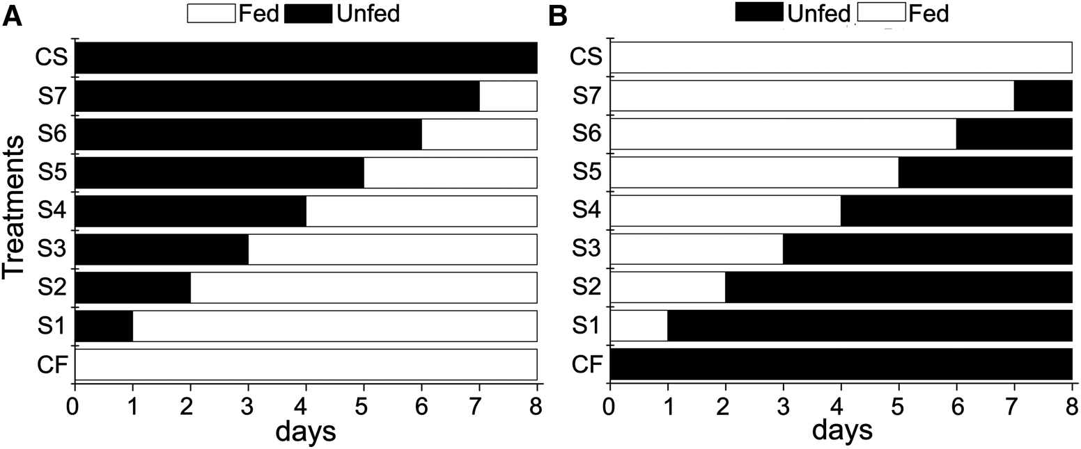
In experiment 1, we tested the effects of starvation on survival, moulting and the carapace length of early zoeal stages (ZI and ZII) of Lysmata vittata. ZI larvae were assigned to the starvation treatments (Figure 1A) consisting of seven different periods of initial starvation and subsequent days of feeding as follows: S1, 1 day starved/7 days fed; S2, 2 days starved/6 days fed; S3, 3 days starved/5 days fed; S4, 4 days starved/4 days fed; S5, 5 days starved/3 days fed; S6, 6 days starved/2 days fed; and S7, 7 days starved/1 day fed. In addition, there were two control groups: continuous feeding (CF) and continuous starvation (CS; Figure 1A). Each treatment consisted of 20 replicates, totalling 180 larvae used in experiment 1. Each replica was concluded when the larvae reached the third larval stage (ZIII), died, or did not moult by the last day of the experiment, i.e. day 8.
Protocol used during the food restriction period in the early larval stages (zoea I and II) of Lysmata vittata. (A) Starvation treatments to determine the point of no return (PNR). Larvae were initially starved (S) for the specified number of days and then fed for the remaining days of the experiment. (B) Feeding treatments to determine the point of reserve saturation (PRS). Larvae were initially fed (F) for the specified number of days and then starved for the remaining days of the experiment. CF, continuously fed control; CS, continuously starved control. (Experimental design based on Paschke et al., Reference Paschke, Gebauer, Buchholz and Anger2004.)

Experiment 2: PRS
In experiment 2, we evaluated the effect of feeding on survival, moulting and the carapace length of early zoeal stages (ZI and ZII) of Lysmata vittata. ZI were assigned to the following feeding treatments consisting of seven different periods of initial feeding and subsequent days of starvation as follows (Figure 1B): F1, 1 day fed/7 days starved; F2, 2 days fed/6 days starved; F3, 3 days fed/5 days starved; F4, 4 days fed/4 days starved; F5, 5 days fed/3 days starved; F6, 6 days fed/2 days starved; and F7, 7 days fed/1 day starved. The same as for the PNR experiments, there were two control groups: CF and CS (Figure 1B). Each treatment consisted of 20 replicates, totalling 180 larvae used in experiment 2. Each replica was concluded when the larvae reached the third larval stage (ZIII), died, or did not moult by the last day of the experiment, i.e. day 8.
Experimental conditions and endpoints
During all the experiments, the larvae were reared individually in plastic containers (80 ml) with 50 ml of artificial seawater. The water used in the experiment was prepared using fresh water purified by a reverse osmosis/DI unit and synthetic sea salt suitable for marine aquariums (Red Sea®). Each larva was considered a replicate, following the same criteria used in previous studies (Gebauer et al., Reference Gebauer, Paschke and Anger2010; Guerao et al., Reference Guerao, Simeó, Anger, Urzúa and Rotllant2012; Pantaleão et al., Reference Pantaleão, Barros-Alves, Tropea, Alves, Negreiros-Fransozo and López-Greco2015; Espinoza et al., Reference Espinoza, Guzmán, Bascur and Urzúa2016; Barros-Alves et al., Reference Barros-Alves, Alves, Antunes, López-Greco and Negreiros-Fransozo2018). The plastic containers were arranged inside a plastic tray (530 × 400 × 120 mm) filled with fresh water and maintained at 25 ± 1°C using thermostats (AT 180–100 W, ATMAN) as a water bath. The plastic containers were cleaned daily and the water was replaced with filtered seawater in both experiments (PNR and PRS). The temperature and salinity were checked daily. In the experiments with feeding, newly hatched Artemia nauplii (BioArtemia®, rate hatching: 80–90%) were provided as food at a density of 2 prey ml−1, and any remaining food was removed the next morning. The larvae were checked daily in the morning to register death or moulting (evidenced by the presence of exuviae). The larvae that reached stage III were conserved in ethyl alcohol 70% and subsequently the carapace length (CL) was measured under a stereomicroscope (Leica M205 C) with the imaging software Leica Application Suite (LAS) version 4.4, with an accuracy of 0.05 mm. The CL was determined from the post-orbital angle to the posterior margin of the cephalothorax.
‘Moulting’ was determined as the percentage of individuals from each treatment that moulted before the end of the experiment (day 8). ‘No moulting’ refers to the percentage of individuals in a certain treatment that did not die and never moulted. ‘Mortality’ was calculated by the percentage of individuals from a certain treatment that died before the end of the experiment, according to methods described by Calvo et al. (Reference Calvo, Tropea, Anger and López-Greco2012). The development time in days (mean ± SD) of stage I to III was calculated for all larvae per treatment. The PNR50 and PRS50 indices were calculated from the mortality data of each treatment. As noted above, mortality was calculated as the percentage of individuals in a given treatment that died before the end of the experiment, and ‘mortality before moulting’ was calculated as the percentage of individuals in a given treatment that died and did not moult. The PNR50 and PRS50 values were calculated by adjusting the sigmoidal Boltzmann model described in the following equation:  $M = {{A_1-A_2} \over {1 + e^{\lpar {x-x_o} \rpar /d_x}}} + A_2$, where M is the percentage of ‘mortality’ + ‘no moulting’ individuals, A 1 is the minimum value of M, A 2 is the maximum value of M, x 0 is the time (days) for M to reach 50%, d x is the time constant, and x is the time (days) of initial starvation (PNR) or feeding (PRS) (Paschke et al., Reference Paschke, Gebauer, Buchholz and Anger2004; Bas et al., Reference Bas, Spivak and Anger2008; Gebauer et al., Reference Gebauer, Paschke and Anger2010). The values of PNR50 and PRS50 were estimated considering the development time of ZI to ZIII. The PRS50/PNR50 ratio constitutes the NVI (for details, see Gebauer et al., Reference Gebauer, Paschke and Anger2010).
Statistical analysis
When data met the model assumptions, parametric tests were used; otherwise, equivalent non-parametric tests were used. The model assumptions of homoscedasticity (Levene's test) and normality (Shapiro–Wilk's test) were initially tested (Zar, Reference Zar2010). The χ2 test was used to test differences in larval ‘mortality’ + ‘no moulting’ between the treatments and the CF control. A one-way ANOVA (parametric) test was used to test differences in the development time, followed by a Tukey's test for multiple comparisons between treatments and the CF control. A linear regression was used to verify differences in the carapace length between treatments and the CF control. Linear regression equations were obtained after log-normal transformation of the data. The level of significance was set at 5% for two-tailed P-values (Zar, Reference Zar2010). The statistical tests used in each experiment are indicated in the legends of Figures 2 and 3. The statistical tests were conducted using Statistica (ver. 7.0, StatSoft, Tulsa, OK, USA).
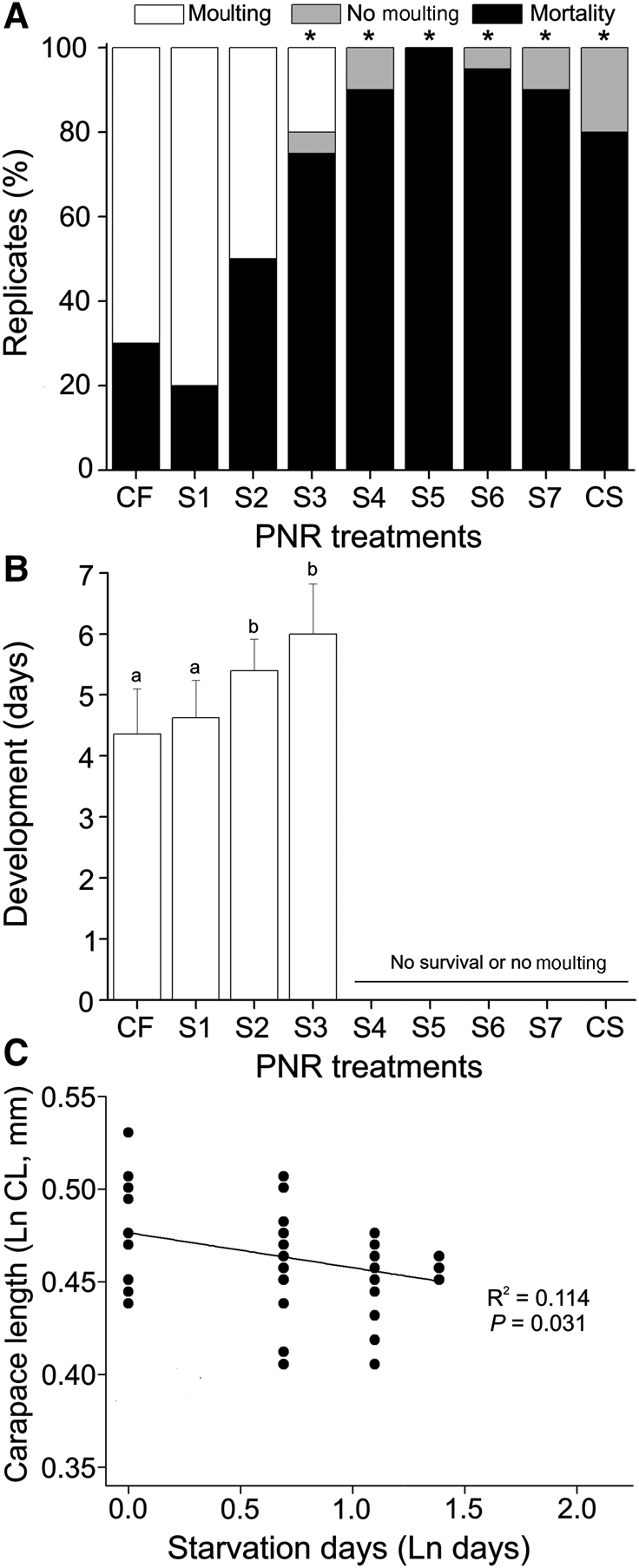
(A) Percentage of moulting, no moulting and mortality; (B) mean (± SD) development time; and (C) carapace length (in mm) of the early larval stages (zoea I and II) of Lysmata vittata in the different point of no return (PNR) treatment groups. The larvae were initially starved (S) for the specified number of days and then fed for the remaining days of the experiment (see text for details). CF, continuously fed control; CS, continuously starved control. Asterisks (*) in (A) indicate significant differences compared with the CF control (χ2, P < 0.05). Different letters in (B) indicate significant differences among the treatments (Tukey, P < 0.05).

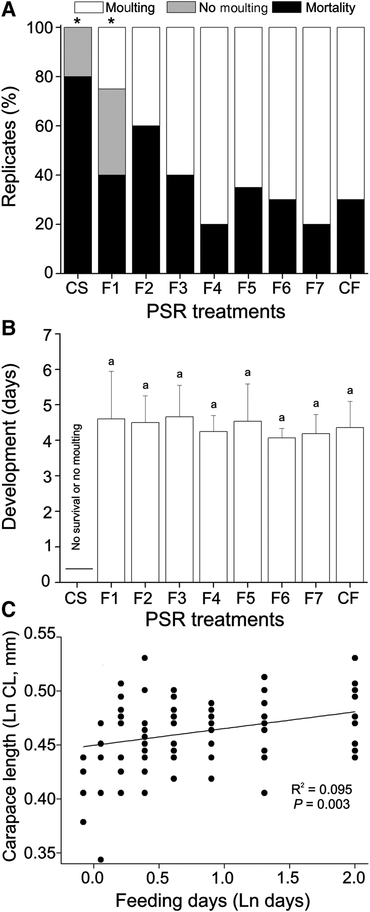
(A) Percentage of moulting, no moulting and mortality; (B) mean (±SD) development time; and (C) carapace length (in mm) of the early larval stages (zoea I and II) of Lysmata vittata in the different point of reserve saturation (PRS) treatment groups. The larvae were initially fed (F) for the specified number of days and then starved for the remaining days of the experiment (see text for details). CF, continuously fed control; CS, continuously starved control. Asterisks (*) in (a) indicate significant differences compared with the CF control (χ2, P < 0.05). Different letters in (b) indicate significant differences among the treatments (Tukey, P < 0.05).

Results
Experiment 1: PNR
Considering the zoea I in the initial starvation experiment, in the CS treatment, 80% died and 20% did not moult, totalling 100% of larvae that did not reach stage III (Figure 2A). In the CF treatment, 70% of larvae moulted to stage III and 30% died. ‘Mortality’ + ‘no moulting’ in the CF treatment was lower than in the other treatments, which obtained values higher than 80% (χ2, P < 0.01), except in the S1 (20%) and S2 (50%) treatments (Figure 2A). The larvae in the CF treatment took 4.36 ± 0.74 (mean ± SD) days to reach stage III (Figure 2B). Only larvae in the CF, S1, S2 and S3 treatments moulted to stage III, and the development time differed between treatments (ANOVA; F = 9.615; P < 0.01). The development time in the S1 treatment (4.63 ± 0.62 days) did not differ significantly from that of the CF treatment (Tukey, P > 0.05), whereas the S2 (5.40 ± 0.52 days) and S3 (6.00 ± 0.82) treatments differed significantly from the CF treatment (Tukey, P < 0.01) (Figure 2B). No larvae reached stage III in treatments S4, S5, S6, S7 and CS (Figure 2A, B). The carapace length (mean ± SD) of the larvae submitted to the CF treatment that reached stage III was 0.61 ± 0.04 mm CL. A correlation was observed between the carapace length and days of starvation in the different treatments, with the carapace length decreasing as the number of days of starvation increased (linear regression; F = 39.31; r 2 = 0.47; P < 0.01) (Figure 2C).
Experiment 2: PRS
Considering the zoea I of the initial starvation experiment, in the CS treatment, 80% died and 20% did not moult, totalling 100% of larvae that did not reach stage III (Figure 3A). In the CF treatment, 70% of larvae moulted to the stage III and 30% died. ‘Mortality’ + ‘no moulting’ in the CF treatment differed only from that recorded in the F1 (75%; χ2 = 3.85, P < 0.05) and CS treatment groups (100%; χ2 = 7.54, P < 0.05) (Figure 3A). The larvae in all treatment groups moulted to stage III, with the exception of those in the CS treatment group (Figure 3A, B). The larvae in the CF treatment took 4.36 ± 0.74 (mean ± SD) days to reach stage III, and the development time did not differ between treatments (ANOVA; F = 1.01; P = 0.43) (Figure 3B). The carapace length of the larvae submitted to the CF treatment that reached stage III was 0.61 ± 0.04 mm CL (mean ± SD). A correlation was observed between the carapace length and days of feeding in the different treatments, with the carapace length increasing as the number of days of feeding increased (Linear regression; F = 9.48; r 2 = 0.09; P < 0.01) (Figure 3C).
Nutritional vulnerability
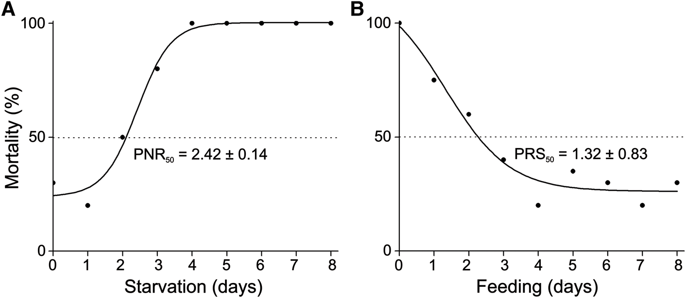
The PNR50 and PRS50 mean (± SD) values of the early larval stages (ZI and ZII) obtained from the sigmoid curves were 2.42 ± 0.14 and 1.32 ± 0.83 days, respectively (Figure 4). From these values, the NVI was estimated to be 0.54.
Adjustment of the sigmoid curve for the estimation of the mean (± SD) time when (A) 50% of the initially starved Lysmata vittata larvae (PNR50) lost the ability to moult to stage III and (B) 50% of the initially fed larvae (PRS50) were capable of moulting to stage III in the different treatment groups.

Discussion
In this study, we tested the hypothesis that larvae of the invasive shrimp Lysmata vittata are able to develop through the early larval stages in the absence of exogenous food, which would contribute to the invasiveness of this species. Therefore, we expected that (1) larvae exposed to different starvation periods would have a similar mortality, development time (in days) and carapace length (in mm); (2) larvae would tolerate long periods of starvation and retain the ability to moult to the next stage (i.e. high PNR value and low PRS value); and (3) there would be a low NVI value (i.e. NVI ≤ 0.5). Our laboratory experiments indicate that L. vittata presents an intermediate level of dependence on exogenous food.
Our results indicated that the damage caused by starvation during the first days of larval development might be irreversible. This suggestion is supported in the present study by larvae of L. vittata submitted to PNR treatments. The larvae that did not feed for 4 consecutive days or more failed to moult to the next stage or died, i.e. the energy deficit caused by the previous starvation period damaged the larval development. Thus, our results also corroborate the study by Calado et al. (Reference Calado, Dionísio and Dinis2007) and Calado et al. (Reference Calado, Dionísio, Bartilotti, Nunes, Santos and Dinis2008b) on other shrimps of the genus Lysmata. Larvae of Lysmata boggessi Rhyne & Lin, 2006 and Lysmata seticaudata (Risso, 1816) that were not fed in the first days of zoea, even if fed later, did not obtain the energy necessary to reach the third stage of development (Calado et al., Reference Calado, Dionísio and Dinis2007).
However, in the present study, a negative influence on the larval size of L. vittata was observed, i.e. larvae submitted to PNR treatments were smaller than larvae exposed to continuous feeding. Periods of feeding restriction have also been shown to negatively affect the growth of Neocaridina davidi (Bouvier, 1904); larvae smaller than those exposed to continuous feeding were observed for the newly hatched stages when exposed to starvation immediately after hatching (Pantaleão et al., Reference Pantaleão, Barros-Alves, Tropea, Alves, Negreiros-Fransozo and López-Greco2015). Other studies have also shown that crustacean larvae subjected to temporary starvation might decrease growth (e.g. Mikami et al., Reference Mikami, Greenwood and Gillespie1993; Calado et al., Reference Calado, Figueiredo, Rosa, Nunes and Narciso2005; Giménez & Anger, Reference Giménez and Anger2005; Figueiredo et al., Reference Figueiredo, Penha-Lopes, Narciso and Lin2008). These studies also showed that the starvation impact on larval development is, in general, directly proportional to the time the larvae are under those conditions (Mikami & Takashima, Reference Mikami and Takashima1993; Giménez & Anger, Reference Giménez and Anger2005). In these cases, as the initial starvation period increases, the efficiency of the feeding activity might decrease, and reserves might not be absorbed or accumulated, thus resulting in smaller larvae even though they were re-fed for a longer period (Liddy et al., Reference Liddy, Phillips and Maguire2003; Stumpf et al., Reference Stumpf, Calvo, Pietrokovsky and López Greco2010). According to Knowlton (Reference Knowlton1974), larvae prioritize their energy for survival and moulting over growth, since in optimal conditions larvae grow, and in sub-optimal or stress conditions, the animals survive and moult but do not grow.
Larvae of L. vittata submitted to PRS treatments moulted to the next stage of development. Therefore, in the CF treatment, 70% of larvae moulted to stage III, different from the observations of Calado et al. (Reference Calado, Dionísio and Dinis2007) for other species of Lysmata in which about 100% of the larvae reached the subsequent stages when fed continuously. Larval survival may be directly related to the nutritional stress undergone by ovigerous shrimps during the period of gonad maturation and embryo development in the wild, since unfortunately this factor cannot be controlled in the laboratory as performed by Calado et al. (Reference Calado, Dionísio and Dinis2007).
Furthermore, larvae fed only in the early days (e.g. F2 and F3) and subsequently exposed to periods of starvation moulted to stage III with a similar time of development and mortality rate when compared with larvae fed continuously. Similar results were observed for L. boggessi and L. seticaudata that also reach the next stage when submitted to PRS treatments (Calado et al., Reference Calado, Dionísio and Dinis2007). For L. boggessi, the lack of dependence on exogenous food during the early larval stages is justified, since in newly hatched larvae, the yolk is distributed in ~10–20% of the cephalothorax (Romero-Carvajal et al., Reference Romero-Carvajal, Turnbull and Baeza2018). However, for L. vittata, the yolk transferred to egg must be completely consumed during the embryonic development, since the larvae hatch without macroscopically visible yolk reserves (Alves et al., Reference Alves, López-Greco, Barros-Alves and Hirose2019).
Therefore, the results of this study agree with our expectations considering that L. vittata is successful as an invasive species, suggesting that the larvae of L. vittata have sufficient energy for their initial development in the absence of exogenous food, despite there not being visible remaining yolk reserves in the newly hatched larvae. According to Anger (Reference Anger2001), the fuel for partial or complete food-independent larval development originates, in general, from remaining parts of the egg yolk, which are commonly deposited in the form of lipid droplets in the hepatopancreas and have been observed in newly hatched larvae of species with some degree of lecithotrophy (Espinoza et al., Reference Espinoza, Guzmán, Bascur and Urzúa2016).
However, the dependence on exogenous food by newly hatched larvae has been recorded for some species of the genus Lysmata, which are considered planctotrophic (e.g. Lysmata amboinensis (De Man, 1888) and Lysmata debelius Bruce, 1983), since these larvae depend on exogenous food to complete their initial development (Calado et al., Reference Calado, Dionísio and Dinis2007), as well as for other crustaceans, such as Maja brachydactyla Balss, 1922 (Guerao et al., Reference Guerao, Simeó, Anger, Urzúa and Rotllant2012), Crangon crangon (Linnaeus, 1758) (Paschke et al., Reference Paschke, Gebauer, Buchholz and Anger2004) and Stenorhynchus seticornis (Herbst, 1788) (Barros-Alves et al., Reference Barros-Alves, Alves, Antunes, López-Greco and Negreiros-Fransozo2018), which also have planctotrophic larvae. For these planctotrophic species, feeding during the first larval stage is crucial to allow larvae to continue their development, since the absence of food during the first larval stage can cause irreversible damage.
In the present study, larvae of L. vittata submitted to continuous starvation from hatching did not reach stage III, whereas the larvae fed on the first days after hatching and subjected to later periods of starvation reached the third stage of development, indicating that these larvae present facultative secondary lecithotrophy (FSL) (see Anger, Reference Anger1989). In larvae with SL, larval prey ingested in the early larval stages contribute to the build-up of energetic reserves that are later catabolized during the starvation periods in the next stage larval, allowing some larvae to moult to the third larval stage (Anger, Reference Anger2001; Calado et al., Reference Calado, Dionísio and Dinis2007). For these caridean shrimps, the energy accumulated through exogenous food ingestion plays a crucial role, since the endogenous reserves from the egg yolk are insufficient to allow starved zoea to advance to the next larval stage (Calado et al., Reference Calado, Dionísio and Dinis2007). Facultative secondary lecithotrophy has been observed only for a small proportion of larvae (~15%) of L. boggessi and L. seticaudata (Calado et al., Reference Calado, Dionísio and Dinis2007). For an invasive shrimp, such as L. vittata, SL is another factor that might contribute to the invasion process, since the larvae can tolerate a period of starvation once they had access to exogenous food after hatching.
The invasive shrimp L. vittata presents an intermediate level of dependence on exogenous food during the initial larval stages (NVI = 0.54), slightly different to a low dependence on exogenous food, which would be the case for an NVI less than or equal to 0.5 (Gebauer et al., Reference Gebauer, Paschke and Anger2010). Other larvae of decapod crustaceans, whose nutritional vulnerability has been determined experimentally, present lower PRS than PNR values (NVI < 1), indicating relatively higher levels of independence on food, such as Neohelice granulata (Dana, 1851) (Giménez, Reference Giménez2002; Bas et al., Reference Bas, Spivak and Anger2008), C. crangon (Paschke et al., Reference Paschke, Gebauer, Buchholz and Anger2004), M. brachydactyla (Guerao et al., Reference Guerao, Simeó, Anger, Urzúa and Rotllant2012), N. davidi (Pantaleão et al., Reference Pantaleão, Barros-Alves, Tropea, Alves, Negreiros-Fransozo and López-Greco2015) and Ucides cordatus (Linnaeus, 1763) (Souza et al., Reference Souza, Simith and Abrunhosa2018). However, some larvae are more dependent on food, with PRS values exceeding the PNR (NVI > 1). This was observed, for example, for larvae of Armases cinereum Bosc, 1802 (Staton & Sulkin, Reference Staton and Sulkin1991), Petrolisthes laevigatus (Guérin, 1835) (Gebauer et al., Reference Gebauer, Paschke and Anger2010), Pleuroncodes monodon (Milne-Edwards, 1837) (Espinoza et al., Reference Espinoza, Guzmán, Bascur and Urzúa2016), Panulirus argus (Latreille, 1804) (Espinosa-Magaña et al., Reference Espinosa-Magaña, Lozano-Álvarez and Briones-Fourzán2017) and S. seticornis (Barros-Alves et al., Reference Barros-Alves, Alves, Antunes, López-Greco and Negreiros-Fransozo2018). In addition, this dependence on exogenous food might be variable between stages of the same species, as recorded for Hyas araneus (Linnaeus, 1758) (Anger & Dawirs, Reference Anger and Dawirs1981), in which zoea I present independence of an exogenous food supply (NVI = 0.38), whereas zoea II present a high dependence (NVI = 1.25).
Overall, our results favour the predictions supporting the success of L. vittata as an invasive species: (1) larvae are able to moult and reach the third larval stage even when exposed to some periods of starvation; (2) the development time in the initial larval stages is not affected by periods of starvation; (3) the size of larvae exposed to periods of starvation might be affected in response to the use of different metabolic pathways, allowing the use of energy for moulting, for example; (4) initial larval stages present an intermediate level of nutritional vulnerability, with a value close to the limit for a low level of nutritional vulnerability (NVI close to 0.5); and (5) initial larval stages present facultative lecithotrophy. Lecithotrophy is common in organisms living in water with lower temperatures and at higher latitudes (Anger et al., Reference Anger, Lovrich, Thatje and Calcagno2004) due to the seasonality of plankton production in this environment resulting in oligotrophic waters, i.e. water with less availability of food at some periods during the year (Thatje et al., Reference Thatje, Calcagno, Lovrich, Sartoris and Anger2003, Reference Thatje, Anger, Calcagno, Lovrich, Pörtner and Arntz2005; Anger et al., Reference Anger, Lovrich, Thatje and Calcagno2004; Calado et al., Reference Calado, Dionísio and Dinis2007). This food independence during the first larval stage might be due to an adaptation of the species to live in environments with a low density of prey, thus, their endogenous reserves are sufficient for the first physiological changes (Thessalou-Legaki et al., Reference Thessalou-Legaki, Peppa and Zacharaki1999). In this sense, facultative primary lecithotrophy (FPL) in L. vittata might be another feature that contributes to the invasive success of this species, in addition to the other factors suggested by Alves et al. (Reference Alves, López-Greco, Barros-Alves and Hirose2019), such as mating system exhibiting ‘protandric simultaneous hermaphroditism’, rapid development of the gonad, short embryonic development and a short reproductive cycle. Therefore, successful larval development in periods of food limitation could be a key factor for the maintenance and establishment of decapod populations. Due to the scarcity of food, developing zoea might face shorter or longer periods of sub-optimal feeding.
Conclusions
Based on our experiments, by imposing gradual periods of initial starvation or feeding (PNR and PRS experiments; Anger & Dawirs, Reference Anger and Dawirs1981), we found that the initial larval stages of Lysmata vittata present an intermediate level (NVI = 0.54) of dependence on exogenous food and that their survival was similar to that recorded for the CF control treatment. Thus, larvae of L. vittata in their early development stages display facultative lecithotrophy. In addition, we observed that larvae exposed to initial starvation (1–3 days) displayed similar development times and mortality to larvae submitted to continuous feeding. Nonetheless, larval size in the third stage of development was affected. Longer periods of starvation might negatively affect the larval development of L. vittata and impair the larvae to reach the third zoeal stage. Therefore, the results of the present study provide more information on the larval development of this species and how different degrees of starvation in its early larval stages might contribute to the establishment of L. vittata in invaded habitats.
Acknowledgements
The authors are grateful to Carmen Regina Parisotto Guimarães and Yana Teixeira dos Reis (University of Sergipe, Department of Biology) for permission to use laboratory equipment. The authors are also grateful to Rafael de Carvalho Santos for his help during the fieldwork. All sampling in this study was conducted according to the applicable state and federal laws.
Financial support
SPBA thanks CAPES (Coordenação de Aperfeiçoamento de Pessoal de Nível Superior, Process number: 88882.314751/2019-01) for research scholarships (Programa Nacional de Pós-Doutorado/Capes—PNPD) linked to the Programa de Pós-Graduação em Ecologia e Conservação de Recursos Naturais (University of Uberlândia) for financial support. ASA thanks FAPESP (Fundação de Amparo à Pesquisa do Estado de São Paulo, Process number: 2017/26792-1) for research scholarships. RCC thanks the Brazilian National Council for Scientific and Technological Development, CNPq (Research Scholarship PQ 306672/2018-9) and FAPESP - Thematic Biota (INTERCRUSTA 2018/13685-5).