Scimitar syndrome, or congenital venolobular syndrome, is a rare (1−3 of the 100 live births) cardiopulmonary malformation that was first described in 1836 by Chassinat. In its classical form, it includes the triad: pulmonary hypoplasia, partial anomalous pulmonary venous return, and abnormal pulmonary arterial supply. It can appear sporadically or in familiar clusters.Reference Halasz, Halloran and Liebow1, Reference Neill, Ferencz, Sabiston and Sheldon2
The main feature of this syndrome is the partial anomalous pulmonary venous return, where part of the entire right lung venous return drains into the inferior caval vein. The irrigation of the low right pulmonary lobe is supplied by an aberrant artery arising from the sub-diaphragmatic aorta or one of its main branches. Despite the findings in most of the cases being on the right lung, in some cases they are described on the left side. Moreover, lung hypoplasia, dextroposition of the heart, and other malformations of the ipsilateral pulmonary artery may be found.Reference Berrocal, Madrid, Novo, Gutierrez, Arjonilla and Gomez-Leon3, Reference Rutledge, Hiatt, Wesley Vick and Grifka4
Associated anomalies are variable and can include cardiac malformations, vascular, and extra-vascular, such as bronchial, diaphragmatic, urinary, and vertebral anomalies.Reference Ruggieri, Abbate, Parano, Distefano, Guarnera and Pavone5
Two forms of presentation are described; the neonatal or infantile form is the most complicated form, coursing with pulmonary hypertension due to the systemic-to-pulmonary fistulae, leading to cardiac failure and high mortality rates; they may require immediate treatment.Reference Bratu, Flageole, Chen, Di Lorenzo, Yazbeck and Laberge7 The adult form, generally asymptomatic or with few respiratory symptoms, is usually discovered in a routine chest X-ray and rarely accompanies pulmonary hypertension.Reference Berrocal, Madrid, Novo, Gutierrez, Arjonilla and Gomez-Leon3
The term scimitar derives from the radiological shadow of the anomalous vein on the chest radiography.Reference Cabrera, Sarrionandia and Idigoras6 Despite the syndrome responding to the previous definition, there are numerous publications that misdefine scimitar syndrome exclusively as a radiological sign, without the other associate alterations forming the triad.
Echocardiography, computerised tomography, and cardio-resonance are used for the detailed morphological study of the heart, vessels, and pulmonary anomalies.Reference Berrocal, Madrid, Novo, Gutierrez, Arjonilla and Gomez-Leon3
Angiography and haemodynamic studies demonstrate the existence of the anomalous vein together with the lung aberrant irrigation, offering a detailed study of the pulmonary and systemic fluxes.
Case report
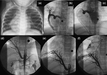
A 6-year-old girl was admitted to our hospital with a history of recurrent respiratory infections since the age of 5 months. Her familiar history was unremarkable. She had been hospitalised on four occasions for pneumonia, three of them being on the inferior right lobe. She was asymptomatic between episodes. All physical findings were normal except for diminished lung sounds in the lower right chest region. The chest radiograph revealed an abnormal shadow running along the right heart border, scimitar sign, with dextro-mesocardia without cardiomegaly (Fig 1a).
Thoracic X-ray showing dextromesocardia and a scimitar sign on the right lung (a). Angiography of the anomalous vein with double drainage to the inferior caval vein (b) and left atrium (c). Abdominal aorta angiography showing an ascendant aberrant artery (d–f).

Echocardiography showed mild dilatation of the right chambers, with normal cardiac function, mild tricuspid regurgitation (gradient 27–30 millimetres of mercury), and abnormal pulmonary vein draining into the inferior caval vein with mild obstruction (gradient 6–8 millimetres of mercury), with no associated intra-cardiac anomalies. There were three pulmonary veins that were normally draining to the left atria.
A computed tomography scan was performed, revealing a communicating vessel (8–9 millimetres) from the right inferior pulmonary vein to the inferior caval vein, at its intra-hepatic portion. The right pulmonary artery was moderately hypoplastic (6 millimetres) with normal main and left pulmonary arteries. Irrigation of the inferior right pulmonary lobe and probably the median lobe was due to a systemic vessel (7 millimetres in its main caliper) arising from the abdominal aorta cephalically to the coeliac trunk. Thoracic aorta was situated normally with normal anatomy.
To describe the respiratory tract anatomy, fibrobronchoscopy was performed. The larynx, trachea, and tracheal carina were normal. The left bronchial tree was normal. The right bronchial tree presented severe dilatation and shortness of the median lobe bronchi, with stenotic right inferior lobe bronchi.
Cardiac catheterisation and angiocardiography were performed. Arising from the inferior caval vein at the infradiaphragmatic level, an anomalous pulmonary vein was observed and selectively catheterised, revealing a right venous collector running along the entire right lung; furthermore, this collector showed, at its middle portion, a wide right pulmonary vein which tortuously drained to the left atrium (Fig 1b and c). Selective angiography of the right pulmonary artery showed three branches. The inferior was hypoplastic and irrigated a small pulmonary area, which the venous return connected, majorly, to the left atrium, and minorly to the inferior caval vein through the anomalous pulmonary vein. Abdominal aorta angiography (Fig 1d, e and f) showed, above the coeliac trunk, a big artery with an ascending portion bifurcating into two branches and irrigating an area of the pulmonary tree with systemic rate pressures (75/45, mean 68 millimetres of mercury), with a unique and small pulmonary venous drainage to the left atria. This area of the pulmonary region did not seem to have connections to the area irrigated by the right pulmonary artery.
Procedure
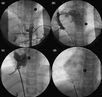
On the one hand, embolisation of the aberrant artery originating from the aorta was performed with a 16 millimetres vascular plug (Amplatzer®, AGA Medical Corporation, Golden Valley, Minnesota, United States of America) obtaining complete occlusion (Fig 2a). On the other hand, pressure in the pulmonary collector vein was measured during occlusion test with a Swanz-Ganz catheter, and because there was no change in the pulmonary vein pressures, we proceed to occlude the caudal segment of the vein abnormally draining to the inferior caval vein using an 18-millimetres Vascular Plug (Amplatzer®; Fig 2c and d). Total procedure time was 145 minutes, and fluoroscopic time was 34 minutes. The patient was discharged the following day on daily low-dose aspirin.
Embolisation of the aberrant artery with a 16-millimetres vascular plug (Amplatzer®) (a). Catheter balloon occlusion of the anomalous vein, showing drainage to the left atrium (b). Occlusion of the anomalous vein with a 18-millimetres vascular plug (Amplatzer®) (c). Final angiography showing complete occlusion (d).

A follow-up time of 3 years shows a patient free of respiratory symptoms. Echocardiography verifies complete regression of the right chambers dilatation, no flow on occluded vessels, and no device migration.
Discussion
Historically, therapeutic management of scimitar syndrome is mainly surgical, when needed.Reference Bratu, Flageole, Chen, Di Lorenzo, Yazbeck and Laberge7 A percutaneous therapeutic approach was first published by Dickinson in 1982 after performing aberrant artery embolisation.Reference Dickinson, Galloway, Massey, Sankey and Arnold8 In most of the cases, aberrant artery embolisation is only a partial cure, as the partial anomalous pulmonary venous return remains abnormal, draining on the inferior caval vein,Reference Lee, Yang and Yang9, Reference Muta, Akagi, Iemura and Kato10 and therefore the patient presents anomalous pulmonary vein return physiology with eventually right chambers dilatation. Indication for closure, in this patient, was based on the fact that she had undergone several respiratory infections. In this particular case, double venous drainage allowed us to also perform occlusion of the anomalous pulmonary vein drainage, leading to complete restoration of normal cardiovascular physiology. It is only in cases of double venous drainage that venous occlusion can be considered. In single venous abnormal drainage, occlusion would be devastating for the patient.
Occlusion of these vessels can be achieved using several occlusion devices: Vascular Plug, PDA I, and II Occluder (Amplatzer®).
Conclusions
Scimitar syndrome is a rare entity responsible for recurrent respiratory pathology and cardiac failure in neonates. Despite most of the cases being only partially treated by aberrant artery embolisation, in certain anatomic variations, a percutaneous approach can completely restore cardiovascular physiology.