Congenital unilateral pulmonary venous atresia remains a diagnostic challenge. Although clinical manifestations of this condition have been well documented, several diagnostic modalities are required to achieve definitive diagnosis. The anomaly can be associated with significant morbidity and mortality.Reference Artero, Serrano, Padín and Serrano1–Reference Pourmoghadam, Moore and Khan3 We report here a patient with atresia of the right pulmonary veins, and discuss the pathogenesis, diagnostic algorithm, and management.
Case Report
A 12 year old male presented with a productive cough and right-sided chest pain. At birth, he had been noted to suffer transient mild respiratory distress, and was subsequently diagnosed with asthma and allergic rhinitis, suffering also 4 episodes of pneumonia.
On this admission, chest radiograph and computed tomographic scan of the chest revealed loss of volume with right lower lobe pneumonia and a small pleural effusion, the heart being noted to occupy the right-side of the chest. His pneumonia was treated, and he was followed as an outpatient. On review, the routine vital signs were normal, and the saturation of oxygen was 100%. Auscultation revealed a soft flow murmur over the mid sternum. Evaluation of the liver and lungs proved unremarkable. Echocardiography confirmed that the heart occupied the right chest, albeit that the apex pointed leftward, and mild hypoplasia was noted of the right pulmonary artery. The right pulmonary veins were not visualized, with review of the previously performed thoracic tomography showing that the right pulmonary veins had not been identified.
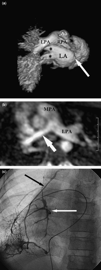
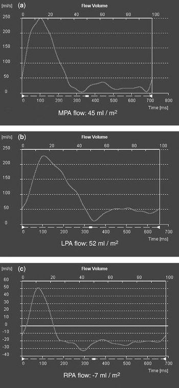
Magnetic resonance imaging demonstrated normal left pulmonary veins, and absence of the right pulmonary veins (Fig. 1a). The first phase suggested an atretic right pulmonary artery (Fig. 1a, but analysis of the second phase showed the artery to be normally formed beyond the hilum and diffusely hypoplastic (Fig. 1b). Phase contrast imaging revealed interesting patterns of flow in the pulmonary trunk and its branches (Fig. 2). The net flow to the pulmonary trunk was 45 ml/m2, with normal flow and no regurgitation (Fig. 2a). The net flow to the left pulmonary artery, of 52 ml/m2, exceeded the flow in the pulmonary trunk, with a pattern of persistent antegrade flow seen during diastole (Fig. 2b). There was minimal antegrade systolic flow jn right pulmonary artery, and a significant amount of holodiastolic retrograde flow, resulting in a net negative flow of −7 ml/m2 (Fig. 2c). We presume this negative volume to be returned to the left pulmonary artery during diastole, due to the absence of effective flow to the right lung.
Magnetic resonance imaging with 3-dimensional surface rendered reconstruction of the heart and great vessels rotated posteriorly and viewed from a superior perspective with the aorta excluded (a) demonstrates the dilated left pulmonary artery and poor opacification of the distal right pulmonary artery. This reconstruction clearly demonstrates the normal left pulmonary veins (asterix) and absent right sided pulmonary venous drainage (white arrow). Only the proximal right pulmonary artery is imaged in this first phase of the angiogram. The axial maximum intensity projection image from the 2nd phase of the magnetic resonance angiogram (b) demonstrates the normal left pulmonary artery and the hypoplastic, though otherwise normal-appearing right pulmonary artery (white arrow). Right pulmonary artery wedge angiography (c) confirms hypoplasia of the parenchymal pulmonary veins with atresia of the hilar right pulmonary veins (white arrow). Also imaged is a varix to a pericardial vein (black arrow). Ao: Aorta, LA: Left atrium, LPA: left pulmonary artery, RPA: right pulmonary artery.

Magnetic resonance phase contrast imaging analysis of pulmonary arterial flow shows (a) normal flow of 45 ml/m2 to the pulmonary trunk (MPA), that flow to the left pulmonary artery (b), at 52 ml/m2, exceeds the flow in the pulmonary trunk, with persistent antegrade flow in diastole, and (c) that net flow in the right pulmonary artery is negative (−7 ml/m2) with holodiastolic retrograde flow. Note the shift of baseline flow.

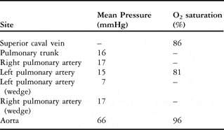
Cardiac catheterization demonstrated a normal cardiac index, and no evidence of a step-up from the superior caval vein to the pulmonary artery. The atrial septum was intact. The pulmonary arterial pressures were within normal limits (Table 1). Angiography confirmed the diffuse hypoplasia of the right pulmonary artery, albeit with no discrete stenosis. No forward flow was noted through the right lung past the capillary phase. Several capillary wedge injections in the right pulmonary artery demonstrated atresia of the right pulmonary veins (Fig. 1c).
Haemodynamic and oxymetric data from cardiac catheterization.

The patient is currently asymptomatic from the cardiac stance, and his reactive symptoms relating to the airways have improved with pharmacological therapy. He remains well, and our management will now be expectant.
Discussion
Isolated congenital unilateral pulmonary venous atresia is a very rare anomaly. To the best of our knowledge, including our patient, 28 have been described to date.Reference Artero, Serrano, Padín and Serrano1–Reference Pourmoghadam, Moore and Khan3 Histopathologic examination of the atretic segment of the veins typically demonstrates medial hypertrophy with intimal fibrosis, without inflammatory reaction.Reference Pourmoghadam, Moore and Khan3 Other congenital cardiac defects, and pulmonary arterial hypertension, are frequent associations.Reference Heyneman, Nolan, Harrison and McAdams2 Definitive diagnosis is often achieved only after multiple diagnostic tests. The absence of a venous connection must be positively identified to ensure that there is no chance of anatomic surgical correction. Respiratory symptoms often result in the patient being identified in infancy or childhood. Clinical features include recurrent pulmonary infections, reactive airways disease, haemoptysis, and exercise intolerance.Reference Artero, Serrano, Padín and Serrano1–Reference Pourmoghadam, Moore and Khan3
It is also often difficult to confirm the diagnosis.Reference Pourmoghadam, Moore and Khan3 We catheterized our largely asymptomatic patient so we could define the pulmonary venous anatomy by means of pulmonary wedge angiography.Reference Pourmoghadam, Moore and Khan3 The magnetic resonance not only defined the anatomic hypoplasia of the right pulmonary artery and absence of right pulmonary veins, but also, the lack of any forward flow to the right lung. The net flow to the right lung was negative, with flow into the left pulmonary artery being greater than that into the pulmonary trunk, alterations of flow previously described in the setting of pulmonary venous obstruction.Reference Masui, Seelos, Kersting-Sommerhoff and Higgins4–Reference Grosse-Wortmann, Hamilton and Yoo6 Aortography failed to demonstrate direct aortopulmonary collateral connections, albeit that small indirect collateral vessels to the right lung and the bronchial circulation likely account for this observation. We recognize that the diagnosis was demonstrated by magnetic resonance. Angiography was undertaken, nonetheless, to be certain that no surgical intervention, short of pneumonectomy, would benefit the patient. Cineangiography differentiated ostial from long segment atresia, and confirmed the futility of surgical intervention.
Our patient presented early with pulmonary symptoms. These responded to medical treatment, and have not to date interfered with his activities of daily living. We believe that the absence of pulmonary arterial hypertension with no concomitant congenital cardiac defects may have prevented the development of cardiac symptoms. We will now monitor the patient closely for the development of pulmonary hypertension or clinical deterioration. In the future he may develop progressive shortness of breath on exertion due to mismatch between ventilation and perfusion.Reference Sutton, Peng and Lock7 Pneumonectomy will be considered if he develops pulmonary dysfunction, pulmonary hypertension, or recurrent pulmonary infections as this has been the advocated for symptomatic patients with this disorder. The procedure eliminates the nidus for both pulmonary infections and dead space ventilation contributing to exercise intolerance.Reference Pourmoghadam, Moore and Khan3 We believe that this should be considered for significantly symptomatic patients with irreversible changes in the pulmonary parenchyma and vasculature. If this diagnosis is made early in life, surgery for restoring the continuity between the atretic veins and the left atrium may be considered.Reference Pourmoghadam, Moore and Khan3
Acknowledgments
We thank Dr David G. Nykanen for his contribution with the cardiac catheterization, images, and editorial advice.