Introduction
While vascular malformations and haemangiomas are relatively common within the central nervous system, they are a rare cause of facial pain.
This case report presents a patient with a venous haemangioma of the mandibular division of the trigeminal nerve (V3).
Case report
A 19-year-old woman presented to a tertiary referral skull base centre with a history of right periorbital pain followed by the development of a progressive, right-sided deficit of V3, which had become complete over a period of six months.
Clinical examination revealed hypoaesthesia in the right V3 dermatome, mild trismus and some wasting of the right masseter muscle.
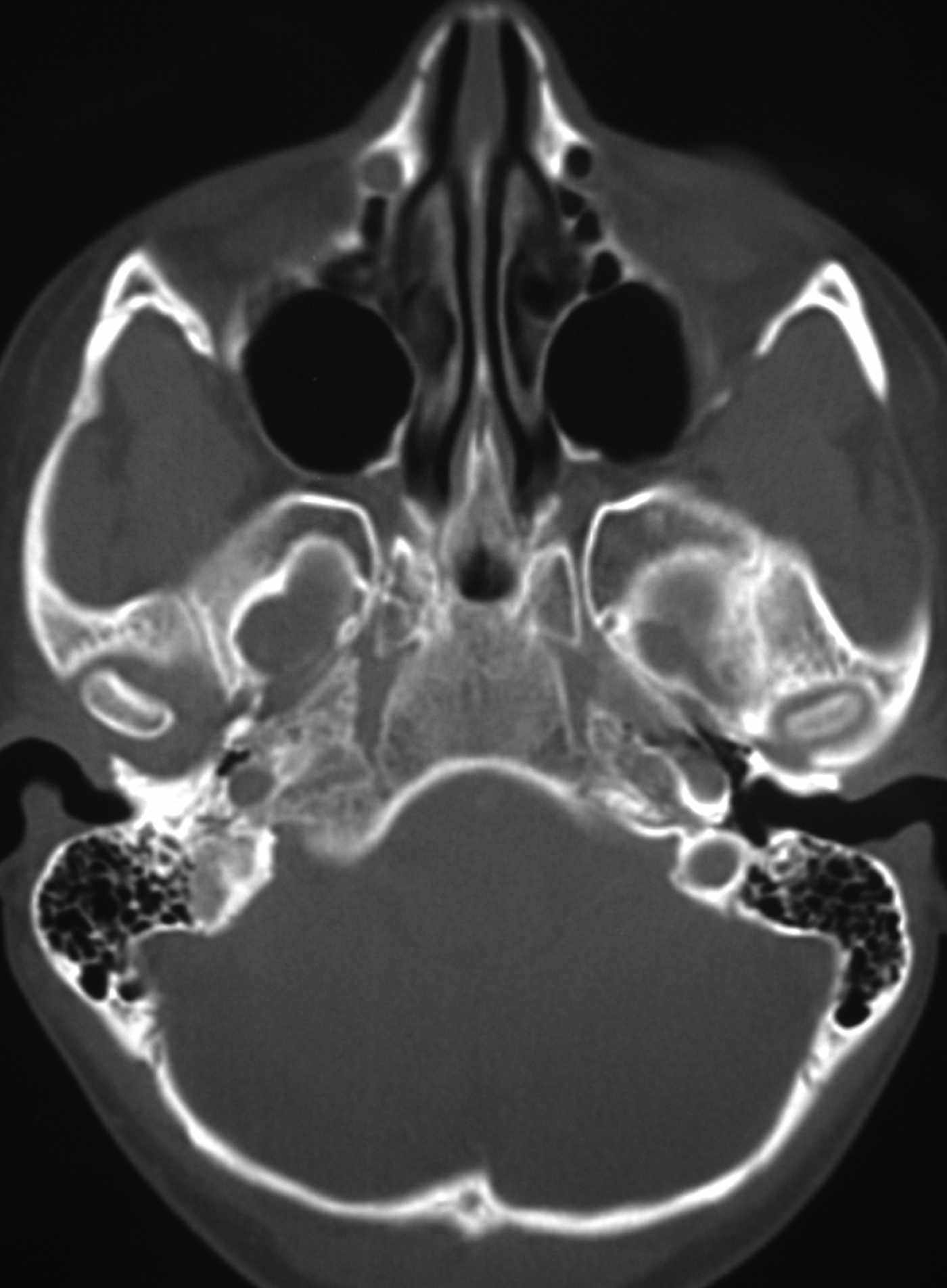
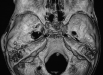
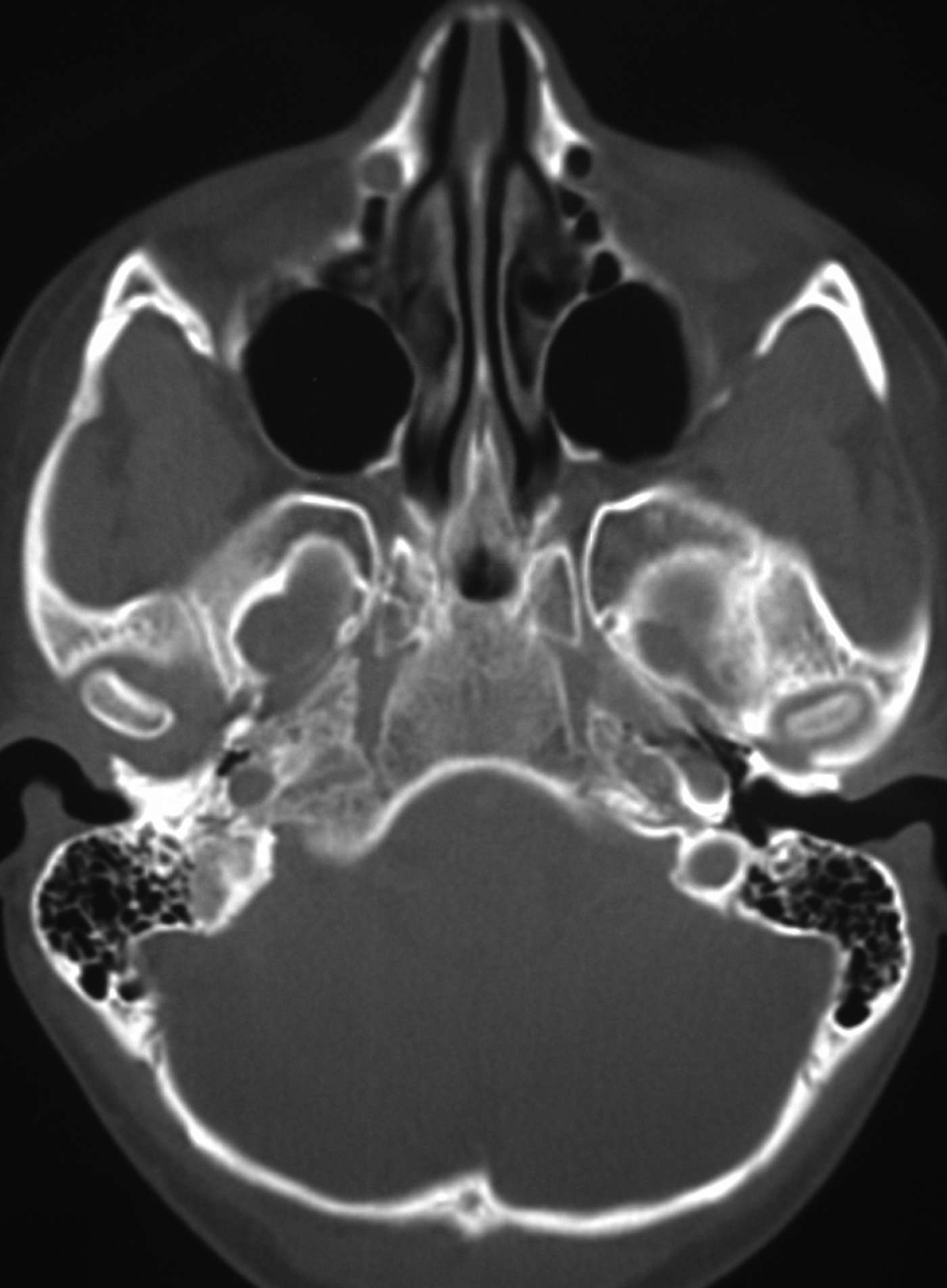
Thin-section computed tomography (CT) with contrast was performed in an axial plane, and images prepared using soft tissue and bone algorithms. A small area of mildly enhancing soft tissue was observed, centred within the foramen ovale. There was smooth enlargement of the foramen ovale, with a sclerotic rim, demonstrable on a three-dimensional rendered image.
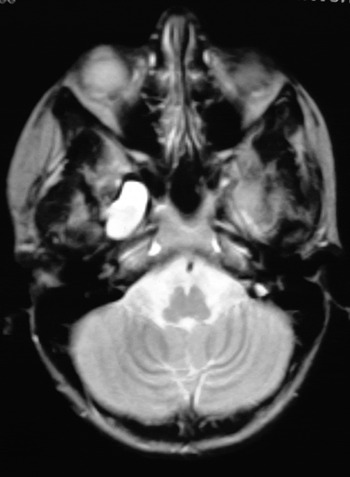
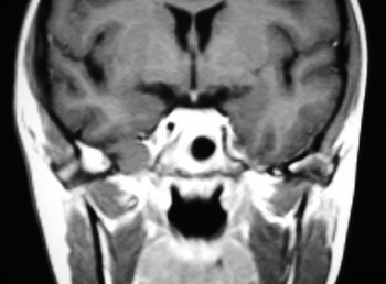
Upon magnetic resonance imaging (MRI), T1-weighted sequences revealed a homogeneous, intermediate-signal lesion which enhanced avidly with gadolinium, while T2-weighted sequences showed marked hyperintensity (Figs 3 and 4). There were no other abnormalities. The key diagnostic points were the location of the lesion within the foramen ovale, with widening of the foramen, and the observed avid enhancement following gadolinium administration (Figs 1 and 2).
Axial computed tomography scan prepared with bone windows, showing widening of the foramen ovale.
Surface-rendered, three-dimensional image of the floor of the middle cranial fossa viewed from above, confirming enlargement of the foramen ovale.

Axial, T2-weighted, magnetic resonance imaging scan showing a lesion of markedly increased signal intensity.

Coronal, T1-weighted, contrast-enhanced magnetic resonance imaging scan, showing homogeneous enhancement.

The differential diagnosis included V3 schwannoma and neurofibroma, meningioma, venous malformation, and haemangioma.
A provisional diagnosis of right V3 schwannoma was made.
Surgery was undertaken via an infratemporal fossa (Fisch) type D approach. A vascular lesion was found filling the foramen ovale, with no obvious nerve separate from the lesion. The lesion was removed en – bloc, after bipolar coagulation.
Histopathological analysis demonstrated a venous haemangioma within the nerve.
The patient made an uneventful recovery, but had residual lower facial hypoaesthesia and mild trismus.
Discussion
Arteriovenous malformations and venous haemangiomas are relatively common in the central nervous system. Arteriovenous malformations and venous haemangiomas that compress the trigeminal nerve at the root entry zone are a recognised cause of trigeminal neuralgia.Reference Karibe, Shirane, Jokura and Yoshimoto1, Reference Maher, Atkinson and Lane2
In our patient's case, the provisional diagnosis of schwannoma was reasonable if inaccurate. Failure to recognise the true nature of the lesion did not alter its clinical management; the same approach would have been undertaken had the provisional diagnosis been a vascular lesion rather than schwannoma.
At the time of writing the patient was pain free, albeit with mild trismus and right lower facial hypoaesthesia.