Introduction
Lacunar stroke is a small (<2 cm) infarction that generally originates from a single perforating artery (Sacco et al., Reference Sacco, Marini, Totaro, Russo, Cerone and Carolei2006). The pathology typically presents as a non-atheromatous thickening of portions of small arteries and/or arterioles, in conjunction with the breakdown of muscular and elastic fibers (Bailey et al., Reference Bailey, Smith, Sudlow and Wardlaw2012). Infarction or arteriolar occlusion can occur at later stages secondary to severe vessel wall damage (Ogata, Reference Ogata1999). Lacunar stroke accounts for approximately 25% of ischemic stroke, and 20% of all strokes (Sudlow and Warlow, Reference Sudlow and Warlow1997). In terms of disease progression and prognosis, lacunar stroke patients have slightly lower rates of stroke recurrence and mortality in the short term (1–2 years) compared to those with non-lacunar stroke (e.g. cortical infarct, hemorrhagic stroke), though the rates between the two tend to converge after the first few years (Sacco et al., Reference Sacco, Marini, Totaro, Russo, Cerone and Carolei2006). Lacunar stroke is accompanied by deficits across a wide range of cognitive domains (e.g. memory, attention, language, processing speed) (Edwards et al., Reference Edwards, Jacova, Sepehry, Pratt and Benavente2013), and the prevalence of post-stroke mild cognitive impairment (MCI) and dementia is 37% (Makin et al., Reference Makin, Turpin, Dennis and Wardlaw2013).
In addition to cognitive impairment, depression may also be associated with lacunar stroke. When taking all types of stroke into account, 33% of stroke patients report depressive symptoms as early as a month after stroke occurrence (Hackett et al., Reference Hackett, Yapa, Parag and Anderson2005). However, the development of depression is not rigidly linked to disease onset. In fact, patients are six times more likely to develop depression even two or more years following the occurrence of their stroke (Whyte et al., Reference Whyte, Mulsant, Vanderbilt, Dodge and Ganguli2004). Importantly, this association is not mediated by disability, which can also contribute to post-stroke depression (Ramasubbu et al., Reference Ramasubbu, Robinson, Flint, Kosier and Price1998). This psychiatric co-morbidity is a significant clinical factor, as the presence of depression in stroke patients raises the risk of mortality three-fold within a decade after stroke (Everson et al., Reference Everson, Roberts, Goldberg and Kaplan1998). Evidence has linked lesion location to the development of post-stroke depression (PSD), offering the possibility that lacunes in certain brain regions may contribute to a greater likelihood of depression. Specifically, some of the brain regions linked to PSD include the left frontal and bilateral prefrontal lobes, left frontal and right occipital lobe (Hutter and Kreitschmann-Andermahr, Reference Hutter and Kreitschmann-Andermahr2013), though these claims have been challenged (Carson et al., Reference Carson2000; Provinciali and Coccia, Reference Provinciali and Coccia2002; Bhogal et al., Reference Bhogal, Teasell, Foley and Speechley2004; Paolucci et al., Reference Paolucci, Gandolfo, Provinciali, Torta, Toso and Group2006; Santos et al., Reference Santos2009b).
An alternate account for PSD posits that the accumulation of vascular lesions such as lacunes, rather than location-specific macrovascular lesions, is a better predictor of the development of depression. A recent study found that interim strokes, transient ischemic attacks (TIA), and possibly white matter hyperintensities (WMH) were more likely to explain the rates of PSD than the severity or laterality of strokes (Brodaty et al., Reference Brodaty, Withall, Altendorf and Sachdev2007). White matter hyperintensities, detected by T2-weighted magnetic resonance imaging (MRI), are the result of white matter lesions (WML) or lacunar stroke (Fazekas et al., Reference Fazekas1993). White matter lesions are thought to develop due to increased blood-brain barrier permeability, which can allow the leakage of blood proteins in the surrounding space and result in vascular inflammation and demyelination (Farrall and Wardlaw, Reference Farrall and Wardlaw2009). The association between WML and depression has been established (Herrmann et al., Reference Herrmann, Le Masurier and Ebmeier2008; Arnone et al., Reference Arnone, Mcintosh, Ebmeier, Munafo and Anderson2012); however, the role of lacunar stroke is less clear.
To our knowledge, this meta-analysis is the first attempt to quantitatively analyze the literature on the association between lacunar stroke and depression, focusing on cohort and case-control studies that recruited patients with lacunar stroke and/or depression. Data from twelve studies were analyzed for the impact of lacunar stroke in general, lacunar volume, thalamic, deep white matter and multiple lacunes on the incidence of depression, as well as the effect of lacunes on the severity of depressive symptoms. Elucidating the links between lacunar stroke and depression will be important for clinical diagnostics and treatment approaches.
Methods
Study selection criteria
Pubmed, MEDLINE and EMBASE were searched for articles written in the English language that assessed the interactions between lacunar stroke and depression using the keywords “depressi*” in combination with either “lacun*,” “microvascular,” “small vessel disease,” or “leukoaraiosis,” up to September 25th, 2013. A caveat of the lacunar stroke patient population is that many have concurrent white matter lesions. Because of the established links between white matter disease and small-vessel ischemic injury (Fernando et al., Reference Fernando2006; Young et al., Reference Young, Halliday and Kril2008; Gouw et al., Reference Gouw2011), lacunar stroke and deep WML were grouped for the purpose of this meta-analysis. However, studies that reported effect sizes adjusted for co-variables mitigated this confound. Studies were restricted to clinical studies of human participants who either harbored lacunes or delineated deep white matter disease and were being assessed for depressive symptoms, or those who presented with depressive symptoms and examined for the presence of lacunes or deep white matter disease. Case studies and reviews were excluded. In order to be included in the meta-analysis, studies had to include a comparison group, patients had to be dementia-free, lacunar stroke identification must have been done using imaging (e.g. MRI, CT) as opposed to neuropathological analysis, the presence of depression must have been established using a validated and commonly used measure (e.g. Diagnostic and Statistical Manual of Mental Disorders (American Psychiatric Association, 1994), Hamilton Depression Rating Scale (Hamilton, Reference Hamilton1960)), and had to either provide an odds ratio (OR), risk ratio (RR), or the appropriate data to allow for the computation of an effects estimate. The rationale behind excluding neuropathological studies was to focus on the psychiatric effects of vascular lesions that are detectable by imaging technologies available in hospitals. If an association is found between lacunar stroke and depression, then detection of these lesions can also have implications for psychiatric treatment. Additionally, autopsy studies are uncommon and the patients in such studies may not be representative of lacunar stroke patients, while neuroimaging is routinely performed in stroke patients. Publication bias was assessed using a funnel plot, and the methodological quality of the studies was assessed using the Newcastle-Ottawa Scale (NOS), which uses a checklist to quantitatively assess studies based on their participant selection, comparability, and outcomes (Wells et al., Reference Wells2011).
Data synthesis and analysis
The study search and data extraction (sample size, study design, patient source, demographics, outcome measures, ORs or RRs or the raw data) were performed by P.E. When reported, effect estimates adjusted for co-variables such as age or gender were used for analysis. However, unadjusted estimates were extracted if adjusted estimates were not reported. If an OR or RR was not reported at all, then an OR or RR was calculated from the raw data. If neither raw data nor an OR/RR was provided, the study was excluded. The log of the ratio measures and their standard errors were calculated using the inverse-variance weighting method, and these estimates were included in the meta-analysis in random-effects models. The mean difference rather than a ratio measure was calculated for lacunar stroke volume and depression severity. While multiple brain regions are implicated in the development or presence of depression (Provinciali and Coccia, Reference Provinciali and Coccia2002), only thalamic and deep white matter lacunes were analyzed here; there was insufficient data in the studies that passed the inclusion criteria to analyze other brain regions. The statistical heterogeneity of the effect estimates of analyses was assessed using the I 2 test, which describes the percentage of variation across the studies which is due to differences between study results rather than chance (Higgins et al., Reference Higgins, Thompson, Deeks and Altman2003). Data entry and analyses were performed using RevMan version 5.2 (the Nordic Cochrane Centre, Copenhagen, Denmark). The methods and findings reported in the present meta-analysis are based on the PRISMA (Moher et al., Reference Moher, Liberati, Tetzlaff, Altman and Group2009) guidelines.
Results
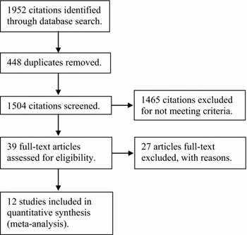
The process of the study search is depicted in Figure 1. The initial search yielded 1,952 citations. After the removal of 448 duplicates, 1,465 citations were excluded for not meeting the criteria outlined above, based on information garnered from their abstracts. Out of the 39 full-text articles reviewed, 27 studies were excluded for not reporting data on depressive symptoms in lacunar stroke patients (Aharon-Peretz et al., Reference Aharon-Peretz, Kliot and Tomer2000; van der Flier et al., Reference Van Der Flier2005; Jokinen et al., Reference Jokinen2006; Ramos-Estebanez et al., Reference Ramos-Estebanez2008; Staekenborg et al., Reference Staekenborg2010; Teodorczuk et al., Reference Teodorczuk2010; Man et al., Reference Man2011; van Norden et al., Reference Van Norden, Van Den Berg, De Laat, Gons, Van Dijk and De Leeuw2011; Xiong et al., Reference Xiong2011; Firbank et al., Reference Firbank2012; Grool et al., Reference Grool2012; Soriano-Raya et al., Reference Soriano-Raya2012; Troisi et al., Reference Troisi2012; Ogawa et al., Reference Ogawa2013; van Norden et al., Reference Van Norden2013; Wolfson et al., Reference Wolfson2013), reporting on the same patients (Grool et al., Reference Grool, Gerritsen, Zuithoff, Mali, Van Der Graaf and Geerlings2013), not reporting data in a usable format (O'Brien et al., Reference O'Brien2006; Jokinen et al., Reference Jokinen2009; Fu et al., Reference Fu2010; Lavretsky et al., Reference Lavretsky2010; Huijts et al., Reference Huijts, Duits, Staals and Van Oostenbrugge2012; Huijts et al., Reference Huijts, Van Oostenbrugge, Rouhl, Menheere and Duits2013; Brookes et al., Reference Brookes, Willis, Patel, Morris and Markus2013), or using neuropathological rather than imaging diagnoses (Thomas et al., Reference Thomas, Perry, Kalaria, Oakley, Mcmeekin and O'Brien2003; Santos et al., Reference Santos2009a; Reference Santos2010). The study characteristics and demographics are summarized in Table 1. Eleven of the 12 included studies were cohort studies, one was a case-control design (Chatterjee et al., Reference Chatterjee, Fall and Barer2010). While longitudinal studies are often considered to be a superior design, only three studies were longitudinal (Lavretsky et al., Reference Lavretsky2008; Grool et al., Reference Grool, Van Der Graaf, Mali, Witkamp, Vincken and Geerlings2011b; White et al., Reference White2011). The mean score on the NOS methodological quality checklist was 7.3, but note that due to variability in the study-checklist compatibility, there is no single maximum score. The NOS score and the maximum for each individual study is depicted in Table 1. The imperfections of the NOS scale have been discussed previously (Stang, Reference Stang2010).
Study characteristics

Abbreviations: atherosclerotic disease (AD), caregiver administered Neuropsychiatric Inventory (CGA-NPI), Centre of Epidemiological Studies Depression Scale (CES-D), cerebral small vessel disease (CSVD), Diagnostic and Statistical Manual of Mental Disorders IV (DSM-IV), depressed without imaging abnormality (DWIA), Geriatric Depression Scale (GDS), Hamilton Depression rating Scale (HDRS), lacunes outside of deep white matter (LODWM), lacunes outside of thalamus (LOT), Montgomery Asberg Depression Rating Scale (MADRS), Minimum Uniform Dataset (MUDS), non-depressed stroke (NDLS), non-lacunar stroke (NLS), Patient Health Questionairre-9 (PHQ-9), subcortical ischemic vascular disease (SIVD), and white matter hyperintensities (WMH).
aNot all studies provided the prevalence of depression, or the data to calculate the prevalence in lacunar stroke patients. Most of them, however, reported the OR for lacunar stroke patients versus comparison groups. Two studies did not provide the prevalence or an OR of general lacunar stroke for depression.
bGrool et al. (Reference Grool2012) did not use cut-off for the PHQ-9; instead, “mood” and “motivational” profiles were derived from the questionnaire. The mood profile analysis was used for this meta-analysis.
Flow chart of study selection process.

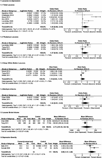
There were a total of 5,029 patients used in the analyses, with a mean age of 65.1±10.5. A random-effects meta-analysis was performed on the contribution of general lacunar stroke (Appelros and Viitanen, Reference Appelros and Viitanen2004; Lavretsky et al., Reference Lavretsky2008; Chatterjee et al., Reference Chatterjee, Fall and Barer2010; Grool et al., Reference Grool, Van Der Graaf, Mali, Geerlings and Group2011a; van Uden et al., Reference Van Uden2011; Kim et al., Reference Kim2013), thalamic (Lavretsky et al., Reference Lavretsky2008; Tang et al., Reference Tang2011; White et al., Reference White2011), deep white matter (Grool et al., Reference Grool, Van Der Graaf, Mali, Witkamp, Vincken and Geerlings2011b; Tang et al., Reference Tang2011; White et al., Reference White2011), multiple (Chen et al., Reference Chen, Chen, Mok, Lam, Wong and Tang2009; White et al., Reference White2011), and volume (Chen et al., Reference Chen, Chen, Mok, Lam, Wong and Tang2009; Tang et al., Reference Tang2011) of lacunes on depression, as well as the effects on depression severity (Komaki et al., Reference Komaki, Nagayama, Ohgami, Takaki, Mori and Akiyoshi2008; Bella et al., Reference Bella2010) (measured using the Hamilton Depression Rating Scale, HDRS (Hamilton, Reference Hamilton1960)). The forest plots of the meta-analyses results is presented in Figure 2. The OR with 95% confidence interval (CI; in parentheses) of depression from lacunar stroke was 1.46 (0.88–2.43), p = 0.15. The lacunar stroke patients were compared to other patients with non-lacunar stroke (cortical infarct, intracerebral hemorrhage) (Appelros and Viitanen, Reference Appelros and Viitanen2004), atherosclerotic diseases (coronary artery disease, cerebrovascular disease, peripheral arterial disease, abdominal aortic aneurysm) (Grool et al., Reference Grool, Van Der Graaf, Mali, Geerlings and Group2011a), WMH (Kim et al., Reference Kim2013), subcortical ischemic vascular disease (Lavretsky et al., Reference Lavretsky2008), cerebral small vessel disease (either with early onset depression or non-depressed) (van Uden et al., Reference Van Uden2011), and non-depressed stroke survivors (Chatterjee et al., Reference Chatterjee, Fall and Barer2010). The OR (95% CI) of depression from thalamic lacunar stroke was 1.37 (0.85–2.20), p = 0.19. Lacunar stroke patients were compared to patients with subcortical ischemic vascular disease (Lavretsky et al., Reference Lavretsky2008) and non-depressed patients with thalamic lacunes (Tang et al., Reference Tang2011; White et al., Reference White2011). Patients with deep white matter (DWM) lacunes had a non significantly higher risk of developing depression (RR = 1.16 (0.85–1.57), p = 0.35) compared to patients with atherosclerotic diseases (Grool et al., Reference Grool, Van Der Graaf, Mali, Geerlings and Group2011a) and non-depressed patients with deep white matter lacunes (Tang et al., Reference Tang2011; White et al., Reference White2011). The OR (95% CI) for depressed versus non-depressed patients harboring multiple lacunes was 1.34 (0.81–2.22), p = 0.25. Lacunar stroke patients with and without depression were compared (Chen et al., Reference Chen, Chen, Mok, Lam, Wong and Tang2009; White et al., Reference White2011). There was no statistical difference between the lacunar volume of depressed versus non-depressed lacunar stroke patients (mean difference (MD) = −4.71 (−351.59–342.18), p = 0.98). Patients with lacunar stroke had non-significantly more severe depressive symptoms: mean difference = 0.96 (−1.57–3.48), p = 0.46. Here, lacunar stroke patients were compared to patients with WMH (Bella et al., Reference Bella2010) and those with major depressive disorder but without imaging abnormalities (i.e. no WML or lacunar stroke) (Komaki et al., Reference Komaki, Nagayama, Ohgami, Takaki, Mori and Akiyoshi2008).
Forest plots of the six analyses. The OR for the general lacunes analysis was 1.46 (0.88–2.43). The OR for thalamic lacunes: 1.37 (0.85–2.20); RR for deep white matter lacunes: 1.16 (0.85–1.57); OR for multiple lacunes: 1.34 (0.81–2.22); MD for lacunar volume: −4.71 (−351.59–342.18); MD for depression severity: 0.96 (−1.57–3.48). All analyses p > 0.05.

The statistical heterogeneity between studies was high among most analyses: I 2 = 87% for general lacunar stroke, 74% for the thalamic lacunar stroke, 78% for the deep white matter, 0% for multiple lacunes, 38% for the lacunar stroke volume, and 51% for the depression severity analyses. Visual interpretation of funnel plots did not indicate obvious publication bias (supplemental Figure S1). A statistical test (e.g. Begg's test (Begg and Mazumdar, Reference Begg and Mazumdar1994)) was not performed due to the low number of studies and the low statistical power it would in turn yield.
Discussion
While stroke has been associated with depression (Hackett et al., Reference Hackett, Yapa, Parag and Anderson2005), the contribution of lacunar stroke is unclear. Twelve studies examining the prevalence of depressive symptoms in patients with lacunar stroke and deep white matter disease were submitted to a random-effects meta-analysis, with separate analyses for the effect of general, multiple, volume, and location specific lacunar strokes on depression prevalence, as well as the effects on depressive symptom severity. None of the analyses produced statistically significant results. Lacunar stroke patients had a non-significantly increased odds of depression compared to other patient groups (see Table 1 for the details of comparison groups by study), at p = 0.15. Similarly, while thalamic, deep white matter, and multiple lacunar strokes contributed to slightly increased depression, neither of these results were significant. Lastly, there was no effect of lacunar volume on depression, and no difference in the severity of depressive symptoms between patient groups.
A number of issues were present with the data, one of which was the variability in the comparison groups. First, the history of depression in patients is not described in most of the studies, though a subset likely did experience prior depressive symptoms. This has the potential to alter the symptoms and pathology of depression patients. Second, none of the studies used healthy control participants, instead comparing lacunar stroke patients with other patients with a variety of diseases. Comparison groups varied even within specific analyses (e.g. the general lacunar stroke analysis contained patients with six different disease types). As each one of the studies had a different comparison groups, the group differences likely differed between studies, thus confounding the meta-analysis. This is exemplified by the high statistical heterogeneity, which limits the conclusions that can be drawn from the analyses. However, it must be appreciated that the comparison groups in these studies had an increased risk of depression compared to healthy controls. Chronic and acute cardiovascular diseases and stroke, lacunar or not, are risk factors for depression. Our data suggest that the risk of depression in lacunar stroke and deep white matter disease is similar to other causes of stroke such as atherosclerotic large artery disease, even though motor outcomes and survival rates are better in lacunar stroke than these other stroke types (Sacco et al., Reference Sacco, Shi, Zamanillo and Kargman1994). By extension, our data also suggest that the risk of depression following lacunar stroke is quite high compared to the healthy population because the prevalence of depression is comparable to that in other cardiovascular diseases.
A second issue with this data is the low number of studies included in the analyses. While the general lacunar stroke analysis had six studies and the deep white matter and thalamic lacunes analyses had three, the rest only contained two studies each. This reduced the power of the statistical analyses, and introduced uncertainty into the results. Additionally, there was insufficient data in the included studies to analyze the impact of other brain regions such as the frontal lobes and subcortical structures including the caudate nucleus, both of which have been implicated in depression (Provinciali and Coccia, Reference Provinciali and Coccia2002; Buijs et al., Reference Buijs, Greebe and Rinkel2012). However, this was a result of the relatively low number of studies available, which was exacerbated in this meta-analysis by the necessary exclusion criteria. This study serves to indicate the relative infancy of this field.
Lastly, studies used different methods of reporting results: some provided raw data from which OR or RR could be calculated, while others provided either adjusted or unadjusted OR, RR, or other forms of statistical measures. This variability presented compatibility problems for analyses, and led to the inability to include certain studies in the analyses due to a lack of usable data. Additionally, some studies did not control for co-variables, while others that performed multivariable analyses differed in the co-variables they included, thus affecting the meta-analyses. The details of co-variables used are presented in supplemental Table S1.
More research is necessary to establish whether the general presence, location-specific, or the accumulation of lacunes and other microvascular lesions can lead to the development of depression. Further, lacunar stroke and deep white matter disease patients need to be examined separately to determine their specific contributions to depression. This meta-analysis hinted that lacunar stroke and deep white matter disease is associated with depression, with rates comparable to other cardiovascular diseases. However, the heterogeneity between studies and the patient populations, the low number of studies analyzed, and the low number of region-specific lesion analyses prevented the formation of any decisive conclusions. From a clinical standpoint, this study had a narrow focus on groups of patients originating from specialized clinical centres, which limits the generalizability of the results. Nevertheless, it indicates the importance for screening patients with microvascular, not just macrovascular pathologies for depressive symptoms, and providing appropriate treatment. Future work can also elucidate whether depressive symptom profiles differ between different vascular pathologies, and whether treating cardiovascular risk factors reduce the risk of lacunar stroke and deep white matter disease, and subsequently depression.
Conflict of interest
None.
Description of authors’ roles
P.E. completed the systematic literature search and review, data extraction and analyses, and wrote the paper and prepared the figures and tables. E.E.S. contributed to the paper preparation, and provided important feedback and edits of the paper. Z.I., C.E.F., E.E.S. and T.A.S. provided important feedback and edits of the paper.
Acknowledgments
This work was supported by a Personnel Award from the Heart and Stroke Foundation of Canada and an Early Researcher Award from the Ontario Ministry of Research and Innovation to T.A.S., the Heather and Eric Donnelly Endowment from St. Michael's Hospital Foundation to C.E.F., and E.E.S. holds the Kathy Taylor Chair in Vascular Dementia at the University of Calgary and receives salary support from Alberta Innovates - Health Solutions and the Canadian Institutes for Health Research.