Case report
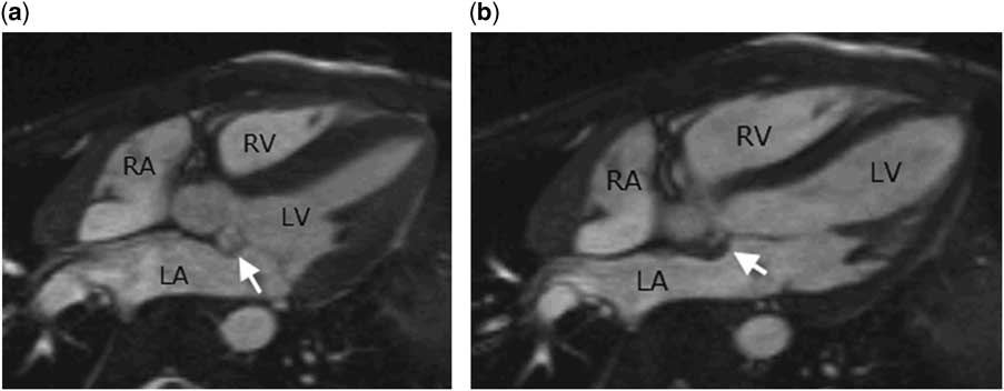
A 12-year-old boy developed two episodes of sharp chest pain while walking or during exercise. His family history, physical examination, and electrocardiograms were unremarkable. Echocardiography revealed a non-pedunculated mass in the left atrium posterior to the aortic root in an otherwise normal heart. Cardiac MRI was requested for characterisation of the nature of the mass. MRI was performed using a 1.5-Tesla MRI system (Avanto, Siemens, Erlangen, Germany). Electrocardiogram-gated, steady-state, free-precession cine imaging and electrocardiogram-gated, respiration-navigated angiography using a blood-pool contrast agent (Gadofoveset: ABLAVAR®; Lantheus Medical Imaging Inc., North Billerica, Massachusetts, United States of America) demonstrated a protrusion of the fibrous; tissue between the insertions of the aortic and mitral valves – namely, intervalvular fibrosa – into the left atrium. At cine imaging, it expanded in systole and collapsed in diastole (Fig 1, Supplementary video 1). No left atrial or ventricular inflow or outflow obstruction or solid mass lesion was identified. The aortic and mitral valves were normal. In addition,24-hour holter monitoring and Bruce exercise test protocol were normal. The patient has not developed any chest pain in the last 6 months and will be on clinical surveillance with clinical follow-up with echocardiography for development of any complication such as rupture of the membrane causing functional mitral regurgitation and arrhythmias.
Left ventricular (LV) three-chamber cine images show the intervalvular fibrosa – between the attachments of the aortic and mitral valves – ballooning into the left atrium (LA) in systole ( a ) and forming a collapsed mass in diastole ( b ). RA=right atrium; RV=right ventricle.

Mitral-aortic intervalvular fibrosa aneurysm is a rare condition that is considered a subtype of annular subvalvular left ventricular aneurysm.Reference Abrahams, Barton, Cockshott, Edington and Weaver 1 , Reference Chesler, Korns, Porter, Reyes and Edwards 2 The mitral-aortic fibrosa is a thin layer of fibrous tissue that separates the posterior portion of the aortic root from the base of the anterior mitral leaflet. Its posterior and lateral limit is defined by the left atrium, the inferior limit by the left ventricular outflow tract, and the upper limit by the pericardium. The disease was initially considered of congenital origin with predominance in African-Americans;Reference Abrahams, Barton, Cockshott, Edington and Weaver 1 however, since then, many cases have been described in other races and is caused by multiple aetiologies such as infective endocarditis, trauma, and surgery, especially valve replacement, questioning the congenital origin.Reference Sudhakar, Sewani, Agrawal and Uretsky 3 – Reference Karalis, Bansal and Hauck 5 The present case did not show any of these acquired aetiological factors and is safely considered to be congenital. MRI, as in this case, provides information about tissue characterisation, including contrast enhancement and anatomical detail for lesions with difficult echo windows.
Multiple complications of the aneurysm have been described, such as perforation with a shunt from the left ventricular outflow tract into the left atrium, or into the pericardial space, causing cardiac tamponade, infection, compression of the coronaries causing ischaemia, primary valvular dysfunctions, or thrombus formation with distal embolisation.Reference Head, Jue and Askren 6 Surgical repair is the treatment of choice in patients with complicated mitral-aortic intervalvular fibrosa aneurysm, and close echocardiographic and clinical follow-up has been proposed for asymptomatic patients.Reference Sudhakar, Sewani, Agrawal and Uretsky 3 , Reference Hasin, Reisner and Agmon 7 In the present case, cine imaging and high-resolution angiograms obtained using a blood-pool agent were diagnostic, whereas echocardiographic characterisation of the lesion was difficult.
Acknowledgements
None.
Financial Support
This research received no specific grant from any funding agency or from commercial or not-for-profit sectors.
Conflicts of Interest
None.
Supplementary Material
To view supplementary material for this article, please visit http://dx.doi.org/10.1017/S1047951116001323