INTRODUCTION
The Japanese skeleton shrimp Caprella mutica Schurin, Reference Schurin1935 (Amphipoda: Caprellidea), originally found in the high sublittoral zone of the low boreal West Pacific (Vassilenko, Reference Vassilenko and Adrianov2006), have successfully been spreading throughout the boreal waters of the northern hemisphere and even reached New Zealand (Willis et al., Reference Willis, Woods and Ashton2009). In Europe, the species was first recorded in The Netherlands, and described as new species Caprella macho Platvoet, de Bruyne & Gmelig Meyling, Reference Platvoet, de Bruyne and Gmelig Meiling1995 (Faasse, Reference Faasse2005), and later in Belgium, Norway, Scotland, Germany, Wales, England, France and Ireland (reviews in Ashton et al., Reference Ashton, Willis, Cook and Burrows2007; Cook et al., Reference Cook, Jahnke, Kerckhof, Minchin, Faasse, Boos and Ashton2007b; Frey et al., Reference Frey, Gartner, Murray and Therriault2009; Turcotte & Sainte-Marie, Reference Turcotte and Sainte-Marie2009; Schückel et al., Reference Schückel, Schückel, Beck and Liebezeit2010). According to the mtDNA tracing, the non-native populations of C. mutica formed, probably, by repeated introductions from multiple native sources (Ashton et al., Reference Ashton, Stevens, Hart, Green, Burrows, Cook and Willis2008; Willis et al., Reference Willis, Woods and Ashton2009).
In its native range, C. mutica was found in quiet bays with algal beds at depth from nearly 0 to 13 m (Arimoto, Reference Arimoto1976; Vassilenko, Reference Vassilenko and Adrianov2006), where it is known to reproduce in summer time (Vassilenko, Reference Vassilenko and Adrianov2006). In laboratory conditions, the species was highly proliferate, ecologically tolerant (Cook et al., Reference Cook, Willis and Lozano-Fernandez2007a) and aggressively competitive (Shucksmith et al., Reference Shucksmith, Cook, Hughes and Burrows2009), revealing it to be a potentially successful survivor in transportation and introduction. In the artificial sites, the species was primarily found on human-made structures like ropes, nets, buoys, aquaculture cages, oil platforms, etc. and often with other non-native species (Ashton et al., Reference Ashton, Willis, Cook and Burrows2007; Cook et al., Reference Cook, Jahnke, Kerckhof, Minchin, Faasse, Boos and Ashton2007b; Frey et al., Reference Frey, Gartner, Murray and Therriault2009; Turcotte & Sainte-Marie, Reference Turcotte and Sainte-Marie2009; Schückel et al., Reference Schückel, Schückel, Beck and Liebezeit2010). The direct effect of the introduction of C. mutica has not yet been observed, though a decrease of mussel farm production was reported since the invasion of the species in Scotland and Canada (Turcotte & Sainte-Marie, Reference Turcotte and Sainte-Marie2009).
This note describes the first record of C. mutica in Sweden. Until recently the caprellid genus Caprella Lamark, 1801 (Caprellidea: Caprellidae) was represented by three native species in Sweden: Caprella ciliata G.O. Sars, 1883, C. linearis (Linnaeus, 1767) and C. septentrionalis Krøyer, 1838 (cf. Boeck, Reference Boeck1870; G.O. Sars, Reference Sars1893; Enequist, Reference Enequist1949; Buhl-Jensen & Fosså, Reference Buhl-Jensen and Fosså1991; Miskov-Nodland et al., Reference Miskov-Nodland, Buhl-Mortensen and Høisæter1999).
MATERIALS AND METHODS
The specimens of Caprella mutica were collected in July 2011 from the piers of Lysekil harbour within the aggregations of invertebrates and macroalgae, composed mainly of bivalves Mytilus and tunicates at depth of 0–2 m (see geographical details in Systematics section). Samples were preserved in 96% ethanol. The total body of a male was photographed and drawn before part of appendages were detached, and put on temporal slide with glycerol to be drawn. The rest of the sample was further inspected without dissection. Body length was measured from the anterior margin of head to the distal margin of pereonite 7. The term ‘seta’ was used for thin, smooth, flexible, articulated cuticular extensions, ‘plumose seta’ for setae with setules along the axis (feather-like setae), ‘robust seta’ for stronger, but not yet spine-like setae, ‘spiniform seta’ for spine-like, clearly articulated extensions, ‘spine’ for robust, sharp, non-articulated structures. The specimens are deposited at the Finnish Museum of Natural History (MZH). The classification of C. mutica follows the latest systematic treatment (Vassilenko, Reference Vassilenko and Adrianov2006).
RESULTS AND DISCUSSION
SYSTEMATICS
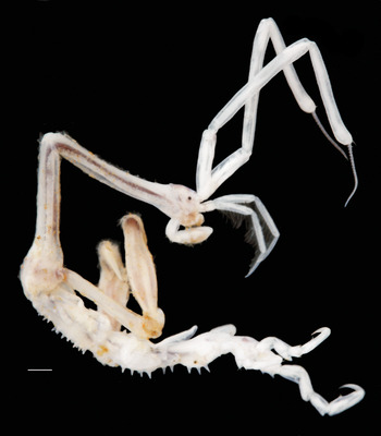
Caprella mutica Schurin, Reference Schurin1935: male, 25.5 mm, from Sweden, Lysekil, entire view (photograph from ethanol fixed specimen). Scale bar: 1 mm.

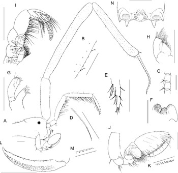
Caprella mutica Schurin, Reference Schurin1935: male, 25.5 mm, from Sweden, Lysekil. (A) Head with antennae 1 and 2; (B) part of cuticle of posterior margin of antenna 1 segment 2; (C) segments of antenna 1 flagellum; (D) posterior seta of antenna 2 peduncular segment 4; (E) distal part of antenna 2 flagellum; (F) labium (left outer plate not shown); (G) maxilla 1, anterior view; (H) maxilla 2, anterior view; (I) maxilliped, posterior view; (J) gnathopod 1, lateral view; (K) posterior margin of gnathopod 1 propodus; (L) dactylus of gnathopod 1, medial view; (M) posterior margin of gnathopod 1 dactylus; (N) distal part of pereonite 7 (pereopod 7 not shown), with rudimentary abdomen and its appendages. Scale bars: A, B, E–J, L & N, 0.5 mm; C & D, 0.25 mm; K & M, 0.1 mm.

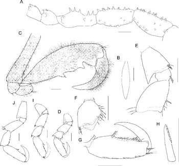
Caprella mutica Schurin, Reference Schurin1935: male, 25.5 mm, from Sweden, Lysekil. (A) Pereonites 3−7 (from right to left); (B) gill of pereonite 3; (C) gnathopod 2, lateral view; (D) pereopod 5, lateral view; (E) basis, ischium and merus (from top to bottom) of pereopod 5, lateral view; (F) carpus of pereopod 5, lateral view; (G) propodus of pereopod 5, lateral view; (H) grasping spiniform seta of pereopod 5 propodus, lateral view; (I) pereopod 6, lateral view; (J) pereopod 7, lateral view. Scale bars: A, B, 1 mm; C, D, E, F, G, I &J, 0.5 mm; H, 0.2 mm.

Caprella mutica Schurin, Reference Schurin1935: 198, figures 1 & 4 (upper); 1937: 27, figures 7 & 8; Utinomi, Reference Utinomi1947: 75; Shchapova et al., Reference Shchapova, Mokievsky and Pasternak1957: 87; Vassilenko, Reference Vassilenko and Bykhovskii1967: 208; Reference Vassilenko1974: 201, figures 118 & 119; 2006: 47, 109, table XV; McCain & Steinberg, Reference McCain and Steinberg1970: 32; Arimoto, Reference Arimoto1971: 18: 1976: 111, figure 59; Marelli, Reference Marelli1981: 655, figure 1c, d, f; Fedotov, Reference Fedotov and Gulbin1987: 40; Faasse, Reference Faasse2005: 23, figure 1; Frey et al., Reference Frey, Gartner, Murray and Therriault2009: 497, figure 2; Schückel et al., Reference Schückel, Schückel, Beck and Liebezeit2010: 87, figures 3 & 4.
Caprella macho Platvoet et al., Reference Platvoet, de Bruyne and Gmelig Meiling1995: 1.
MATERIAL EXAMINED
Four adult males, 20–25.5 mm, one subadult male, 15.5 mm, Skagerrak Strait, Sweden, Lysekil, southern guest boat harbour, 58°27′N 11°44′E, 27 July 2011, coll. Laakkonen (MZH 112030).
DESCRIPTION
Head merged with pereonite 1, leaving only shallow dorsal fissure. Eye small and round. Head and pereonite 1 smooth. Pereonites 1 and 2 and gnathopod 2 covered by fine plumose setae. Pereonite 1 nearly as long as pereonite 2, and about as long as or slightly longer than pereonites 3 and 4 together. Pereonite 2 with only 1 posterolateral spine on each side. Pereonite 3 only slightly longer than pereonite 4; both nearly cylindrical, with 7–9 blunt straight dorsal, 2–4 lateral and 4–7 marginal spines. Gills spindle-shaped, about as long as and half as wide as corresponding pereonites. Pereonite 5 anteriorly and posteriorly narrowed, with 5 pairs of dorsal, 2 anterolateral and 1 posterolateral (pre-pereopodal) sharp straight spines. Pereonites 6 and 7 about equally long, and together about as long as pereonite 5, dorsally convex, each with 1 pair of sharp, posteriorly directed dorsal spines and 1 posterolateral spine on each side.
Antenna 1 0.6–0.7 times as long as body; peduncle segment 1 0.4 times as wide as head, with rare short setae; peduncle segments 2 and 3 with rare short setae in duplexes and multiple cuticular scales, each bearing microseta at anterior base; peduncle segment 3 slightly widening distally, also with anterodistal pair of robust setae with subapical setule and multiple short posterodistal setae; ratio of peduncle segment lengths 1:2:1.5–1.7; ratio of peduncle segment widths 1.3–1.4:1.1–1.2:1; flagellum thin, 17–25-segmented, 0.7–0.8 times (1.1 in subadult male) as long as and 0.25–0.3 times as wide as peduncle segment 3; each flagellar segment, except for terminal, with anterodistal seta and posterodistal bunch of setae and aesthetasc. Antenna 2 0.3–0.4 times as long as antenna 1, not reaching segment 3 of antenna 1 peduncle (reaching in subadult male); peduncle 0.5 times as wide as peduncle of antenna 1; ratio of peduncle segment lengths 1:1.3:5:5.7; ratio of peduncle segment widths 1:1:1.1:1; flagellum 2-segmented, 0.7 times as long as peduncle segment 4; flagellum segment 2 0.2 times as long as segment 1; peduncle segment 2 with 1–2 posteroproximal setae and 1 group of posterodistal setae; peduncle segments 3 and 4 and flagellum segment 1 with many posterior bunches of long simple setae in duplexes and long distally serrated setae with subapical setule, and short anterior setae; peduncle segment 4 and flagellum segment 1 also with short posterior setae between bunches long setae; posterodistal setae of flagellum segment 1 with stronger serration; flagellum segment 2 with 2 (including terminal) anterior duplexes of setae, 2 apical robust serrated setae with subapical setule and 2 apical simple setae.
Mandibles without palp: labium with well-developed inner lobes, covered by dense setules. Maxilla 1 without outer plate; inner plate distally with strong, roughly serrated spiniform setae with 1–4 lateral denticles; palp distally widened, with multiple anterior setae, 6 distal strong spiniform setae and tooth-like setae. Maxilla 2 plates with multiple distal setae.
Maxilliped: basis, ischium and merus about equally long, with multiple long anterior setae. Basis endite with plumose posteromedial and medial setae and 5 medial tooth-like spiniform setae. Ischium endite as long as and about as wide as basis endite, with about 10 medial tooth-like spiniform setae and simple setae. Carpus, the largest segment, with multiple long medial setae. Propodus slightly shorter than carpus, with multiple long posterior, posteromedial and anteromedial setae. Dactylus without unguis, covered by multiple combs of fine robust setules.
Gnathopod 1: basis 2.2 times as long as wide, with distal anterolateral process, nearly as long as ischium, and 1 anteromedial long seta. Basis anterior margin with 2 proximal scales with setule; posterior margin with 2 scales in distal half and distal marginal bunch of long setae. Basis process with 4 scales with setule. Ischium with posterodistal bunch of long setae. Merus about as long as ischium, with 1 posteroproximal scale with setule, 2 posterior bunches of long setae and multiple long distomedial and anteromedial setae. Carpus with anteromedial long setae, group of 3 anterodistal setules and posterior lobe, covered by multiple long setae. Propodus almond-shaped, tapering distally, with rare anterior scales, bearing setule, posteroproximal pair of strong spiniform setae, long medial, distomedial and posterolateral setae, distolateral bunch of short setae; posterior margin with nearly quadrangular serration and short setae. Dactylus with short anterior, posterolateral and posteromedial setae, subrectangular bilobate posterior serration and multiple medial crests of spinules.
Gnathopod 2: jointed nearly at posterior margin of pereonite 2. Basis thin and long, 0.7 times as long as pereonite 2, with short anterodistal process. Propodus elongate, nearly as long as basis; posterior margin with 3 processes. Proximal process with apical spiniform seta. Median (poison) process, separated by long gap with convex margin from proximal, slightly longer than proximal. Distal (grasping) process about as long as median. Dactylus strong, curved; posterior margin with broad grasping tubercle.
Pereopods: stout; posterior margin of segments covered by microscales. Basis with large posterior, triangular process, reaching merus. Ischium with posterior inflation and anterodistal bunch of stout, blunt and slightly hooked setae. Merus posterodistally lobate; posterior margin with distal bunch of setae. Carpus anterior margin with two lines of numerous robust setae, some of them blunt and slightly hooked. Propodus, the longest segment; posterior margin with bunches of 1–3 long setae; anterior margin with pair of median, posterodistally serrated, spiniform grasping setae, slightly shifted to proximal half of segment, numerous short setae along distal sharp edge, distal lobe and bunch of long setae. Dactylus stretching slightly beyond median spiniform setae, with rare, short setae. Pereopod 5 longer than pereonite 5; ratio of segment lengths 2.5:1:3:2.5:6:4; ratio of segment width 2:1.9:3:2.4:1. Basis with anterodistal bunch of setae. Merus anterior margin with 3 distal bunches of setae, distally less setose. Carpus posterior margin with distal bunch of setae. Pereopod 6 slightly larger than pereopod 5; ratio of segment lengths 2.7:1:3.4:3.1:6:4; ratio of segment widths 1.9:1.7:2.4:2.4:1. Basis with anterodistal pair of robust, blunt, distally hooked setae. Merus with 2 distal pairs of blunt, distally hooked setae. Carpus posterior margin with 2 distal bunches of setae. Pereopod 7 slightly larger than pereopod 6; ratio of segment lengths 3.8:1:4.3:3.6:6:4. Basis with anterodistal pair of robust, blunt, distally hooked setae. Merus anterior margin with 1 predistal seta. Carpus posterior margin with 4 bunches of setae.
Penes: smooth, about as long as uropods. Uropod with pair of anterolateral setae. Posturopodal tubercle with a number of short and long setae.
REMARKS
The caprellid specimens from Sweden clearly possess the diagnostic characters of C. mutica: head and pereonite 1 without spines; pereonite 2 only with posterolateral spine on each side; pereonites 1 and 2 together with gnathopods 2 covered by dense hair-like setae; posterodistal (grasping) process of gnathopod 2 propodus rather prominent, about as long as posteromedian (so called poison) process. The large (more than 20 mm long) specimens from Sweden differ from those illustrated originally (Schurin, Reference Schurin1935) and subsequently found and illustrated from the native range (Vassilenko, Reference Vassilenko1974, Reference Vassilenko and Adrianov2006) by (1) longer pereonites 1 and 2, which exceed the combined length of pereonites 3 and 4 (shorter than combined length of pereonites 3 and 4 in the native range); and (2) flagellum of antenna 1 0.7–0.8 times as long as peduncle segment 3 (against 1.1–1.5 times as long as segment 3 in the native range). The holotype of C. macho (junior synonym of C. mutica (Faasse, Reference Faasse2005)) described from The Netherlands (Platvoet et al., Reference Platvoet, de Bruyne and Gmelig Meiling1995) also seems to fit the variation pattern of the Swedish specimens. However, the flagellum of antenna 1 in 15 mm male from Sweden was longer than peduncle segment 3, probably indicating that this character varies depending on specimen maturity. Further detailed comparison of C. mutica from around the range is necessary to clarify possible taxonomic value of the described differences.
Marelli (Reference Marelli1981) synonymized Caprella acanthogaster humboldtiensis Martin, Reference Martin1977 with C. mutica, based on the fact that some paratype males had setose head, pereonites 1 and 2 and gnathopod 2; and pereonite 2 with one pair of dorsal spines. However, rather the absence of dorsal spines on pereonite 2 and absence of setae on head are diagnostic features of C. mutica. Besides, the originally illustrated male of C. a. humboldtiensis (Martin, Reference Martin1977, figure 3A) has three pairs of dorsal spines (from one to three in the description (Martin, Reference Martin1977, p. 154)). Therefore, we propose the revalidation of Caprella acanthogaster humboldtiensis Martin, Reference Martin1977 stat. rev., which differs from C. acanthogaster acanthogaster Mayer, 1890 by the presence of setae on head, pereonites 1 and 2 and gnathopod 2, and well-formed distal triangular grasping process of gnathopod 2 propodus.
DISTRIBUTION
Low boreal West Pacific (native range): the Sea of Okhotsk, the Sea of Japan (Schurin, Reference Schurin1935, 1937; Utinomi, Reference Utinomi1947; Shchapova et al., Reference Shchapova, Mokievsky and Pasternak1957; Vassilenko, Reference Vassilenko and Bykhovskii1967, Reference Vassilenko1974; McCain & Steinberg, Reference McCain and Steinberg1970; Arimoto, Reference Arimoto1971, Reference Arimoto1976; Fedotov, Reference Fedotov and Gulbin1987). Introduced range: coasts of North America, North Europe and New Zealand (reviewed with details in Ashton et al., Reference Ashton, Willis, Cook and Burrows2007; Cook et al., Reference Cook, Jahnke, Kerckhof, Minchin, Faasse, Boos and Ashton2007b; Frey et al., Reference Frey, Gartner, Murray and Therriault2009; Turcotte & Sainte-Marie, Reference Turcotte and Sainte-Marie2009; Schückel et al., Reference Schückel, Schückel, Beck and Liebezeit2010). Newly recorded in the eastern part of Skagerrak Strait, Sweden.
HABITAT
Natural: high sublittoral species found on algal beds (detailed account of algal species in Vassilenko, Reference Vassilenko and Adrianov2006) at depth from nearly 0 to 13 m (Arimoto, Reference Arimoto1976; Vassilenko, Reference Vassilenko and Adrianov2006). Artificial: on man-made constructions in association with other non-native species (Ashton et al., Reference Ashton, Willis, Cook and Burrows2007; Cook et al., Reference Cook, Jahnke, Kerckhof, Minchin, Faasse, Boos and Ashton2007b; Frey et al., Reference Frey, Gartner, Murray and Therriault2009; Turcotte & Sainte-Marie, Reference Turcotte and Sainte-Marie2009; Schückel et al., Reference Schückel, Schückel, Beck and Liebezeit2010). In Sweden found on pier constructions among the mussel Mytilus sp. and tunicate aggregations.
Caprella mutica was found in the harbour area in Sweden, where it most probably could have been artificially introduced from other man-made constructions and habitats. The record of C. mutica among the settlements of benthic invertebrates probably indicates that the species can occupy not only artificial structures, but also natural habitats.
ACKNOWLEDGEMENTS
Fieldwork took place during a project funded by a grant from the Academy of Finland to Risto Väinölä (No. 127471), who also made valuable comments about the manuscript. We thank Petteri Lehikoinen for the fieldwork assistance. Marco Faasse kindly provided us with a copy of his paper, and Marjatta Mikkonen helped to find other rare literature. We appreciate important comments of two anonymous referees.