Introduction
According to our present knowledge, neuropeptides and biogenic amines are the most important secretions of neuroendocrine cells. The role of the central nervous system (CNS) neurotransmitters, which appear to be the mediator of many control mechanisms in mammals (Weiner and Ganong, Reference Weiner and Ganong1978), is little understood in other vertebrate groups except for mammals. The neuroendocrine system is an integral component of the neuronal apparatus of the brain, a major portion of which is concentrated in the hypothalamus. The hypothalamic monoamines dopamine (3,4-dihydroxyphenylalanine), noradrenalin and serotonin have profound effects on the release of mammalian pituitary hormones, presumably in most cases by modulating the release of the hypothalamic hormone (Meites et al., Reference Meites, Simpkins, Bruni and Advis1977). The hypothalamus is of paramount importance as an ‘integration center’ for the various influences on pituitary function. The hypothalamus apparently responds to the exteroceptive and interoceptive stimuli by altering the secretion of certain neurotransmitters and release hormones. Ideally, a study of the control of pituitary function should not only show the effects of any particular influence on hormone secretion but also demonstrate the changes in the metabolism of neurotransmitters and release of inhibiting hormones.
Dopamine (DA) is reported to be synthesized largely in CNS. At the time of its discovery in the brain, DA was proposed to act only as an intermediate in the biosynthesis of norepinephrine and epinephrine. However, in 1958 DA was demonstrated to be present in the brain in concentrations similar to that of norepinephrine, suggesting that DA not only serves as a precursor but might also function as a neurotransmitter in its own right. When it was discovered that striatum contained 70% to 80% of the brain DA and that depletion of striatal DA was important in the pathogenesis of Parkinson’s disease, it rapidly became a subject of intense interest among neuropharmacologists. Injections of DA into the third ventricle also result in an increase of peripheral plasma testosterone. This rise in testosterone level was also noted after intraventricular DOPA administration. So the effect of exogenous DA was similar to that of the endogenous amine synthesized in brain after the injection of its precursor, possibly reaching the brain via the blood–brain barrier.
A prominent neurotransmitter affecting prolactin (PRL) secretion is DA. DA inhibits PRL release from anterior pituitary cells in vitro in chicken, pigeon and turkey (Harvey et al., Reference Harvey, Chadwick, Border, Scanes and Phillips1982; Hall and Chadwick, Reference Hall and Chadwick1983 and Reference Hall and Chadwick1984; Fehrer, Reference Fehrer1984) and also in vivo in turkeys (El Halawani et al., Reference El Halawani, Youngren, Silsby and Phillips1991). DA-binding sites in the anterior pituitary of bantam hens decrease during incubation, when PRL is high (Macnamee et al., Reference Macnamee, Sharp, Lea, Sterling and Harvey1986). The dopaminergic D2 receptor agonist quinpirole inhibits VIP-induced PRL secretion and PRL mRNA level when incubated with turkey anterior pituitary cells (Xu et al., Reference Xu, Proudman, Pitts, Wong, Foster and El Halawani1996), suggesting the presence of inhibitory D2 DA receptors within the pituitary. Central infusion of DA into the turkey brain at low concentration results in significant increase in plasma PRL (Youngren et al., Reference Youngren, Chaiseha and El Halawani1998).
Egg size represents a key, hormonally mediated, life-history trait, which varies markedly among species (from 0.25 g in bee hummingbirds, Mellisuga helenae, to 1500 g in the ostrich, Struthio camelus). Most of the variation in egg size within species is due to difference in the mean egg size produced by individual females, i.e. it is independent of variation in environmental, nutritional and social conditions (Reed, Reference Reed2000). Experimental manipulation of hormone levels can be a powerful tool to understand not only the physiological basis of phenotypic variation in hormonally mediated traits but also the ecological and evolutionary consequences of this variation (Sinervo and Basolo, Reference Sinervo and Basolo1996; Ketterson and Nolan, Reference Ketterson and Nolan1999). Hormones of the hypothalamo–pituitary–gonadal (HPG) axis regulate ovarian function in birds and other oviparous vertebrates. Our knowledge of this system (Norris, Reference Norris1997; Williams, Reference Williams1999) allows for the identification of several ‘candidate’ hormones that might be involved in the regulation of egg-size variation; principally, the gonadotropins, follicle-stimulating hormone (FSH) and luteinizing hormone (LH), and the steroid hormones (estradiol and progesterone). It is clear that certain hormonal manipulations (in particular antiestrogens and FSH) can modify expression of the egg-size phenotype while maintaining the range of inter-individual variation in this trait. Several studies have shown that manipulation of egg size results in a corresponding, often compensatory, adjustment in clutch size.
Currently available neurotransmitters suffer from a number of serious adverse effects. As a consequence, there continue to be a high demand for new alternatives, preferably with least side effects. So, herbal medication is the most commonly used alternative. Moreover, herbs are known for their safety, efficacy, cultural acceptability and lesser side-effects. However, their safety and efficacy need to be further evaluated by experimental and clinical studies. To date, a large number of traditional plants have been reported but only a small number of these has received scientific and clinical evaluation to assess their efficacy. The efficacy of a herb should be first tested on an animal model before their extrapolation on human subject.
Mucuna pruriens is a rich source of l-dihydroxyphenylalanine (l-DOPA, a precursor of DA), a compound chiefly used in treating the symptoms of Parkinson’s disease (Lubis and Sastrapradha, Reference Lubis and Sastrapradha1981; Parikh et al., Reference Parikh, Doshi, Sawant and Salunkh1990; Zhang et al., Reference Zhang, Su, Xu and Tang1991; Su et al., Reference Su, Tang, Xu and Zhang1992; Lorenzetti et al., Reference Lorenzetti, MacIsaac, Arnason, Awang and Buckles1998). It is widely believed that M. pruriens significantly influences the DA metabolism in the stritonigral tract and improves relief from Parkinson’s symptoms in rats and humans (Manyam, Reference Manyam1995; Office de Vulgarisation Pharmaceutique (OVP), 1995). The importance of this particular study is not that Mucuna is an alternative to l-DOPA, rather it is that compound occurring naturally in plants for example, may contain biologically active components that can be isolated, tested and used to provide safer and better treatment for many diseases. In view of our interest in the neurotransmitter-induced developmental and reproductive changes of avian species, the present study was designed for a high food value bird, Japanese quail, Coturnix coturnix japonica. Experiments were preformed in both male and female quail following feeding of M. pruriens seed powder (MPSP, natural source of l-DOPA) and its comparison with that of synthetic l-DOPA.
Material and methods
Mucuna pruriens
M. pruriens, commonly known as velvet bean, contains l-DOPA. Mucuna has been used for ages in India to treat Parkinson’s disease. l-DOPA is used to make DA, an important brain chemical involved in mood, sexuality and movement. Studies in rats show M. pruriens supplements influence DA content in the cortex of the brain. M. pruriens has antioxidant properties. Parkinson’s disease has low prevalence in India except in the small Parsi community. Parkinson’s disease has been known in India since ancient days and the powder of M. pruriens seeds has been used for its treatment.
Japanese quail
The Japanese quail was used first as a singing bird and then as an egg producer. It later became popular as a laboratory animal, because of its small body size, little food consumption and rapid maturation. The Japanese quail belongs to the order Galiformes, genus Coturnix and species japonica. The scientific designation for Japanese quail is Coturnix japonica. Adult females have pale breast feathers that are spackled with dark colored spots. Adult males have uniform dark rust red feathers on the breast and cheek. Sex differences in plumage colors appear at about 3 weeks of age.
Hatching eggs were collected from the breeding cages of Japanese quail maintained in the bird room of animal house of our department and loaded in the incubator. Suitable incubation environment was selected to hatch quail eggs successfully. Dry bulb thermometer was set at 37.5°C and wet bulb at 29.3°C and 32.2°C in the incubation and hatching compartments, respectively. Chicks were hatched successfully on the 18th day of incubation. Chicks were kept in 24 h light for the first 1 week (before starting the experiment). After 1 week, chicks were transferred to LD12 : 12 till the end of the experiments. These photoperiodic conditions were maintained as recommended by Panda et al. (Reference Panda, Ahuja, Shrivastava, Singh, Agarwal and Thomas1987) (the chicks up to 2 weeks of age may be grown under 24 h light period and then 12 h photoperiod up to 6 weeks of age for adequate and normal growth of the quail). Special chick ration, cool and clean water were provided at all times.
Experiments
One-week-old (sexually immature) quail chicks were divided into three groups of 36 birds each. Group I provided with normal diet and served as control. Groups II and III (experimental groups) were provided with food mixed with l-DOPA (a DA precursor, 50 mg/15 g food, M.W. 197.19; Batch No. J01Z/1592/2807/62/Product No. 47079, S.D. Fine Chemicals Ltd, Mumbai, India) and MPSP (480 g/kg food), respectively. At the age of 3 weeks (when birds were sexually distinguished), Group I was divided into two sub-groups IA (male) and IB (female) of six birds each. Similarly Group II was sub-divided into IIA (male) and IIB (female) of six birds each. Group III was sub-divided into IIIA (male) and IIIB (female) of six birds each. Birds of all the groups were maintained in LD 12 : 12. Observations were made up to the age of 5 weeks. For the purpose of observation of egg laying, size and weight in control groups IA and IB they were maintained together by marking them with a particular identification (numbering tag in the leg). Similar grouping was done in both experimental groups.
Male
After 5 weeks of l-DOPA and MPSP feeding, male birds (control-IA and experimental-IIA and IIIA) were anesthetized with ether. Blood was collected directly from the left ventricle of the heart with a heparinized syringe and 20 gauge needles. Blood was centrifuged and plasma (50 μl) with a small drop of 0.1% sodium azide was stored at −20°C until assayed. Plasma concentrations of LH and testosterone were measured by radioimmunoassay (RIA) by micromodification of the method developed by Chastel et al. (Reference Chastel, Barbrand, Weimerskirch, Lormee, Lacroix and Tostain2005) and validated for Japanese quail plasma (Qureshi, Reference Qureshi2008). The sensitivity (i.e. 90% B/BO) of the LH assay was 0.10 ng/ml (2 pg/tube). Inter- and intra-assay variation was 13.5% (n = 6) and 8.8% (n = 8), respectively. The lower limit of detectability of the testosterone assay (i.e. the sensitivity of the assay, 90% B/BO) was 0.06 ng/ml (0.12 pg/tube). Inter- and intra-assay variation was 15.4% and 9.2%, respectively (n = 8). Volume of cloacal gland and testes were calculated by measuring the long and short axes (with the help of a scale and a divider) and by using Bissonet formula 4/3 πab 2 (where a = 1/2 of the long axis and b = 1/2 of the short axis). Growth of body was assessed by body weight (BW, in g). Testes were removed and processed for histological studies.
Female
Regular observations were made on egg laying by female birds (control-IB and experimental-IIB and IIIB). Eggs laid were weighed and volume was also taken with the help of the Bissonet formula. After 5 weeks of l-DOPA and MPSP feeding, birds of each group were weighed and anesthetized with ether. The procedure for blood collection was the same as in male quail for the assay of LH and testosterone. Plasma PRL was measured by RIA (Boswell et al., Reference Boswell, Sharp, Hall and Goldsmith1995) and validated for Japanese quail plasma (Qureshi, Reference Qureshi2008). The sensitivity of the assay was less than 50 pg/tube. The intra- and inter-assay coefficients of variation were 3% and 9% (n = 5), respectively. Reproductive organs (uterus, ovary, oviduct and ovarian follicles) were removed by dissecting birds and weighed. Follicular sizes were measured with the help of a divider and a scale. Numbers of follicles were counted.
Animal housing, care and application of experimental procedures were in accordance with institutional animal ethic guidelines. Values from all the above groups were expressed in terms of mean ± s.e. and statistical analysis was done with the help of Student’s t-test (Bruning and Kintz, Reference Bruning and Kintz1977).
Results and discussion
Male
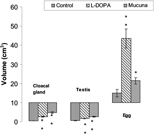
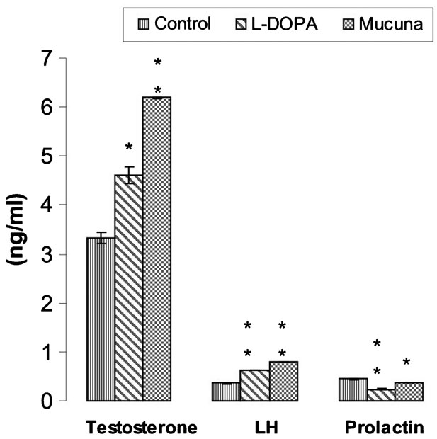
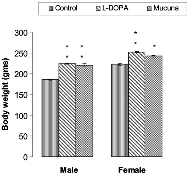
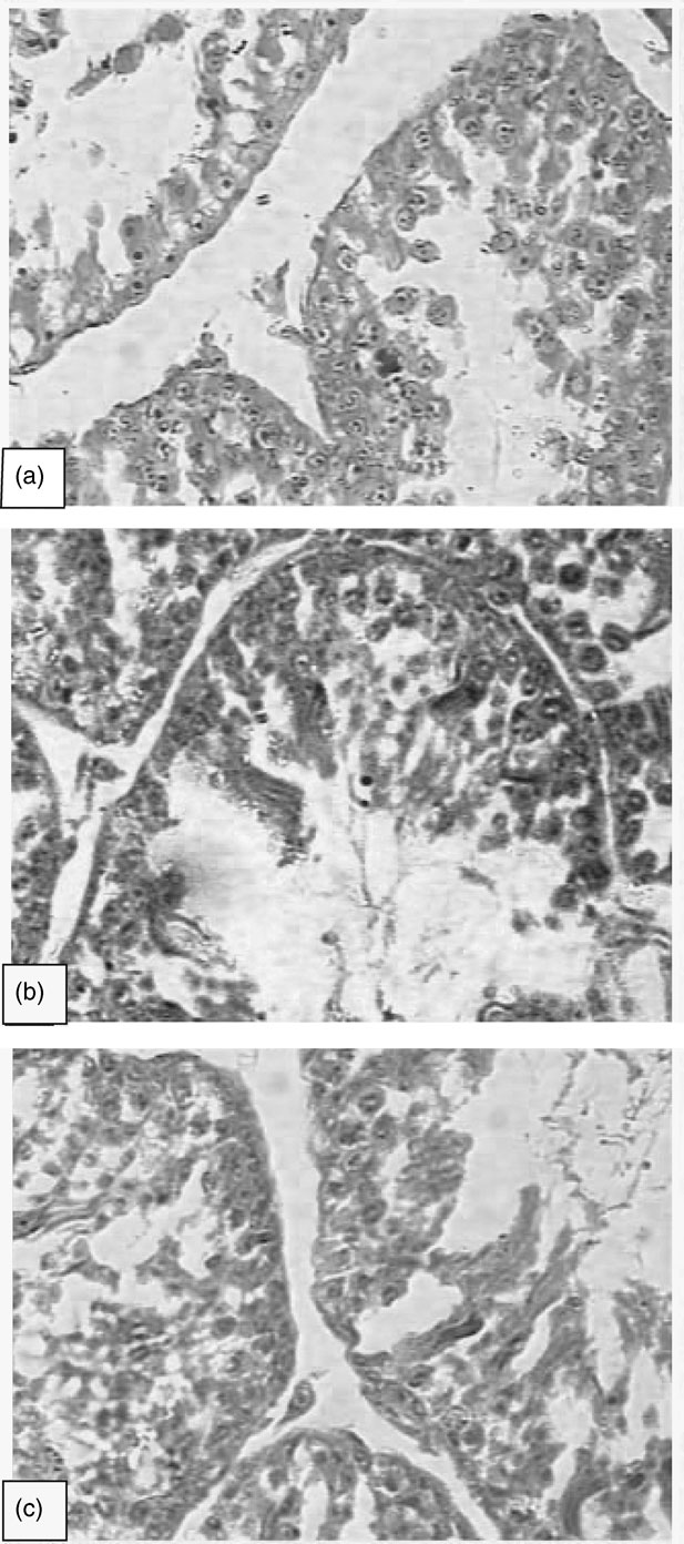
Cloacal gland (an androgen-dependent accessory sex organ), testicular volume (Figure 1), plasma testosterone and LH level (Figure 2) and BW (Figure 3) of l-DOPA- and MPSP-fed group showed significantly (P < 0.001) higher values when compared to the control. The size of the cloacal gland correlated with plasma LH and testosterone level and testicular size. Histologically, the testis of control bird, sacrificed at the 5th week, showed moderate activity while the testis of l-DOPA- and MPSP-fed birds showed full breeding condition. Enlarged seminiferous tubules had some stages of spermatogenesis with abundant spermatozoa (Figure 4).
Effect of l-DOPA and Mucuna pruriens seed powder feeding on cloacal gland, egg and testicular volume of Japanese quail. Values are mean ± s.e. Significance of difference from control (*P < 0.05, **P < 0.001; Student’s t-test).

Effect of l-DOPA and Mucuna pruriens seed powder feeding on plasma testosterone, LH and prolactin level of Japanese quail. Values are mean ± s.e. Significance of difference from control (*P < 0.01, **P < 0.001; Student’s t-test).

Effect of l-DOPA and Mucuna pruriens seed powder feeding on the body weight of male and female Japanese quail. Values are mean±s.e. Significance of difference from control (*P < 0.01, **P < 0.001; Student’s t-test).

(a) Photomicrograph showing the moderate condition of T.S. of testis of the control group; 40 ×. (b) Photomicrograph of T.S. of testis of the l-DOPA-fed group showing hyperactivity and full breeding conditions with stages of spermatogenesis. Spermatozoa and active Leydig cells are clearly seen; 40 ×. (c) Photomicrograph of T.S. of testis of the Mucuna pruriens seed powder-fed group showing more prominent reproductive conditions; 40 ×.

Female
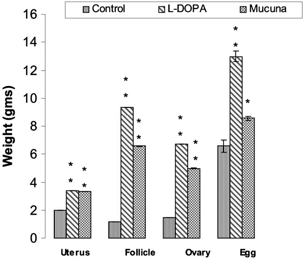
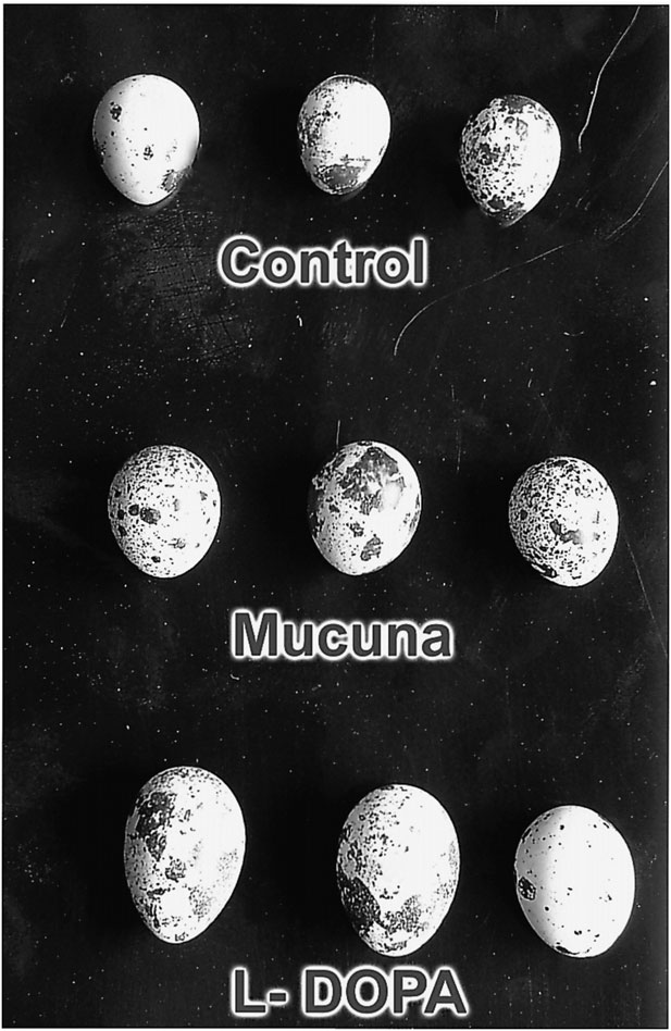
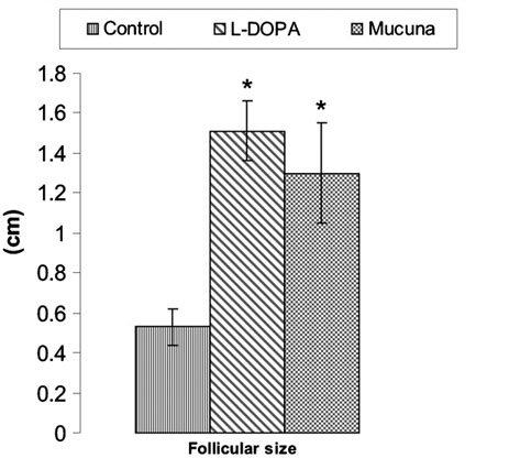
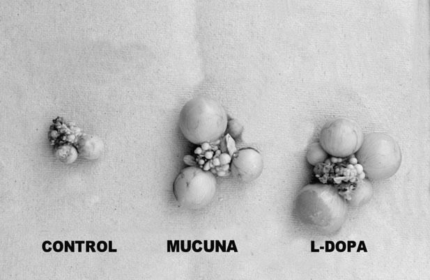
Maturation of female reproductive organs (uterus, follicles and ovary) of l-DOPA- and MPSP-fed group showed significantly (P < 0.001) higher values when compared to the control (Figure 5). The l-DOPA-fed group started egg laying at the 3rd week. The MPSP-fed group also started laying eggs at the 3rd week. However, in the control group, first egg laying was noticed at the 5th week. Eggs laid at the 5th week were collected from all the groups (female, control-IB and experimental-IIB and IIIB) and comparisons were made. The size and weight of the eggs laid by l-DOPA- and MPSP-fed quails were significantly higher (P < 0.001) than those of the control group (Figures 1, 5 and 6). The follicular size was also significantly greater (P < 0.01) in the l-DOPA- and MPSP-fed group than that of the control (Figures 7 and 8). Similarly, the BW of experimental groups (IIB and IIIB) showed significantly higher values (P < 0.05) when compared to the control (IB) (Figure 3). In contrast to BW and reproductive organs in female, plasma PRL level was significantly (P < 0.001) lowered in both l-DOPA- and MPSP-fed group (Figure 2) in comparison to control.
Effect of l-DOPA and Mucuna pruriens seed powder feeding on weight of eggs, follicles, ovary and uterus of female Japanese quail. Values are mean ± s.e. Significance of difference from control (*P < 0.01, **P < 0.001; Student’s t-test).

Photograph showing significant (P < 0.01) difference of the egg size between the control, l-DOPA and Mucuna pruriens seed powder-fed group.

Effect of l-DOPA and Mucuna pruriens seed powder feeding on the size of follicles in female Japanese quail. Values are mean ± s.e. Significance of difference from control (*P < 0.01, Student’s t-test).

Photograph showing significant (P < 0.01) difference of follicular size between the control, l-DOPA and Mucuna pruriens seed powder-fed group.

It is widely believed that M. pruriens influences the DA system; however, there may be other factors involved. A study was designed to elucidate the long-term effect of M. pruriens on the neurotransmitters in various regions of the rat brain. M. pruriens was mixed with rat chow and fed daily ad libitum to rats for 52 weeks. Controls received no M. pruriens. The rats were sacrificed at the end of 52 weeks and the neurotransmitters were analyzed in the cortex, hippocampus, substantia nigra and striatum. Oral administration of M. pruriens had a significant increase on DA content in the cortex with no significant effect on levodopa, norepinephrine or DA, serotonin and their metabolites. The failure of M. pruriens endocarp to significantly affect DA metabolism in the striatonigral tract along with its ability to improve Parkinsonian symptoms in the 6-hydroxydopamine animal model and humans may suggest that its anti-Parkinson’s effect may be due to components other than levodopa or that it has a levodopa-enhancing effect (37 min) (Katzenschlager et al., Reference Katzenschlager, Evans, Manson, Patsalos, Ratnaraj, Watt, Timmermann, Van der Giessen and Lees2004; Manyam et al., Reference Manyam, Dhanasekaran and Hare2004).
Brain monoamines are reported to upregulate to some extent the activity of the hypothalamic factor, which in turn regulates the pituitary gonadotropins (Lu et al., Reference Lu, Huang, Chen, Kurez, Miduszewski and Meites1977). In general, hypothalamic norepinephrine (NE) is known to stimulate whereas serotonin is found to inhibit the release of gonadotropin (Meites et al., Reference Meites, Simpkins, Bruni and Advis1977). NE has been reported to elicit the release of hypothalamic decapeptide, gonadotropin-releasing hormone (GnRH), which in turn causes the reduction in hypothalamic catecholamine (CA) content, and an increase in serotonin turnover occurs with the progress of age in male (Simpkins et al., Reference Simpkins, Muller, Huang and Meites1977) and female rats (Huang and Meites, Reference Huang and Meites1975), showing gradually decreasing reproductive activities. High serotonin concentration in certain areas of the brain (hypothalamus, midbrain and medulla) has also been reported in sexually regressed scotosensitive hamsters. This seasonal reproductive condition may be induced by tryptophan feeding in photorefractory hamsters and is associated with high serotonin concentration and turnover rate (Wilson and Meier, Reference Wilson and Meier1983). Retardation in reproductive activity in aging rat is also correlated with hypothalamic deficiency of catecholamine and can be corrected with administering l-DOPA, epinephrine or improniazid (Quadri et al., Reference Quadri, Kledzik and Meites1973). Maintenance of estrous cycle in aging female rat is also reported to be influenced by centrally acting drug like l-DOPA, which alters the catecholamine level (Forman et al., Reference Forman, Sonntog, Miki and Meites1980) and scotosensitivity in Syrian hamster by tryptophan feeding (Wilson and Meier, Reference Wilson and Meier1983).
It has been reported that elevating the level of the amino acid l-DOPA in the blood stream of poultry (by incorporation in the food) has the effect of reducing the cholesterol content in the eggs produced by such poultry and also has the effect of reducing the ratio of saturated to unsaturated fatty acids in the eggs produced by such poultry. It has also been reported that the injections of DA into the third ventricle of rat reduced serum PRL values and increased PIF activity in the portal circulation. A single systemic injection of DA failed to alter serum PRL concentration in rat (Shaar and Clemens, Reference Shaar and Clemens1974). But it was also shown that the infusion of relatively large doses of DA via a carotid arotic cannula depressed PRL (Shaar and Clemens, Reference Shaar and Clemens1974). Systemic injection of l-DOPA, the precursor of DA, was found to inhibit PRL release in rats (Shaar and Clemens, Reference Shaar and Clemens1974). There is evidence that l-DOPA acts to depress this release only after being converted to DA (Jimenez et al., Reference Jimenez, Voogt and Carr1978). DA agonists (apomorphine) have also been shown to inhibit PRL release (Mueller et al., Reference Mueller, Simpkins, Moore and Meites1976). Administration of drugs that depress hypothalamic DA activity results in an increase of PRL secretion. Injection of α-methyl-p-tyrosine, α-methyl-m-tyrosine or methyldopa, all inhibitors of catecholamine synthesis, promote the elevation of serum PRL (Koch et al., Reference Koch, Lu and Meites1970). The agonists of DA have also been reported to inhibit TSH and stimulate GH release. Pulsatile GH secretion persists with an increased number of secretory bursts in rat with a complete hypothalamic island produced by sectioning with a Halasz knife. This technique depletes the hypothalamic NE content without affecting the DA content (see review Weiner and Ganong, Reference Weiner and Ganong1978).
Other studies in the field of avian reproduction have made an interesting suggestion that the seasonality of reproduction in many more avian species may be determined by specific temporal relationship between serotonin and DA neurotransmitter precursors (5-HTP and l-DOPA). Testicular and ovarian development and fat deposition are stimulated or inhibited after daily injections of 5-HTP and l-DOPA at the interval of 12 or 8 h, respectively, in the white-throated sparrow Zonotrichia albicollis (Miller and Meier, Reference Miller and Meier1983), Japanese quail C. coturnix japonica (Chaturvedi and Meier, Reference Chaturvedi and Meier1988), the migratory red headed bunting, Emberiza bruniceps (Chaturvedi and Bhatt, Reference Chaturvedi and Bhatt1990), non-migratory spotted munia, Lonchura punctulata (Chaturvedi and Prasad, Reference Chaturvedi and Prasad1991; Prasad et al., Reference Prasad, Thapliyal and Chaturvedi1992), Lal munia Estrilda amandava (Chaturvedi et al., Reference Chaturvedi, Bhatt and Prasad1994) and Indian weaver bird Ploceus philippinus (Prasad and Chaturvedi, Reference Prasad and Chaturvedi2006). Injections of 5-HTP and l-DOPA given at different intervals showed marked changes in certain blood and serum constituents (Prasad and Thakur, Reference Prasad and Thakur2005; Prasad et al., Reference Prasad, Thakur and Qureshi Taj2006), hormonal level (Prasad and Roy, Reference Prasad and Roy2003; Prasad et al., Reference Prasad, Alone and Roy2003) and neurosecretory materials of the median eminence (Prasad and Rathore, Reference Prasad and Rathore2004).
The effect of MPSP feeding in Japanese quail is evident from the results of the present experiments. Earlier studies also revealed that the dopaminergic activity, as one of the circadian oscillators, may influence neuroendocrine–gonadal axis (Bhatt and Chaturvedi, Reference Bhatt and Chaturvedi1993). It is suggested that high levels of DA resulting from DOPA feeding possibly helped to induce and maintain reproductive conditions. DOPA feeding is reported to cause significant increase in brain DAs (Wilson and Meier, Reference Wilson and Meier1989). The brain monoamines have been reported to modulate pituitary gonadotrophin release (Meites et al., Reference Meites, Simpkins, Bruni and Advis1977). From the finding of the present study, it may be suggested that MPSP feeding induced the synthesis of DA in the brain and high dopaminergic activity affected the neuroendocrine–gonadal axis, resulting in changes in the body growth, reproductive conditions (cloacal gland, testicular volume, egg laying, egg weight, egg size, uterus, follicles and ovary) and plasma hormonal levels (LH, testosterone and PRL) in the sexually immature, high food value bird, Japanese quail, C. coturnix japonica.
Acknowledgement
Financial support from UGC, Government of India, New Delhi, in the form of Research Award Scheme (no. F.30-1/2004 (SA-II)) to SKP is gratefully acknowledged.