Fundamental principals of insulin gene therapy approach
Data from the 2011 National Diabetes Fact Sheet (released 26 January 2011) indicate that an estimated 25.8 million children and adults in the US have diabetes, and the rate of new diagnoses is rapidly increasing. For patients with type-1 diabetes (T1D), immune system-mediated destruction of the pancreatic beta cells removes the body of the only cells capable of making insulin that regulates blood glucose. Thus, survival depends on the life-long delivery of insulin via injection or pump. The first genetically engineered, synthetic human insulin for the treatment of T1D was produced 33 years ago (Ref. Reference Teuscher1). In recent years, there has been an increased interest in developing new methods of insulin therapy that decrease the need for exogenously delivered synthetic insulin. It was first shown in 1983 that a nonbeta cell, specifically mouse pituitary corticotroph cells (AdT20), could synthesise proinsulin, store it in secretory granules after generating smaller fragments and release it upon stimulation as insulin-like substances (Ref. Reference Moore2). Neuroendocrine cells are very similar to beta cells in terms of prohormone convertase expression (PC1/3 and PC2) and the presence of a regulated secretory pathway, but these cells lack glucose responsiveness because they do not express glucokinase (GK) and glucose transporter-2 (GLUT2). The fact that these cells secrete adrenocorticotropic hormone involved in glucocorticoid synthesis, which inhibits insulin action, eliminates their future utility as a treatment modality for diabetes. Consequently, recombinant insulin production in nonbeta cells in vivo has become a major research interest alternative to insulin injection and/or pancreatic islet cell transplantation.
The objective of insulin gene therapy is to genetically manipulate nonbeta cells to synthesise, store and secrete insulin-like pancreatic beta cells. One advantage of using nonbeta cells instead of beta cells is that they are resistant to the beta cell-specific autoimmune response present in T1D patients (Ref. Reference Dong and Woo3). Moreover, an insulin gene transfer system should possess a number of key features to increase the chance for successful therapy. First, the system should provide an effective gene delivery method to the target organ. Second, the target organ should have glucose-sensing machinery, especially GLUT2 and GK, and the necessary biochemical processing components to convert proinsulin to mature insulin. Third, the target cells should have storage capacity and a regulated secretory pathway inducible by glucose. Finally, transcription of the insulin transgene should be induced during hyperglycemia and inhibited during hypoglycemia.
Considering the requirement for an effective gene delivery method, viral and nonviral delivery systems have been tested in insulin gene transfer experiments. Although viral and nonviral vector delivery systems have a number of their own advantages and disadvantages, viral vectors are superior in transducing a variety of target cells (Refs Reference Wong, Hawthorne and Manolios4, Reference Hughes5, Reference Bagley6). Of the available viral vectors, lentivirus is the vector of choice for nonbeta cell targeted insulin mediated gene therapy, in terms of safety, efficacy and prolonged gene expression (Refs Reference Nett, Sollinger and Alam7, Reference Kobinger8). Lentiviral vectors can transduce both dividing and nondividing cells, so they are preferable to retroviruses, which can only infect dividing cells (Ref. Reference Zhang9). Adeno associated virus (AAV) is also becoming popular. Unlike the first generation of adenovirus, which is highly immunogenic and toxic at high doses (Refs Reference Doerschug10, Reference Barbu, Akusjarvi and Welsh11, Reference Zhang12), AAV provides long-term gene expression without generating toxicity (Ref. Reference Sanlioglu13). Recombinant AAV (rAAV) does not, however, have the ability for site-specific integration like wild-type AAV, which inserts at a specific locus in human chromosome 19 (Ref. Reference Sanlioglu and Engelhardt14). rAAV also cannot accommodate transgenes larger than 4.7 kb (Refs Reference Sanlioglu15, Reference Sanlioglu, Benson and Engelhardt16). On the other hand, since last-generation adenovirus vectors also known as helper-dependent or gutless adenovirus are totally devoid of viral encoding sequences they can accommodate large piece of DNA. In addition, they are able to mediate high levels of transduction in vivo and provide sustained levels of transgene expression with negligible toxicity compared with first- and second-generation adenovirus vectors (Refs Reference Alba, Bosch and Chillon17, Reference Palmer and Ng18).
Insulin gene therapy involving unregulated but sustained levels of insulin gene expression
Initial trials of experimental insulin gene delivery methods involving viral vectors date back nearly 20 years. The portal vein delivery of a Moleney murine leukaemia virus-based retroviral vector encoding rat preproinsulin 1 cDNA into partially hepatectomised rat liver (70%) achieved 5–15% liver transduction and persistent gene expression for at least 6 months (Ref. Reference Kolodka19). The low-level hepatic insulin gene expression ameliorated fasting hyperglycemia, glycogen breakdown, triglyceride deposition, ketoacidosis and streptozotocin (STZ)-induced diabetic death without causing hypoglycemia. Despite the protection against ketoacidosis, uncontrolled insulin gene expression risks the development of hypoglycemia as demonstrated in a study involving implantation of human fibroblasts genetically engineered to release mature human insulin (Ref. Reference Falqui20). Furthermore, insulin produced in the liver exhibits less activity because of the absence of proinsulin processing machinery. Intriguingly, administration of PC2- and PC3-encoding adenoviral vectors to liver failed to enhance proinsulin cleavage contrary to what is observed in muscle tissue (Ref. Reference Auricchio21). Since exogenously delivered PC2 and PC3 activities are masked by proteases present in the constitutive secretory pathway of liver cells, further modification of the transgene is needed for expression and secretion of fully functional insulin in liver.
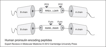
Cells having the constitutive secretory pathway use furin endopeptidase to efficiently cleave secretory peptides at specific amino acid residues (Ref. Reference Vollenweider22). Thus, dibasic amino acid residues (Arg-X-Lys-Arg) introduced at BC and CA junctions of insulin permit proinsulin cleavage into mature insulin by furin endopeptidases (Fig. 1). Nonhepatoma cell lines, such as HEK293 (Ref. Reference Groskreutz, Sliwkowski and Gorman23) and myoblast cell line (Ref. Reference Simonson24), transiently transfected with modified human insulin cDNA with furin recognition sequence produced biologically active insulin indicating successful restoration of post-translational processing of proinsulin. The fact that recombinant adenovirus-mediated overexpression of PC2 or PC3 facilitated conversion of proinsulin to insulin in rat insulinoma cells further supports these results (Ref. Reference Irminger, Meyer and Halban25). In another approach, a hepatocyte cell line (H4-II-E) was infected with replication-defective adenovirus carrying the human proinsulin cDNA with the dibasic PC recognition sequence mutated to a tetra-basic furin cleavage site to test the extent to which hepatocytes could constitutively synthesise proinsulin and secrete mature insulin (Ref. Reference Short26). Injection of this viral construct into mice resulted in insulin gene expression mainly in the liver, and transiently reversed diabetes. Similarly, livers of STZ-induced diabetic nude rats were infected with an adenovirus engineered to express rat preproinsulin under the control of the human elongation factor 1-alpha (EF1-alpha) promoter to assess the benefit of sustained hepatic insulin production on glycemic control (Ref. Reference Dong27). In this setting, sustained basal expression of plasma insulin significantly reduced both plasma glucagon and glucose levels in diabetic animals. Liver pathology (glycogen breakdown and lipid deposition) associated with T1D was also reduced through constitutive hepatic insulin gene expression at low levels. However, all treated animals developed severe hypoglycemia upon fasting because of the constitutive insulin gene expression.
Human proinsulin encoding peptides. The top peptide is the wild-type human proinsulin sequence cleavable by prohormone convertases (PC1/3 or PC2), which are only expressed in beta cells, K cells, L cells and some endocrine cells. The bottom peptide is the modified human proinsulin with tetrabasic furin endopeptidase processing sites.

In contrast to hepatocytes that lack the convertases necessary for processing prohormones into their fully active forms, lung tissue has an abundant supply of neuroendocrine secretory cells that naturally express the PCs. Liposome-mediated transfer of a plasmid containing the rat insulin gene driven by a Cytomegalovirus (CMV) promoter was tested in the lung (Ref. Reference Tanaka28). Multiple transfers yielded a significant increase in insulin gene expression in lung alveolar epithelial cells, as well as the processing and secretion of insulin protein, which lowered hyperglycemia and ketoacidosis in STZ-induced diabetic mice without any adverse effect. Nonviral vectors (such as plasmids) are nonimmunogenic and have fewer side effects than viral vectors, but repeated administration is required to sustain a therapeutic effect.
rAAV vectors provide much longer transgene expression compared with first- and second-generation adenoviral vectors. Recently, the efficacy of long-term rAAV-mediated insulin gene therapy was demonstrated using rAAV encoding a furin-modified human insulin under the control of CMV promoter (Ref. Reference Park29). Although transient normoglycemia was observed in animals treated with a recombinant adenovirus encoding furin-modified human insulin, rAAV delivery into the portal vein resulted in normoglycemia over 90 days post-treatment in STZ-induced diabetic rats. In addition, sustained insulin production reduced HbA1c levels and improved glucose tolerance without any adverse events. Similarly, Elsner et al. recently tested the therapeutic efficacy of a lentiviral vector encoding furin-cleavable human insulin in autoimmune-diabetic rats and STZ-diabetic rats (Ref. Reference Elsner30). Blood glucose concentrations were normalised in the treated animals throughout the observation period (about a year), suggesting diabetes could be reversed by a somatic gene therapy approach involving insulin release from nonendocrine cells. The limited capacity of nonbeta cell to process proinsulin to insulin can be overcome using furin recognition sequence-modified human insulin cDNA; however, coupling insulin expression to metabolic demand, which varies throughout the day, is another major obstacle to overcome.
Insulin gene therapy for enabling physiologic control of insulin gene expression
Glucose regulates many genes in pancreatic beta cells and hepatocytes, making it a potent regulator of insulin production. Beta cells and hepatocytes possess GLUT2 and GK activities for responding to alterations in blood glucose. Glucagon and cyclic AMP (cAMP) induce Phosphoenolpyruvate carboxykinase (PEPCK) promoter activity in hepatocytes, whereas PEPCK promoter activity is downregulated by insulin. Thus, proinsulin DNA engineered to include a furin recognition site driven by the PEPCK promoter creates a regulatable system for targeting hepatocytes as insulin-producing surrogate cells (Ref. Reference Lu31). Introduction of such a construct into primary cultured rat hepatocytes using a recombinant adenovirus resulted in mature human insulin production. The use of basal glucose responsive promoters, such as the promoters for rat S-14 or L-PK, in place of PEPCK is less optimal because of their weak promoter activity in liver (Ref. Reference Lu31).
Since constructs with natural glucose regulated promoter-driven insulin expression yield weak transcriptional activities causing failure to suppress nonfasting hyperglycemia, liver-targeted insulin gene therapy can occur using synthetic promoters generated from the combination of HNF-1, C/EBP binding elements and GlRE in conjunction with an L-PK promoter (Ref. Reference Han32). Intravenous administration of a recombinant adenovirus expressing furin-cleavable insulin under the control of a synthetic promoter corrected hyperglycemia and improved glucose tolerance in STZ-induced diabetic nonobese diabetic–severe combined immunodeficiency (NOD–SCID) mice. These results suggest that artificial promoters can be used to regulate glucose-responsive insulin production for liver-directed insulin gene therapy. Constitutive insulin expression via CMV promoter eventually causes death because of hypoglycemia, but constructs containing a glucose responsive enhancer/liver-specific promoter activity result in mild hypoglycemia. In addition, there is still a delay in restoring normoglycemia, as demonstrated by glucose tolerance tests suggesting the necessity of further improvement in the vector design to achieve a negative regulatory response.
As an alternative to using PEPCK and L-PK promoters and the negative response by insulin, a liver-specific albumin promoter with glucose inducible regulatory elements (GIRE) from S14 (Ref. Reference Shih and Towle33) can be used to confer glucose responsiveness of insulin gene expression modified for furin cleavage in liver cells (Ref. Reference Alam and Sollinger34). Adenovirus-mediated glucose regulated insulin secretion in liver improved glucose tolerance yielding fasting normoglycemia. However, a prolonged lag period of insulin synthesis and insufficient production were blamed for failure to correct nonfasting hyperglycemia. To generate a novel glucose responsive promoter active in hepatocytes but negatively regulated by insulin, carbohydrate-responsive elements from the rat L-PK gene were cloned into the insulin-suppressive basal promoter of the rat insulin-like growth factor binding protein-1 (rIGFBP-1) gene (Ref. Reference Thule, Liu and Phillips35). The use of a glucose-insulin sensitive promoter with the modified recombinant insulin cDNA in the context of an adenoviral vector system generated a construct capable of successfully mediating glucose-inducible human insulin secretion from rat primary hepatocytes in vitro. In addition, hepatic insulin gene therapy improved glycemia, induced a beneficial adipocytokine profile and enhanced insulin sensitivity preserving endothelium-dependent vascular function in diabetic rats (Ref. Reference Thule36).
The conversion of glucose-6-phosphate to glucose is catalysed by glucose-6-phosphatase (G6Pase), an enzyme mainly responsible for controlling glucose output from liver. As diabetic patients display increased glucose output from the liver causing fasting hyperglycemia, the G6Pase promoter (Ref. Reference Argaud37) has been used to regulate insulin gene expression in rat hepatoma cell lines. The G6Pase promoter is well suited for this function because it is highly active under diabetic conditions and negatively regulated by insulin (Ref. Reference Chen38). Glucose-stimulated (but self-limiting) insulin production can be achieved using an adenoviral vector encoding genetically engineered human insulin gene under control of the G6Pase promoter, but transgene expression was low in liver because of the negative feedback by insulin. Thus, incorporation of glucose responsive enhancers to generate stronger promoters is advised as a new approach to produce adequate insulin expression for lowering blood glucose under diabetic setting. For this purpose, a hybrid G6Pase promoter linked to intronic enhancers of the aldolase B gene was used to achieve auto-regulated insulin gene expression in liver (Ref. Reference Chen, Meseck and Woo39). Adenoviral delivery of this modified insulin gene to the liver significantly reduced nonfasting hyperglycemia without causing fasting hypoglycemia in STZ-induced diabetic nude rats.
Several glucose responsive insulin promoters have also been evaluated for their potential to provide glucose-regulated hepatic insulin gene expression. Human insulin promoter (HIP) was originally designated as a beta cell-specific promoter, and it directed strong gene expression in multiple hepatocyte cell lines (Ref. Reference Burkhardt40). The HIP can be used to drive glucose-regulated insulin gene expression in liver especially when AAV is used as a gene delivery vehicle, since AAV forms circular intermediates before integration (Refs Reference Duan41, Reference Sanlioglu, Duan and Engelhardt42). Multimerised rat insulin promoter (RIP) fused to rat proinsulin cDNA with furin cleavage sites cloned into Epstein–Barr virus (EBV)-based plasmid vectors were injected through tail-vein into STZ-induced diabetic mice (Ref. Reference Yasutomi43). Although RIP-directed insulin gene expression in liver resulted in glucose-regulated insulin gene expression, the level of the expression was not sufficient to correct fasting hyperglycemia in diabetic mice.
The rAAV-mediated liver transduction is generally low, therefore, a number of different methods to enhance rAAV-mediated insulin gene delivery to liver have been tested. For example, the effect of calcium phosphate (CaPi) on rAAV-mediated insulin therapy in diabetic animals was investigated using human insulin gene under the control of the PEPCK promoter (Ref. Reference Yang and Chao44). Although rAAV administration by itself significantly decreased blood glucose levels of diabetic animals, mixing of rAAV with calcium phosphate augmented the hypoglycemic effect of rAAV-mediated insulin gene transfer as well. A rAAV vector harbouring a furin-mutated insulin gene under the control of 410-bp rat insulin 1 promoter was complexed with polyethylenimine (PEI) to investigate the feasibility of glucose-modulated insulin gene therapy for diabetes (Refs Reference Hsu and Yang45, Reference Hsu, Kotin and Yang46). rAAV-transduction of Huh7 cells, a human hepatoma cell line with several endocrine pancreatic cell-like features, resulted in the secretion of insulin in response to high glucose levels. Furthermore, use of insulin secreting agents [dibutyryl cAMP, theophylline and forskolin] enhanced glucose-regulated rAAV-mediated transgene expression demonstrating that metabolic regulation can be coupled to hormonal regulation for a better induction of insulin biosynthesis. In addition, glucose challenge stimulated secretion of human insulin and significantly lowered hyperglycemia in the STZ-induced diabetic mice. However, a lag time of 30 min was needed to induce insulin secretion demonstrating slow kinetics of transcriptional regulation. Nevertheless, none of the animals treated with rAAV–PEI complex manifested hypoglycemia. Although PEI treatment enhanced rAAV-mediated transduction in liver, it can cause membrane damage inducing apoptosis. Furthermore, rAAV encoded transgene products can be immunogenic based on the route of delivery (Ref. Reference Brockstedt47). Thus, immunosuppression of individuals might be necessary to prevent immunity against AAV vectors, which would otherwise limit the efficacy of the treatment (Refs Reference Flotte48, Reference Sun49).
Insulin gene therapy with pharmacological control of insulin gene expression
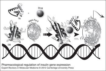
The liver is the preferred organ for producing insulin (via gene therapy) once the pancreas can no longer produce insulin, even though physiologic glucose-mediated control of insulin expression is not fully restored in the liver. Owing to the kinetics of regulation, glucose responsive promoters achieved limited success in regulating blood glucose levels in diabetic animals. For example, insulin gene transcription and translation require several hours to be effective, long after the onset of hyperglycemia. Transcription repression required for shutting down insulin gene expression after achieving normoglycemia also takes time. Consequently, patients may experience prolonged hyperglycemia or hypoglycemia. As constitutive insulin secretion from liver is associated with hypoglycemia, pharmacological stimulation of insulin transcription is tested as a novel approach to gain control of insulin secretion. In this scenario, a pharmacological agent controls a recombinant transcription factor that induces insulin gene expression (Fig. 2). For example, rapamycin facilitates heterodimer formation between the immunophilin FK506 Binding Protein (FKBP) and the lipid kinase homologue FKBP rapamycin-associated protein (FRAP). FKBP DNA binding is mediated by the ZFHD1 domain, and the rapamycin interacting domain of FRAP is joined to the transcription activation domain of the p65 subunit of necrosis factor-κB. Such a rapamycin-based regulatory system was tested to manage adenovirus-mediated transcription of the furin-cleavable proinsulin gene in diabetic nude mice (Ref. Reference Auricchio21). Adenoviral vectors encoding either wild-type proinsulin (hIns-wt) or a modified proinsulin cleavable by the ubiquitously expressed protease furin (hIns-M3) in association with each of the two beta cell-specific proinsulin convertases, PC2 and PC3, were used to test the therapeutic efficacy and consequence of mature insulin production in liver. Intravenous administration of the adenovirus vector expressing furin-cleavable proinsulin generated higher levels of mature insulin in liver compared with concurrent administration of adenovirus encoding wild-type proinsulin and proinsulin convertases. These data suggest that high-level expression of the mature form of insulin can occur by infecting the liver with an adenovirus encoding a furin cleavable form of proinsulin. Although endogenous PC2 or PC3 expression is not detected in the liver, high levels of furin expression is present. To test the efficacy of the rapamycin inducible system, adenoviral vectors encoding the rapamycin-regulated transcription factor (Ad-CMV-TF1) and an hIns-M3 transgene located downstream of the 12 binding sites for the regulated transcription factor (Ad-Z12-hInsM3) were co-administered into diabetic mice. Insulin secretion from mouse liver was induced only in the presence of rapamycin resulting in the lowering of blood glucose and halting of insulin gene expression upon drug withdrawal. These results suggest pharmacological regulation of insulin gene transcription in the liver to be a promising alternative for controlling glucose homoeostasis in diabetic animals (Fig. 2). Although transcriptional control of insulin expression using rapamycin allows dose-dependent induction of circulating levels of insulin alleviating hyperglycemia in diabetic animals, optimum levels of insulin expression required 12 h to generate. Although the approach may not be useful to mimic postprandial acute rise of insulin, it may be helpful to obtain therapeutic baseline levels of insulin expression, an effect similar to that of long-acting insulin.
Pharmacological regulation of insulin gene expression. The model proposes the initial expression of two recombinant transcription factors carrying a DNA-binding domain (DB) or a transcription activation domain (TA). The administration of rapamycin then leads to the two transcription factors forming a heterodimer resulting in the activation of RNA polymerase II, thereby inducing insulin gene expression.

Current obstacles and future prospects in insulin gene therapy
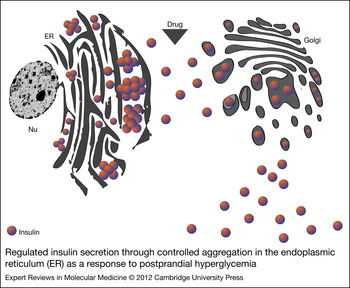
Hepatocytes constitutively secrete proteins, making it difficult to achieve an instant release of sufficient amounts of insulin to deal with the postprandial glucose excursions. Recombinant insulin designed to modulate postprandial glucose levels must be produced quickly in bursts following signal recognition. As insulin is stored in secretory granules in beta cells until their release, regulation of insulin secretion is necessary to achieve physiologically controlled insulin production by gene delivery. Thus, direct control of secretion provides faster regulation of blood glucose than transcriptional control. Regulation of insulin secretion through controlled aggregation in the endoplasmic reticulum (ER) is suggested as a novel complementary method to achieve controlled insulin release (Ref. Reference Rivera50). In this model, engineered proinsulin is retained in the ER of constitutive secretory cells (Fig. 3). After induction by an orally administered small molecule, insulin is processed and subsequently released quickly in relatively large amounts since proinsulin is engineered to carry a signal sequence attached to conditional aggregation domains (CAD) – regions necessary to retain target molecules in the ER. In addition, furin cleavage sites are included within the proinsulin encoding sequence to allow post-translational modification of proinsulin. In this scenario, the ER is intended to perform the function that is normally carried out by secretory granules in specialised secretory cells. Administration of a small drug leads to the dissociation of proinsulin aggregates and their translocation to Golgi. Following processing of proinsulin in Golgi, mature insulin is quickly released in abundance such that insulin is detectable in blood within 15 min of drug administration and peaks by 2 h leading to a 90% decrease in blood glucose levels. Circulating insulin decreases and glucose levels start to rise up again upon drug withdrawal. Therefore, kinetics of insulin secretion in this model closely resembles that observed with natural insulin secretion. Ultimately, glucose-stimulated insulin gene expression combined with a drug-induced system has great potential to simulate pancreatic beta cell function.
Regulated insulin secretion through controlled aggregation in the endoplasmic reticulum (ER) as a response to postprandial hyperglycemia. Modified insulin peptides accumulate within the ER as protein aggregates. Administration of a synthetic drug causes disaggregation of the insulin peptides, facilitating their passage to Golgi. Insulin is released via the constitutive secretory pathway. This system was originally designed for direct pharmacological control of protein secretion to generate fast and transient delivery of therapeutic proteins. Nu, Nucleus.

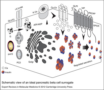
Controlled transcription and translation of proinsulin, presence of a regulated secretory pathway and inductive secretion are the major notable features of the pancreatic beta cells (Fig. 4). Although hepatocytes possess glucose-sensing machinery composed of GK and GLUT2, they are deprived of the regulated secretory pathway. In addition, neuroendocrine cells can store insulin in secretory granules and manifest a regulated secretory pathway, but they lack a glucose-sensing mechanism. Thus, neither hepatocytes nor neuroendocrine cells can be ideal beta cell surrogates. In contrast, K cells present in duodenum and jejunum are claimed to be outstanding targets for the synthesis and secretion of insulin in a glucose-controlled manner because of GK, GLUT2 and PC expressions just like beta cells (Ref. Reference Dong and Woo3). The fact that transgenic animal studies involving K cell-specific promoters driving human proinsulin gene expression are refractory to STZ treatment (Ref. Reference Cheung51) suggests that glucose homoeostasis can successfully be controlled despite the absence of beta cells (Ref. Reference Corbett52). However, K cell progenitor cells must be targeted to achieve long-term gene expression owing to rapid turnover of the intestinal epithelial cells. Only 1% of the intestinal epithelial cell population consists of enteroendocrine cells, so the isolation and identification of an enteroendocrine cell target for insulin production remains to be accomplished. Nonetheless, promising results have been reported using viral vectors, such that rAAV2 vector successfully transduced and genetically altered enteroendocrine (NCI-H716) L-cells exhibiting a regulated insulin response in a co-culture environment despite the presence of a large number of enterocytes (Caco-2 or T84) (Ref. Reference Tang and Sambanis53). The fact that human insulin gene trapped in chitosan nanoparticles delivered to cells of the gastrointestinal tract through lavage and coloclysis decreased the fasting blood glucose of STZ-induced diabetic rats further supports the possibility of using enteroendocrine cells for insulin production (Ref. Reference Niu54).
Schematic view of an ideal pancreatic beta cell surrogate. Glucose transporter 2 (GLUT2) and glucokinase (GK) act as cellular glucose sensors. Prohormone convertase (PC1/3 and PC2) converts proinsulin to insulin. In addition, a beta cell substitute should possess a regulated secretory pathway allowing insulin storage in the cytoplasm and release upon stimulation. G protein coupled receptor (GPCR) expression is also desired to respond to glucagon-like peptide 1 (GLP-1), vasoactive intestinal peptide (VIP), and pituitary adenylate cyclase-activating polypeptide (PACAP). Key players of glucose-mediated insulin secretion are depicted. In brief, when blood glucose level rises above 5.5 mM, glucose enters into the cell through GLUT2 and gets phosphorylated by GK. Glycolysis increases the ATP/ADP ratio activating K channels (K Ch) leading to membrane depolarisation. Opening of Ca2+ Channels (Ca Ch) results in Ca2+ influx and fusion of insulin containing vesicles with the plasma membrane. Molecules with incretin effect (GLP-1, VIP and PACAP) also stimulate insulin release through GPCR.

Ideally, a beta cell substitute (Fig. 4) should also be expected to express glucagon-like peptide 1 (GLP-1) receptors important in regulating postprandial insulin secretion in the presence of glucose (Ref. Reference Halban55), and neuronal input is required to control glucose metabolism through pituitary adenylate cyclase-activating polypeptide (PACAP)- and vasoactive intestinal peptide (VIP)-induced pathways under metabolic stress. Insulin is released within minutes after beta cell stimulation, under normal conditions, through the discharge of secretory granules already present in the cytoplasm, making for very rapid kinetics of insulin secretion. Insulin secretion from a surrogate cell, which relies on transcriptional activation, takes place in hours rather than minutes. Turning off the signal through transcriptional inhibition also is a very slow process despite the shorter half-life of preproinsulin mRNA in surrogate beta cells (less than 6 h) than actual beta cells (about 24 h). Nevertheless, insulin secretion is expected to stop in minutes following the signal withdrawal. Since beta cell-specific factors are involved in controlling preproinsulin mRNA stability and processing, gene therapy vectors encoding preproinsulin cDNA should be carefully designed considering all the above-mentioned situations to allow proper regulatory control in surrogate beta cells. Although somatic cell-targeted insulin gene therapy with C-peptide (Ref. Reference Wahren, Kallas and Sima56) have a great potential for use (Ref. Reference Elsner30), future studies are needed to demonstrate its effectiveness compared with the current treatment modalities such as insulin regimens and islet cell transplantation (Refs Reference Kahraman57, Reference Shapiro58).
Insulin gene therapy- versus gene therapy-induced beta cell generation
Pancreas development and the maturation of beta cells require a pancreatic and duodenal homeobox 1 (PDX-1) protein, also known as insulin promoter factor 1 (Ref. Reference Collombat59). In addition, PDX1 transcription factor is central to regulation of islet cell function and islet restricted insulin gene expression. One plausible alternative to nonexocrine tissue expression of insulin via gene therapy is the generation of insulin-secreting beta cells from other somatic cell types through gene transfer (Ref. Reference Yi, Liu and Izpisua Belmonte60). Hepatocytes and acinar pancreatic cells originate from the same germ layer, the primitive foregut endoderm, so a recombinant adenovirus carrying PDX-1 gene (Ad-CMV-PDX-1) was delivered into the livers of mice to test the extent to which exogenous PDX-1 expression (gain-of-function) in nonislet tissue would be successful in reprogramming hepatocytes into ‘beta cell like’ cells in vivo (Ref. Reference Ferber61). PDX-1 gene expression stimulated endogenous insulin 1 and 2 genes and PC1/3 generating biologically active insulin sufficient to ameliorate hyperglycemia in STZ-induced diabetic mice (Ref. Reference Ferber62). Intriguingly, despite the transient nature of gene expression provided by adenovirus vectors, Ad-CMV-PDX-1 recombinant adenovirus injection prevented STZ-induced hyperglycemia in Balb/c mice for at least 8 months after the gene transfer (Ref. Reference Ber63). The fact that recombinant adenovirus-mediated PDX-1 gene therapy ameliorated hyperglycemia in cyclophosphamide-accelerated diabetes in NOD (CAD-NOD) mice also supports this therapeutic option (Ref. Reference Shternhall-Ron64). Co-expressing PDX-1 with one or several other pancreatic transcription factors, including musculoaponeurotic fibrosarcoma oncogene homolog A (MAFA), neurogenin3 (NGN3) and/or NEUROD1 augmented the efficiency of reprogramming of liver cells to islet beta cell-like clusters (Refs Reference Kaneto65, Reference Tang66, Reference Wang67). Furthermore, by using PDX-1 and soluble factors adult human liver cells were successfully transformed into functional insulin-producing cells (Ref. Reference Sapir68). The fact that hyperglycemia can be ameliorated for a prolonged period of time when these transdifferentiated cells get transplanted under the renal capsule of diabetic mice has raised the possibility of using adult human liver tissue as an alternative for autologous beta-cell-replacement therapy. Apart from liver tissue, reprogramming acinar exocrine cells into beta cells has been successful using agonists of the JAK2/STAT3 signalling pathway (epidermal growth factor and leukaemia inhibitory factor) (Ref. Reference Baeyens and Bouwens69). Similarly, re-expressing key developmental regulators of three transcription factors (NGN3, PDX1 and MAFA) in vivo resulted in transdifferentiation of pancreatic exocrine cells into beta cell-like clusters in adult mice (Ref. Reference Zhou70). Lastly, the ability to directly reprogram adult somatic cells into endocrine pancreatic lineage was tested using a developmentally unrelated ectodermal tissue, human skin keratinocytes (Ref. Reference Mauda-Havakuk71). PDX-1-treated keratinocytes were reprogrammed and acquired beta cell-like functions, such as insulin production, processing and secretion in response to elevated glucose levels.
Beta cell neogenesis can result from the budding of islet endocrine cells from ductal epithelium during embryogenesis. Ngn3 activation in the budding ducts of developing pancreas induces differentiation of epithelial progenitors into islet cells. With this in mind, ectopic expression of Ngn3 in adult human duct cells produced a gene expression profile indicating reprogramming of duct cells into islet endocrine cell type (Ref. Reference Swales72). Collectively, these results suggest that gene therapy-induced transdifferentiation of beta cells might be useful in the production of autologous surrogate beta cells suitable for correcting islet cell function impaired in diabetic patients as an alternative to insulin gene therapy.
Acknowledgements
This work is supported by grants from Akdeniz University Scientific Research Administration Division and the Scientific and Technological Research Council of Turkey (TUBITAK-112S114). The authors declare that there is no duality of interest associated with this manuscript.