The clinical course of children with dilated cardiomyopathy is highly variable, ranging from complete recovery to death or cardiac transplantation.Reference Towbin, Lowe and Colan1 Clinicians have long sought indicators that might, in an individual patient, aid outcome prediction and, if necessary, permit timely listing for transplantation. To date, no single marker has been found. Traditionally,Reference Azevedo, Santos, Banesi Filho, Castier, Tura and Amino2 and continuing today,Reference Towbin, Lowe and Colan1 indices of left ventricular systolic function have featured prominently in risk stratification in dilated cardiomyopathy. There is, however, increasing evidence that the right ventricle plays a vital role in the patients’ clinical status as well as their probability of survival.Reference La Vecchia, Varotto, Zanolla, Spadaro and Fontanelli3 Complex geometry and limited acoustic windows make the right ventricle not amenable to echocardiographic volumetry. This may be one of the reasons why so few investigators have addressed the impact of right ventricular function on either symptom progression or survival in dilated cardiomyopathy.
Magnetic resonance imaging has been proved to be particularly useful for the quantification of right ventricular volumes and function in patients with congenital cardiac disease.Reference Buechel, Dave and Kellenberger4 We wished to determine the value of magnetic resonance imaging as a tool for the assessment of ventricular function in children and adolescents with dilated cardiomyopathy. Specifically, we were interested in whether the reported abnormalities in right ventricular function that have been shown in adults with dilated cardiomyopathy by X-ray ventriculographyReference La Vecchia, Varotto, Zanolla, Spadaro and Fontanelli3, Reference Unterberg, Dacian and Rudolph5 and echocardiographyReference Sun, James and Yang6 are also seen by magnetic resonance imaging. In addition, we sought to find out whether a population of children with dilated cardiomyopathy that is oligosymptomatic may already exhibit changes in right ventricular function and whether these are independent of left ventricular function. Finally, we wanted to investigate whether any alterations in right ventricular size and function are clinically important.
Methods
After approval from the institution’s ethics review board, we prospectively screened the echocardiograms of all children attending the cardiac function and cardiomyopathy clinic at our institution between March, 2007 and March, 2008. Children with either a dilated left ventricle, defined by M-mode left ventricular end-diastolic dimension with a Z-score of greater than 2, or an ejection fraction of less than 55%, or both, were invited to take part, and written informed consent was obtained from those who agreed to participate. Patients with congenital cardiac disease including abnormal origin of the coronary arteries, clinical evidence of acute phase myocarditis, or a recent change in clinical status were excluded.
Universally accepted normal values for cardiac magnetic resonance imaging volumetry have not yet been established in children. These values vary according to institutional practices for image acquisition and analysis. Therefore, we collected normative control data from children undergoing a clinically indicated magnetic resonance imaging study because of a family history of arrythmogenic right ventricular cardiomyopathy, and in whom this disease was subsequently excluded. All control participants were clinically asymptomatic, had normal electrocardiograms and echocardiograms, and no abnormalities were detected on the clinical interpretation of their magnetic resonance imaging.
Magnetic resonance imaging protocol
Magnetic resonance imaging scans were performed on a 1.5 Tesla scanner (Signa Excite, General Electrics Healthcare, Milwaukee, United States of America), with identical protocols used for both the dilated cardiomyopathy patients and the controls. For ventricular volumetry, a short-axis cine stack was acquired during breath hold, using a steady-state free precession gradient echo sequence with minimum echo and repetition times, flip angle of 45 degrees, bandwidth of 31.25 kilohertz, one excitation, views (lines) per segment to allow for 20 true reconstructed phases per cardiac cycle, 5–6 millimetres of slice thickness, 10–12 slices, gap adjusted to cover both ventricles, and in-plane spatial resolution of 1.5–2.5 millimetres. For late gadolinium enhancement, a gradient-echo sequence with an inversion pre-pulse was used to image five short-axis and two four-chamber slices after 10 minutes of contrast administration, with the following settings: minimum echo and repetition times of 3.1 and 6.7 milliseconds, respectively, flip angle at 20 degrees, bandwidth of 31.25 kilohertz, one excitation, 24 views per segment, trigger delay set to target diastole, 8–10 millimetres of slice thickness, and 1.5–3.0 millimetres of in-plane spatial resolution. Phase-contrast velocity mapping of the mitral and tricuspid valve inflows was performed parallel to the atrioventricular groove, using a retrospectively electrocardiography-gated segmented K-space technique with the following parameters: 8–10 milliseconds of repetition time, 3.5–5 milliseconds of echo time, flip angle of 15 degrees, bandwidth of 31.25 kilohertz, 1–2 views per segment, adequate to achieve 20 true phases per cardiac cycle, free breathing, two excitations, velocity-encoding range of 150 centimetres per second, 5 millimetres of slice thickness, in-plane resolution of 1.5–2.5 millimetres. A dedicated workstation (Mass Analysis and CV Flow, Medis Medical Imaging Systems, Leiden, The Netherlands) was used for volumetric and flow analysis. The study readers (LGW and SJY) were blinded to the results of the other investigations.
On the insertion of the intravenous cannula for the administration of gadolinium, blood was drawn for the analysis of N-terminal pro-brain natriuretic peptide levels (Modular Analytics, Roche Diagnostics, Laval, Quebec, Canada). N-terminal pro-brain natriuretic peptide is a cardiac hormone with diuretic and vasodilatory properties.Reference Koch and Singer7 It is secreted mainly by the ventricles as a response to pressure and volume overload. In adults and children, N-terminal pro-brain natriuretic peptide is used to monitor patients with congestive cardiac failure.Reference Williams, Ng, O’Brien, Taylor, Wright and Tan8
Clinical assessment, exercise testing, and echocardiography
The clinical records of children with dilated cardiomyopathy were examined to establish health status as recorded at the closest clinic visit to magnetic resonance imaging (always within 2 months). We determined the QRS duration from an electrocardiogram recorded within 6 months of magnetic resonance imaging. The results of the most recent metabolic exercise test on file for each patient were also reviewed. All patients were examined with the Bruce protocol and their results related to published normal values.Reference Washington, van Gundy, Cohen, Sondheimer and Wolfe9 Medical records were examined again at the start of manuscript preparation to establish whether the specified endpoints, death, or listing for cardiac transplantation had occurred during follow-up.
A clinical echocardiogram performed within 6 months and closest to the date of the magnetic resonance imaging scan was reviewed by one of the investigators (SLR) who was blinded to the magnetic resonance imaging findings. Left ventricular end-diastolic and end-systolic diameters were measured by M-mode and used to estimate ejection fraction. Chamber diameters were standardised to the body surface area as Z-scores on the basis of published normative data.Reference Daubeney, Blackstone, Weintraub, Slavik, Scanlon and Webber10 The results of pulsed wave Doppler of atrioventricular valve inflow, mitral and tricuspid annular tissue Doppler velocities, and the semi-quantitative degree (none to severe) of atrioventricular valve regurgitation were recorded.
Statistical analysis
Descriptive analyses are presented as means with standard deviations, or, where data distribution was skewed, as medians with ranges.
Parametric unpaired two-tailed Student’s t-tests were used to compare results between patients and control groups and between sub-groups within the dilated cardiomyopathy cohort. Linear regression was performed to assess for associations between variables. Results with a p-value less than 0.05 were considered significant.
Results
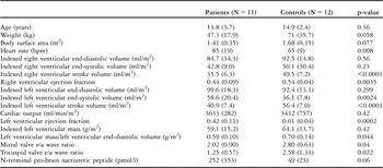
We enrolled a total of 11 patients, including seven patients with idiopathic dilated cardiomyopathy, two with anthracycline-induced dilated cardiomyopathy, and one with sickle cell disease. One patient had a primary diagnosis of left ventricular non-compaction. At the time of magnetic resonance imaging, nine out of ten patients were asymptomatic (New York Heart Association Class 1) and one had mild symptoms on exertion (Class 2). Until March, 2009, two patients had undergone cardiac transplantation for intractable cardiac failure (3.3 and 15.3 months after magnetic resonance imaging, respectively). All other patients remained in New York Heart Association Class 1, during a mean follow-up period of 12.9 months (7.0–22.0 months). The patient demographic data and the results of magnetic resonance imaging and N-terminal pro-brain natriuretic peptide analysis in dilated cardiomyopathy patients and controls are summarised in Table 1. To test whether one ventricle was more severely affected, we related the patients’ ejection fractions to the mean ejection fraction in the control group – left ventricular ejection fraction is 0.61 and right ventricular ejection fraction is 0.54. The mean decrease in the ejection fraction in the dilated cardiomyopathy group compared to the ejection fraction of the ipsilateral ventricle in the control group was 30.3% for the right ventricle and 18.7% for the left ventricle (p = 0.06).
Demographic data, magnetic resonance imaging, and N-terminal pro-brain natriuretic peptide results for patients and normal controls.

Results are presented as mean and standard deviation (standard deviations are given in brackets)
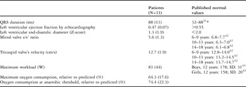
Results of the echocardiography and exercise test in the dilated cardiomyopathy patients are presented in Table 2. The median times between the echocardiogram and magnetic resonance imaging and between the exercise test and magnetic resonance imaging were 2.5 months (0.2–6.0 months) and 2.0 months (0–9.5 months), respectively. All but one patient who had mild tricuspid regurgitation had either no or trivial tricuspid regurgitation. Two patients had mild mitral regurgitation. The other nine had no mitral regurgitation.
Echocardiographic, exercise test, and electrocardiogram results for patients with dilated cardiomyopathy.

Results are presented as mean and standard deviation (standard deviations are given in brackets)
*2nd–98th percentile in normal children aged 12–18 years
Three patients had echocardiographic evidence of left ventricular non-compaction and two of them had a primary diagnosis of dilated cardiomyopathy. These and three other patients, two with idiopathic dilated cardiomyopathy and one with anthracycline-induced dilated cardiomyopathy, had either definitive or highly suspicious magnetic resonance imaging findings of left ventricular non-compaction. No differences between these patients and those without left ventricular non-compaction were found regarding N-terminal pro-brain natriuretic peptide, exercise tolerance, right and left ventricular systolic function, or ventricle size. Only the patient who had a primary diagnosis of left ventricular non-compaction showed evidence for fibrosis on the late gadolinium enhancement images (Fig 1).
Bi-ventricular non-compaction short-axis projection of the right and left ventricles in a patient with bi-ventricular non-compaction. (a) The image acquired in the double inversion recovery “black blood” technique shows the hypertrabeculated left ventricular myocardium. (b) The late gadolinium enhancement image shows a rim of bright signal outlining the left and right ventricular endocardium. Whether this represents fibrosis or originates from a slow-flow artefact is unclear; LV, left ventricle; RV, right ventricle.

Correlation of magnetic resonance imaging and echocardiographic findings
M-mode echocardiography estimates of left ventricular ejection fraction did not differ significantly from the magnetic resonance imaging measurements, the correlation between them being almost statistically significant (r = 0.59, p = 0.06). Tricuspid annulus velocity, as measured by tissue Doppler echocardiography, correlated with right ventricular ejection fraction as measured by magnetic resonance imaging (r = 0.60, p = 0.05). Mitral valve annulus velocity, on the other hand, did not correlate with left ventricular ejection fraction.
Relationship of left ventricular to right ventricular function indices
Left ventricular ejection fraction by magnetic resonance imaging correlated inversely with left ventricular end-diastolic volume and mass, indexed to body surface area (r = −0.74, p = 0.0095, r = −0.67, p = 0.023, respectively). Indexed left ventricular mass correlated positively with left ventricular end-diastolic volume (r = 0.76, p = 0.0061). Right ventricular ejection fraction and end-diastolic volume were negatively correlated (r = −0.78, p = 0.0043). Left ventricular ejection fraction did not correlate with right ventricular ejection fraction (r = 0.16, p = 0.64), but there was a positive correlation between left and right ventricular end-diastolic volumes (r = 0.71, p = 0.015).
Correlation of left and right ventricular volume and function with brain natriuretic peptide and exercise capacity
Right ventricular ejection fraction and indexed end-diastolic volume both correlated with N-terminal pro-brain natriuretic peptide (r = −0.67, p = 0.03, r = 0.65, p = 0.04, respectively). The right ventricular ejection fraction did not correlate with the predicted maximum oxygen consumption (r = 0.30, p = 0.40) but with the predicted oxygen consumption at the anaerobic threshold (r = 0.67, p = 0.049). The correlation of right ventricular end-diastolic volume with the anaerobic threshold almost reached statistical significance (r = −0.64, p = 0.06). Neither left ventricular ejection fraction nor left ventricular end-diastolic volume correlated with N-terminal pro-brain natriuretic peptide, maximum oxygen consumption, or oxygen uptake at the anaerobic threshold.
Diastolic function
Patients with dilated cardiomyopathy had lower e/a wave ratios for both mitral and tricuspid valve inflow when compared to normal controls, as measured by magnetic resonance imaging. A lower e/a wave ratio in the left ventricle correlated strongly with a lower e/a ratio in the right ventricle (r = 0.89, p = 0.0072).
Patients with a lower mitral valve e/a ratio had a lower left ventricular ejection fraction (r = 0.90, p = 0.0062), both measured by magnetic resonance imaging. For the right ventricle, no association was found between tricuspid valve e/a ratio and right ventricular ejection fraction (r = 0.49, p = 0.26).
A higher mitral valve inflow e wave to annular tissue Doppler e’ velocity ratio was associated with a lower right ventricular ejection fraction (r = 0.73, p = 0.01) and a larger right ventricular end-diastolic volume (r = 0.80, p = 0.0033). The mitral valve e/e’ ratio did not correlate with left ventricular end-diastolic volume or ejection fraction. Neither the mitral valve e/e’ ratio, nor the e/a ratio correlated with N-terminal pro-brain natriuretic peptide, absolute or predicted maximum oxygen consumption, or oxygen consumption at the anaerobic threshold, respectively.
Discussion
This is the first detailed description of the right ventricular size and function, using magnetic resonance imaging in patients, children or adults, with dilated cardiomyopathy. The echocardiographic assessment of the right ventricular systolic function and size is problematic.Reference Anavekar, Gerson, Skali, Kwong, Yucel and Solomon11, Reference Ommen, Nishimura and Appleton12 In our cohort, systolic tricuspid annulus velocity by ultrasound predicted the right ventricular ejection fraction by magnetic resonance imaging and may be a useful surrogate of the right ventricular systolic function.Reference Miller, Farah, Liner, Fox, Schluchter and Hoit13 Although echocardiographic estimates of left ventricular ejection by M-mode are widely accepted, the correlation between magnetic resonance imaging and echocardiographic left ventricular ejection fraction were far from perfect because M-mode echocardiography is only an approximation of the true ejection fraction. This is especially true in dilated cardiomyopathy patients who often have abnormal left ventricular geometry and regional systolic dysfunction, as has been shown by fluoroscopic ventriculography.Reference Boudoulas, Ruff, Fulkerson and Lewis14
Contractile dysfunction affects both ventricles
We were able to show, for the first time, that children with dilated cardiomyopathy not only have an impaired left ventricular systolic function, but also a significantly reduced right ventricular ejection fraction. In the right ventricle, as in the left ventricle, loss of systolic function and dilation go together. The patients investigated here had clinically mild disease, and left ventricular ejection fraction was, on average, only mildly to moderately reduced. Therefore, the impaired right ventricular function is an early phenomenon in dilated cardiomyopathy. Early changes in left and right ventricular geometry and size precede clinical manifestations in carriers of one of the dilated cardiomyopathy genes.Reference Koikkalainen, Antila and Lotjonen15 Whether right ventricular failure in dilated cardiomyopathy is due to primary involvement in the underlying myocardial disease or secondary to either adverse “parallel” ventricular interactions,Reference Weber, Janicki, Shroff and Fishman16–Reference Santamore and Dell’Italia18 or a series effect from left ventricular failure resulting in increased right ventricular afterload, is indistinguishable. The fact that in patients with relatively mild disease, the right ventricular ejection fraction trended towards being more severely reduced than the left ventricular ejection fraction, that is, (30.3% for the right ventricle and 18.7% for the left ventricle, as compared to normal, leads us to speculate that the right ventricle is primarily involved in the disease, and not solely as a “victim” of poor left ventricular function.Reference Rominger, Bachmann, Pabst and Rau19 Nevertheless, a cascade effect of elevated left ventricular filing, left atrial and pulmonary arterial pressure may contribute to the dysfunction of the right ventricle, which, beyond the immediate newborn period, is vulnerable to increases in afterload.Reference Belik and Light20 In fact, restrictive ventricular physiology, as indicated by a higher ratio of early transmitral flow velocity to early diastolic velocity of the mitral annulus,Reference Lindqvist, Calcutteea and Henein21 has been identified as a powerful predictor of a clinical outcome in patients with dilated cardiomyopathy.Reference Galrinho, Branco and Soares22 In our cohort, this ratio was associated with right ventricular dysfunction and enlargement.
Expectedly, lower left ventricular ejection fraction was associated with larger left ventricular end-diastolic volume, as both reduced systolic function and increased ventricular size are hallmarks of the disease. Whereas in this mildly affected cohort, neither of the two ventricles was significantly enlarged, the stroke volumes of both ventricles were significantly smaller in the dilated cardiomyopathy patients than in the controls. It is conceivable that early changes during compensated dilated cardiomyopathy manifest as a decrease in contractility rather than chamber dilation. The patients in our cohort compensated the decrease in stroke volume with an increase in their heart rate so that they were able to maintain their cardiac output. Myocardial mass per millilitre of enclosed volume was smaller in the patient group than in the control group, confirming that dilated cardiomyopathy is a dilatory process with a decrease of myocardial mass, relative to ventricular size.Reference Brooks, Schinde, Bateman and Gallagher23 There was an inverse relationship between left ventricular ejection fraction and mass, indicating that increased myocardial tissue mass does not equate with improved contractile performance, and reflecting unfavourable remodelling. Knaapen et alReference Knaapen, Gotte and Paulus24 showed a disproportionate increase in myocardial fibrosis in patients with dilated cardiomyopathy, with an inverse correlation to left ventricular contractile function.
Diastolic function
This cohort displayed impaired relaxation of both the left and the right ventricles as indicated by lower-than-normal magnetic resonance imaging e/a wave ratios across the mitral and tricuspid valves. In the left ventricle, diastolic dysfunction was strongly associated with systolic dysfunction, as has been described in adults with dilated cardiomyopathy, using tissue Doppler imaging.Reference Mohammed, Mertens and Friedberg25 Whereas a causal relationship between the two is speculative, disturbances of diastolic function, via impaired filling and recoil, may well hamper systolic performance. Alternatively, as both consume energy, they may be the result of sub-optimal substrate supply and metabolism. Interestingly, the degree of impaired relaxation correlated between the two ventricles.
Right ventricular dysfunction matters
The clinical and prognostic significance of right ventricular dysfunction and dilation in children with dilated cardiomyopathy has not been thoroughly investigated. In adults with dilated cardiomyopathy, higher right ventricular ejection fraction by fluoroscopic angiography independently predicts transplant-free survival, whereas the left ventricular ejection fraction does not.Reference La Vecchia, Varotto, Zanolla, Spadaro and Fontanelli3, Reference Juilliere, Barbier, Feldmann, Grentzinger, Danchin and Cherrier26, Reference de Groote, Millaire and Foucher-Hossein27 Adults with dilated cardiomyopathy are three times more likely to die in the next 4 years if their right ventricle is dilated, as shown by echocardiography.Reference Sun, James and Yang6 Paediatric data are scarce. In a study of 148 patients with idiopathic dilated cardiomyopathy, Azevedo et alReference Azevedo, Banesi Filho, Santos, Castier and Tura28 found that, on average, the right ventricles of those who died of the disease were almost twice as big as those of the survivors. Friedman et alReference Friedman, Moak and Garson29 showed that in paediatric dilated cardiomyopathy patients enlarged right ventricles are predictive of poor outcome, and systolic right ventricular area change predicts mortality or the need for cardiac transplant.Reference McMahon, Nagueh and Eapen30
The number of patients in our study with adverse outcomes, that is, cardiac transplantation, or clinical deterioration was too small to analyse for risk factors. New York Heart Association class may be too blunt a tool to describe the clinical situation of patients with compensated cardiac failure. Therefore, we used N-terminal pro-brain natriuretic peptide and exercise tolerance as surrogates for the degree of cardiac failure and prognosis.Reference Williams, Ng, O’Brien, Taylor, Wright and Tan8 Earlier studies found a correlation of N-terminal pro-brain natriuretic peptide and lower left ventricular ejection fraction with the Ross score of cardiac failure in younger children.Reference Mir, Marohn, Laer, Eiselt, Grollmus and Weil31 In the present study, N-terminal pro-brain natriuretic peptide correlated with lower right ventricular ejection fraction and larger end-diastolic volume, but not with left ventricular ejection fraction or end-diastolic volume. These findings suggest that early right ventricular dysfunction is an important regulator of natriuretic peptide production in cardiac failure. Similarly, an association of brain natriuretic peptide with indexed right ventricular end-diastolic volume has been previously shown in children with repaired congenital cardiac disease and right ventricular volume overload.Reference Trojnarska, Szyszka and Gwizdala32
Maximum oxygen consumption during standardised exercise testing reflects intrinsic cardiac reserve and has prognostic value in children and adults with dilated cardiomyopathy.Reference Guimaraes, d’Avila, Camargo, Moreira, Lanz and Bocchi33 We found an association of right ventricular ejection fraction and anaerobic threshold. Similarly, Di Salvo et alReference Di Salvo, Mathier, Semigran and Dec34 found a correlation, albeit weak, between radionuclide right ventricular ejection fraction, but not left ventricular ejection fraction, and oxygen consumption during cardiopulmonary exercise testing in patients with advanced cardiac failure. No correlation manifested between magnetic resonance imaging markers of the right or left ventricular diastolic function and either N-terminal pro-brain natriuretic peptide or exercise testing.
Myocardial morphology on magnetic resonance imaging
An interesting feature of this cohort is the presence of features of left ventricular non-compaction in five of the cases, by magnetic resonance imaging criteria, half of which were not considered to have this condition by echocardiographic criteria. Magnetic resonance imaging appears to be a more sensitive tool for the detection of myocardial non-compaction than echocardiography.Reference Alhabshan, Smallhorn, Golding, Musewe, Freedom and Yoo35, Reference Duncan, Brown and Worthley36 This finding also supports the concept of overlap between left ventricular non-compaction and dilated cardiomyopathy.Reference Murphy, Thaman and Blanes37 Many patients with dilated cardiomyopathy have or will develop imaging findings of apical left ventricular hypertrabeculation. On the other hand, ventricular dilation and dysfunction are frequent phenomena in what is considered primary left ventricular non-compaction.Reference Murphy, Thaman and Blanes37
Children with mild dilated cardiomyopathy do not appear to exhibit the characteristic mid-wall late gadolinium enhancement that can be seen in up to one-third of adults with this condition, in whom it has prognostic implications.Reference Assomull, Prasad and Lyne38, Reference Villuendas and Kadish39 Fibrosis of the non-compacted myocardium, as detected by magnetic resonance imaging has been described recently.Reference Iwashima, Ishikawa and Ohzeki40 The patient with a primary diagnosis of left ventricular non-compaction had an area along the left and right ventricular endocardium wall that was suspicious, but not conclusive for sub-endocardial fibrosis.
Limitations
We enrolled patients with relatively stable dilated cardiomyopathy. Ideally, however, the magnetic resonance and echocardiographic studies would have been conducted on the same day, ensuring similar contractility and loading conditions. This study was underpowered for multivariate analysis. The fact that we did not detect any differences between the groups or associations of certain variables does not exclude their possibility in a larger cohort with more advanced disease. Tighter correlations may have been achieved if the various examinations had all been performed on the same day.
In conclusion, we were able to show that right ventricular contractility is impaired in children with dilated cardiomyopathy. Right ventricular dysfunction impacts on their clinical condition. Echocardiographic assessment of left ventricular ejection fraction in patients with dilated cardiomyopathy is inaccurate. Therefore, we recommend magnetic resonance imaging as a routine diagnostic and follow-up tool in this disease. Longitudinal studies are needed to evaluate the prognostic significance of right ventricular size and function for children with dilated cardiomyopathy.