


Introduction
Biological invasions are often accompanied by changes in host–parasite interactions, including parasite loss (enemy release), acquisition of native parasites (spillback), or introduction of novel parasites (spillover) (Simberloff Reference Simberloff2011; Capinha et al. Reference Capinha, Essl, Seebens, Moser and Pereira2015; Schoeman et al. Reference Schoeman, Kruger, Secondi and Du Preez2019). Despite the ecological importance of these processes, parasitological data on invasive reptile species in Eastern Europe remain limited.
A common wall lizard, Podarcis muralis (Laurenti, 1768), is the most widely distributed representative of the genus (Arnold et al. Reference Arnold, Arribas and Carranza2007; Sillero et al. Reference Sillero, Bonardi, Corti, Creemers, Crochet, Ficetola, Kuzmin, Lymberakis, Pous, Sindaco, Speybroeck, Toxopeus, Vieites and Vences2014). Originally native to the sub-Mediterranean region, the species was mainly found in continental Europe, particularly in western and southern regions with its natural range including France, Belgium, Luxembourg, Germany, Switzerland, Italy, and across the Balkans, including Bulgaria, Romania, Greece, and particular areas of Türkiye, generally avoiding the far northern and southern areas (Cogălniceanu et al. Reference Cogălniceanu, Rozylowicz, Székely, Samoilă, Stănescu, Tudor, Székely and Iosif2013; Jablonski Reference Jablonski2011; Kowalik et al. Reference Kowalik, Skawiński, Boesl, Celiński, Jablonski and Kolenda2024; Romano et al. Reference Romano, Sansone, Cacace and Biancolini2023). Later on, P. muralis has undergone substantial range expansion, establishing populations in Northern Europe and even North America (Burke et al. Reference Burke, Goldberg, Bursey, Perkins and Andreadis2007; Gassert et al. Reference Gassert, Schulte, Husemann, Ulrich, Rödder, Hochkirch, Engel, Meyer and Habel2013; Oskyrko et al. Reference Oskyrko, Laakkonen, Silva-Rocha, Uller, Carretero, Nekrasova and Marushchak2019).
The only known stable population of P. muralis in Ukraine has been recorded within the city of Reni, Odesa region (Matveev et al. Reference Matveev, Kukushkin and Sokolov2013; Oskyrko et al. Reference Oskyrko, Nekrasova and Marushchak2018, Reference Oskyrko, Laakkonen, Silva-Rocha, Uller, Carretero, Nekrasova and Marushchak2019). Molecular phylogenetic analyses indicating that Ukrainian populations belong to lineages widespread across Central and South-Eastern Europe (and thus point to multiple introduction sources) support the hypothesis that the species was initially introduced via the Danube River trade (Oskyrko et al. Reference Oskyrko, Laakkonen, Silva-Rocha, Uller, Carretero, Nekrasova and Marushchak2019).
Parasite surveys of P. muralis have been conducted within both its native and invasive ranges. In a comprehensive study from Spain, eight helminth species were reported, including the trematode Plagiorchis molini Lent et Freitas, 1940, cestodes of the genera Oochoristica Lühe, 1898 and Mesocestoides Vaillant, 1863, the nematodes Skrjabinodon medinae (García-Calvente, 1948) Specian et Ubelaker, 1974, Spauligodon carbonelli Roca et Garcia-Adell, 1988, Skrjabinelazia pyrenaica Roca et Garcia-Adell, 1988, Skrjabinelazia sp., and Oswaldocruzia filiformis (Goeze, 1782) (Garcia-Adell and Roca Reference Garcia-Adell and Roca1988). A subsequent study of 66 specimens from Bulgaria recorded Mesocestoides spp., Spauligodon extenuatus (Rudolphi, 1819), and Skrjabinelazia hoffmanni Li, 1934 (Kirin Reference Kirin2002). Several specimens of the cestode Mesocestoides litteratus (Batsch, 1786) were reported from two of 16 studied lizards in the French Pyrenees (Feiner et al. Reference Feiner, Uller and Aubret2016). The most recent study, based on 39 specimens from Türkiye, identified five helminth species: Plagiorchis elegans (Rudolphi, 1802) Braun, 1902, Mesocestoides sp., Skrjabinelazia hoffmanni, Skrjabinodon medinae, and Spauligodon saxicolae Sharpilo, Reference Sharpilo1961 (Yıldırımhan and Sümer Reference Yıldırımhan and Sümer2019). Within the invasive range, data from Ohio (USA) showed the presence of Mesocestoides sp. and the nematodes Physaloptera sp. and Acuariidae gen. sp., all of which were found as larval stages, suggesting trophic transmission pathways (Burke et al. Reference Burke, Goldberg, Bursey, Perkins and Andreadis2007).
The second species, Balkan wall lizard Podarcis tauricus (Pallas, 1814), has a much more restricted distribution and has therefore been studied less frequently. Its natural range includes the southern part of Ukraine, south-eastern Romania, south-eastern Hungary, Macedonia, Bulgaria, Greece, western Türkiye, Albania, and southern Moldova (Jablonski Reference Jablonski2011; Stoev Reference Stoev2000; Uhrin et al. Reference Uhrin, Havaš, Minařík, Kodejš, Bugoš, Danko, Husák, Koleska and Jablonski2016). The species prefers open, dry habitats such as steppes, meadows, forest edges, and shrublands, and in Ukraine is found in the Crimea Peninsula, Kherson, Mykolaiv, and Odesa regions (Kotenko Reference Kotenko2005; Kotenko and Fedorchenko Reference Kotenko and Fedorchenko1991; Kukushkin Reference Kukushkin2007; Oskyrko et al. Reference Oskyrko, Lysenko, Obikhod, Yakovliev and Haidash2024). Available data on its helminth fauna remain fragmentary and largely based on historical records. Schad et al. (Reference Schad, Kuntz and Wells1960) listed two nematode species, namely Oswaldocruzia skrjabini Travassos, 1937 and Skrjabinelazia taurica Sypliaxov, 1930, from material collected in Türkiye. In a comprehensive monograph on reptilian parasites, Sharpilo (Reference Sharpilo1976) reported several helminths from P. tauricus, including S. taurica, as well as larvae of nematodes Physocephalus sexalatus (Molin, 1860) and Agamospirura minutissima Sharpilo, Reference Sharpilo1976 (but see below). The cestode Nematotaenia tarentolae Lopez-Neyra, 1944 was recorded as an adult parasite, while two acanthocephalans, Prosthorhynchus transversus (Rudolphi, 1819) and Sphaerirostris teres (Westrumb, 1821), were reported as larval stages.
Recent studies on invasive lacertid lizards in Ukraine (Darevskia armeniaca and D. dahli) have demonstrated that introduced species can participate in both spillover and spillback processes, altering local parasite communities (Svitin et al. Reference Svitin, Marushchak, Dmytriieva, Dupak, Greben, Nechai and Syrota2025). However, comparable data for Podarcis muralis in Ukraine are currently lacking.
In the present study, we aimed to characterise the helminth fauna of both the invasive P. muralis and the native P. tauricus in southern Ukraine, based on newly collected material. In order to provide a broader temporal perspective on its parasite fauna, we also analysed historical collection data on P. tauricus from multiple localities in southern Ukraine.
Materials and methods
Lizards were collected manually from two localities in southern Ukraine. A total of nine specimens of P. muralis were collected in the urban environment of the city of Reni (Odesa region) in August 2021 (coordinates: 45°25’48"N 28°17’24"E). The second sample consisted of 20 specimens of P. tauricus that were collected within the natural environment at the border of the Tarutyns′kyy Steppe Landscape Reserve (Odesa region) in April 2025 (coordinates: 46°20’46"N 29°15’28"E). After capture, the lizards were carefully transported in plastic containers to the parasitology lab in the I. I. Schmalhausen Institute of Zoology NAS of Ukraine for further examination. More information on each host specimen is provided in Supplementary file 1.
In the laboratory, lizard specimens were humanely euthanised by intracranial injection of 10% lidocaine. All procedures were conducted in accordance with institutional and national ethical guidelines for the use of animals in research.
Each host individual was examined for helminths following standard parasitological methods (Justine et al. Reference Justine, Briand and Bray2012). All internal organs were removed and placed in separate Petri dishes containing 0.9% saline solution. The body cavity, muscles, the internal surface of the skin, and the internal organs were examined separately. Organs were examined individually under a stereomicroscope to ensure thorough parasite recovery. All parasites were transferred to new Petri dishes, washed in 0.9% saline, and fixed according to their taxonomic group. Nematodes were fixed in hot 70% ethanol, while cestodes were first killed in hot water to ensure muscle relaxation and then fixed in 70% ethanol. Acanthocephalans were excysted and placed in tap water for approximately one hour to allow full evagination of the proboscis, which is essential for accurate morphological identification, and subsequently fixed in 70% ethanol. All collected specimens were added to the parasite collection of the Department of Parasitology of the I. I. Schmalhausen Institute of Zoology NAS of Ukraine under accession numbers PM [2; 4–9] for specimens from P. muralis and PT [4–9; 11–13; 15–20] for specimens from P. tauricus.
Morphological identification and photomicrography were performed using ZEISS Axio Imager M1 and AmScope T690B light microscopes. Nematodes were cleared in lactophenol for 15–30 minutes and examined as temporary mounts. Cestodes were stained with iron acetocarmine following Georgiev et al. (Reference Georgiev, Biserkov and Genov1986), dehydrated in an ascending ethanol series, cleared in clove oil, mounted in Canada balsam, and examined as permanent slides. Acanthocephalans were washed in distilled water, and subsequently, temporary slides mounted in Berlese’s medium were prepared.
Parasite identification was based on standard morphological keys and original species descriptions (Lisitsyna Reference Lisitsyna2019; Lisitsyna and Greben Reference Lisitsyna and Greben2015; Sharpilo Reference Sharpilo1976). General occurrence and abundance of helminths were estimated by calculating the prevalence and intensity of infection, and the mean abundance, according to the definitions of Bush et al. (Reference Bush, Lafferty, Lotz and Shostak1997).
The dataset visualisation was performed in the R environment (version 4.2.1; R Core Team, 2024). For each helminth species, parasitological descriptors (infection prevalence, infection intensity – mean, median, minimum, and maximum – and mean abundance) were calculated in Microsoft Excel 2019. The resulting summary table was then imported into R, where data manipulation was carried out using functions from the dplyr package. Parasitological descriptors were used to construct a bubble plot using the ggplot2 package; the mean abundance of each helminth species was shown, with bubble size proportional to the prevalence value.
Results
Of nine P. muralis individuals examined, seven (77.8%) were found to be infected with at least one helminth specimen. Two species of helminths were recovered: the acanthocephalan Centrorhynchus globocaudatus (Zeder, 1800) was represented by two cystacanths, and the nematode Spauligodon saxicolae Sharpilo, Reference Sharpilo1961 was commonly found in the intestines of lizard specimens. For the convenience of comparison, the findings from the current study as well as literature data are summarised in Supplementary file 2.
Spauligodon saxicolae Sharpilo, Reference Sharpilo1961 ( Figure 1 )
Spauligodon saxicolae Sharpilo, Reference Sharpilo1961, photomicrographs. A – female, lateral view; B – male, ventral view; C – male caudal end, ventral view, ca – caudal alae, cp – caudal papillae, gc – genital cone; D – female tail, lateral view, s – spines; E – fragment of female body at mid-body level, e – eggs. Scale bars: A = 1 mm; B–E = 100 μm.

Figure 1. Long description
A five-panel set of photomicrographs labeled A through E.
Panel A shows a full-length female nematode in lateral view, appearing as a long, slender, curved translucent body with a thin, pointed tail. A vertical scale bar represents 1 millimeter.
Panel B shows a full-length male nematode in ventral view, which is shorter and thicker than the female, tapering at both ends. A vertical scale bar represents 100 micrometers.
Panel C provides a high-magnification ventral view of the male caudal end. Labels include: c a pointing to the lateral caudal alae; c p pointing to the small caudal papillae at the base of the tail; and g c pointing to the central genital cone. A vertical scale bar represents 100 micrometers.
Panel D shows the female tail in lateral view, tapering to a long, fine point. Arrows labeled s point to three distinct, small spines along the dorsal surface of the tail. A vertical scale bar represents 100 micrometers.
Panel E shows a fragment of the female mid-body. The interior is densely packed with numerous oval-shaped eggs, indicated by arrows labeled e. A vertical scale bar represents 100 micrometers.
Site of infection: intestine.
Intensity: 1–25 (mean – 6, median – 4).
Prevalence: 78% (seven of nine specimens were infected).
Remarks. The species was identified based on several morphological traits, including three pairs of caudal papillae (two pairs on narrow bursal lobes and one pair anterior to the bursa; Figure 1B, C), a well-developed genital cone and reduced spicule in males (measuring 39–51 μm; N = 4), as well as the presence of cuticular spines on the female tail (Figure 1C, D). Egg length ranged from 118 to 120 μm and width from 30 to 40 μm (n = 10) (Sharpilo Reference Sharpilo1961, Reference Sharpilo1976) (Figure 1E). Spauligodon saxicolae is commonly reported from numerous Eurasian lizards, with more than 15 known host species, including P. muralis (Sharpilo Reference Sharpilo1976; Yıldırımhan and Sümer Reference Yıldırımhan and Sümer2019). In Ukraine, the species has previously been recorded only from Darevskia saxicolae (reported as Lacerta saxicolae) from Crimea (Sharpilo Reference Sharpilo1961, Reference Sharpilo1976).
Centrorhynchus globocaudatus (Zeder, 1800) (Figure 2A)
Centrorhynchus globocaudatus (Zeder, 1800) and Oochoristica tuberculata (Rudolphi, 1819), photomicrographs. A – C. globocaudatus, cystacanth, lateral view; B – O. tuberculata, scolex, ventral view; C – O. tuberculata, proglottids. Scale bars: A–C = 100 μm.
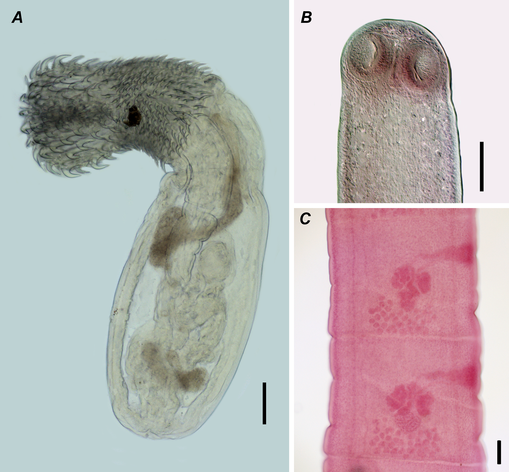
Figure 2. Long description
A three-panel set of photomicrographs.
Panel A, located on the left, shows a lateral view of a Centrorhynchus globocaudatus cystacanth. The organism has a prominent, curved anterior proboscis densely covered in numerous small, dark, recurved hooks. The body is translucent and elongated, containing internal structures including a dark central mass and coiled tubular organs. A vertical black scale bar representing 100 micrometers is positioned at the bottom right of the panel.
Panel B, at the top right, shows a ventral view of the scolex of Oochoristica tuberculata. The head is rounded and features two visible circular suckers at the apex. The tissue is granular and light gray. A vertical black scale bar representing 100 micrometers is located to the right of the specimen.
Panel C, at the bottom right, shows a stained pink section of Oochoristica tuberculata proglottids. Two rectangular segments are visible, each containing a central cluster of darker pink, lobed reproductive organs and smaller granular structures. A vertical black scale bar representing 100 micrometers is placed at the bottom right corner.
Site of infection: body cavity (encysted cystacanths).
Prevalence and intensity: one of nine lizard specimens (11%) was infected with two cystacanths.
Remarks. Found cystacanths were identified as C. globocaudatus based on the following morphological characters: 34 longitudinal rows of hooks with 20–21 hooks in each; of these 6 hooks are large with powerful roots, 2–3 transitional hooks with cross-shaped root processes, and the following hooks are spiny with shield-shaped root processes. The species commonly parasitises birds of prey as definitive hosts and terrestrial arthropods as intermediate ones, while various terrestrial vertebrates may serve as paratenic hosts (Lisitsyna and Greben Reference Lisitsyna and Greben2015; Lisitsyna Reference Lisitsyna2019; Amin et al. Reference Amin, Heckmann, Dallarés, Constenla and Rubini2020). To the best of our knowledge, there were no previous records of Centrorhynchus species in wall lizards; however, numerous findings of Centrorhynchus cystacanths from amphibians and reptiles have been recorded throughout the Palaearctic (Düşen et al. Reference Düşen, Kumlutaş, Ilgaz, Avci and Yakagül2016).
Of the 20 freshly collected P. tauricus, 15 were found to be infected with at least one parasite species (75%). The parasite load included the cestode Oochoristica tuberculata (Rudolphi, 1819) and the aforementioned nematode S. saxicolae.
Oochoristica tuberculata (Rudolphi, 1819) (Figure 2B, C)
Site of infection: intestine.
Prevalence and intensity: one of 20 studied lizards (5%) was infected with 11 cestode specimens.
Remarks. Collected specimens were identified based on distinctive morphological features including the morphology of the scolex (absence of armament and a rostellum; the suckers 95–100 μm × 82–90 μm in size), the position of the genital atrium (in the anterior third of the lateral proglottid margin), and the position (in the posterior part of the median field of the proglottid) and number (31–35) of the testes (Figure 2C). The species is widely distributed in the Old World and occurs primarily in lizards, and only rarely in snakes (Birlik et al. Reference Birlik, Yıldırımhan, Ilgaz and Kumlutaş2018; Sharpilo Reference Sharpilo1976; Yildirimhan et al. Reference Yıldırımhan, Goldberg and Bursey2006). In Ukraine, it has previously been recorded from Lacerta agilis L. and Eremias arguta (Pallas, 1773) (Sharpilo Reference Sharpilo1976). The present finding represents the first record of O. tuberculata in P. tauricus.
Spauligodon saxicolae Sharpilo, Reference Sharpilo1961
Site of infection: intestine.
Intensity: 1–39 (mean – 11, median – 7).
Prevalence: 75% (15 of 20 specimens were infected).
Remarks. Collected nematode specimens were morphologically identical to those collected from P. muralis, including details of male and female caudal end morphology and egg size.
In addition to newly collected material, historical collection data (a handwritten database of parasites identified by Viktor Sharpilo) from the Department of Parasitology (I. I. Schmalhausen Institute of Zoology) were analysed (Supplementary file 1). These data included 116 specimens of P. tauricus collected between 1951 and 1990 from southern Ukraine (Odesa region, Kherson region, and Crimea).
In the entire host sample of 116 lizards, 21 (18.1%) were infected with at least one helminth specimen. A total of nine helminth species were recorded in the entire sample (Table 1; Supplementary file 1). Of these, four species (Skrjabinelazia taurica Sypliaxov, 1930, Spauligodon sp., Centrorhynchus sp., and an unidentified nematode larva) were each represented by a single specimen. Another species was also found in only one lizard, but it reached a high abundance of 70 nematode larvae identified as Agamospirura minutissima Sharpilo, Reference Sharpilo1976. However, many spirurate larval stages that were previously identified as Agamospirura spp. in Ukrainian vertebrates were later re-identified as belonging to other genera or even families (Syrota and Kharchenko Reference Syrota and Kharchenko2015). Thus, we prefer not to use that name but rather leave these nematodes as Spirurida gen. sp.
Collection data on helminths of 116 Podarcis tauricus collected from various areas of southern Ukraine during 1951–1990

Table 1. Long description
The table contains five columns: Parasite species, Prevalence percent, Intensity min-max, Intensity mean and [median], and Mean abundance. The data is categorized into three taxonomic groups:
1. Nematoda:
* Spauligodon saxicolae: 4.3 percent prevalence, intensity 5-17, mean and median 11, mean abundance 0.46.
* S. taurica: 0.9 percent prevalence, intensity 1, mean and median 1, mean abundance 0.01.
* Spauligodon sp.: 0.9 percent prevalence, intensity 1, mean and median 1, mean abundance 0.01.
* Physocephalus sexalatus: 1.7 percent prevalence, intensity 1-18, mean 9.5 and median 10, mean abundance 0.16.
* Spirurida gen. sp.: 0.9 percent prevalence, intensity 70, mean and median 70, mean abundance 0.6.
* Nematoda gen. sp.: 0.9 percent prevalence, intensity 1, mean and median 1, mean abundance 0.01.
2. Cestoda:
* Oochoristica tuberculata: 1.7 percent prevalence, intensity 2-4, mean and median 3, mean abundance 0.05.
3. Acanthocephala:
* Sphaerirostris picae: 7.8 percent prevalence, intensity 1-7, mean and median 2, mean abundance 0.17.
* Centrorhynchus sp.: 0.9 percent prevalence, intensity 1, mean and median 1, mean abundance 0.01.
A footnote indicates that for species with a superscript ‘a’, only a single parasite specimen was recorded in the entire host sample.
a Only a single parasite specimen was recorded in the entire host sample.
The most prevalent parasite among the other species in the historical dataset was the acanthocephalan Sphaerirostris picae (Rudolphi, 1819), recorded in nine hosts from multiple localities. Two species from the present study were also listed in the collection data. Spauligodon saxicolae was found in five lizards, and O. tuberculata was found in two lizards collected in two localities in Crimea.
Discussion
The parasite list for all studied lizards included ten parasite species, the great majority of which were recorded with low prevalence and low mean abundance (Figure 3). Previous surveys of helminths in native populations of P. muralis from Spain (Garcia-Adell and Roca Reference Garcia-Adell and Roca1988), Bulgaria (Kirin Reference Kirin2002), and north-western Türkiye (Yıldırımhan and Sümer Reference Yıldırımhan and Sümer2019) have reported higher helminth species richness than that observed in our study. All authors found the cestode Mesocestoides sp. and nematodes of the genera Skrjabinodon Inglis, 1968, Skrjabinelazia Sypliaxov, 1930, and Spauligodon Skrjabin, Schikhobalova et Lagodovskaja, 1960, suggesting that they form the core components of the helminth assemblages of P. muralis. Additionally, the trematode P. molini was reported in lizards from Spain and Türkiye.
Bubble-plot visualisation of helminth abundance and prevalence from three samples: Podarcis muralis, P. tauricus modern data, and P. tauricus historical data.
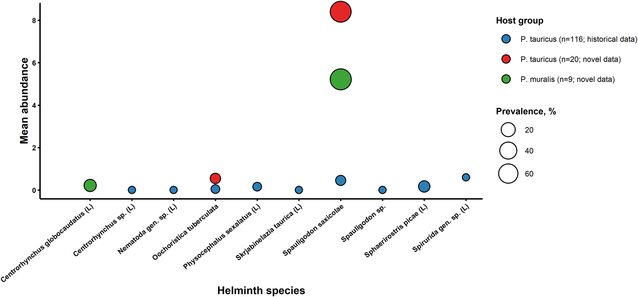
Figure 3. Long description
The bubble plot uses the X-axis for Helminth species and the Y-axis for Mean abundance ranging from 0 to 8. A legend on the right identifies three Host groups: P. tauricus historical data (blue), P. tauricus novel data (red), and P. muralis novel data (green). Bubble size represents Prevalence percentage, with a scale showing 20, 40, and 60 percent.
From left to right along the X-axis:
* Centrorhynchus globocaudatus (L): One medium green bubble at abundance 0.2.
* Centrorhynchus sp. (L): One small blue bubble near 0.
* Nematoda gen. sp. (L): One small blue bubble near 0.
* Oochoristica tuberculata: One small blue bubble at 0 and one small red bubble slightly above it.
* Physocephalus sexalatus (L): One small blue bubble near 0.
* Skrjabinelazia taurica (L): One small blue bubble near 0.
* Spauligodon saxicolae: Three bubbles vertically aligned. A small blue bubble near 0.5, a large green bubble at abundance 5.2, and the largest red bubble at abundance 8.5.
* Spauligodon sp.: One small blue bubble near 0.
* Sphaerirostris picae (L): One small blue bubble near 0.2.
* Spirurida gen. sp. (L): One small blue bubble near 0.6.
In the present study, only nine specimens of P. muralis from a single locality were examined; however, despite the limited sample size, two helminth species were detected, indicating that even recently established or localised populations may harbour parasite communities. Of these, the nematode S. saxicolae has been previously reported from this host as well as from other lacertid lizards in Ukraine, whereas the acanthocephalan C. globocaudatus has not previously been recorded in P. muralis. Thus, the present finding represents a novel host record and expands the known host range of this acanthocephalan. However, Centrorhynchus sp. was previously listed for this host in the collection data of the I. I. Schmalhausen Institute of Zoology and for other lizards in Ukraine (Sharpilo Reference Sharpilo1976), it is possible that some of those earlier records correspond to C. globocaudatus, given the known difficulties in identifying larval acanthocephalans to species level.
In the current study, we did not find Mesocestoides and P. molini, and only S. saxicolae was observed among nematodes. Such depauperate helminth assemblages of P. muralis may be at least partially explained by its occurrence in an industrial, highly transformed environment in the studied region. In Ukraine, P. muralis was recorded in the area of the trading port of Reni (Odesa region), where it inhabited ruined buildings downstream along the Danube River. In this highly transformed port environment, P. muralis occurred syntopically with Lacerta viridis (Red Book of Ukraine), whereas in more natural biotopes on the outskirts of the city, it was observed in proximity to the native P. tauricus (Figure 4). Such spatial association with native lacertid lizards may facilitate the incorporation of the invasive species into local trophic and parasite-transmission networks. Such urban habitats may lead to the restriction of lizards’ diet and thus limit trophic transmission pathways for certain helminth species that require aquatic invertebrates as intermediate hosts (Plagiorchis sp.) or predatory mammals as definitive hosts (Mesocestoides sp.).
Photographs of Podarcis species from Odesa region, Ukraine: A – P. muralis from the building wall; B – P. tauricus from the tree bark in the natural environment.

Nevertheless, the detection of C. globocaudatus cystacanths in P. muralis, even though at low prevalence and abundance, suggests that non-native wall lizards can still become integrated into local trophic networks. Given that definitive hosts of Centrorhynchus are typically birds of prey, the occasional presence of cystacanths in lizards may facilitate parasite transmission across trophic levels. Similar patterns have been reported for other reptiles and amphibians in the Palaearctic region (Düşen et al. Reference Düşen, Kumlutaş, Ilgaz, Avci and Yakagül2016).
Spauligodon saxicolae was the most prevalent helminth species in both P. muralis and P. tauricus, infecting the majority of examined individuals. Its high prevalence and relatively high intensity indicate that it is a core component of the helminth community in lacertid lizards in southern Ukraine. Since this nematode has been previously reported from P. muralis and related lacertid hosts in the region, its occurrence in the invasive population does not allow a clear distinction between spillover and spillback processes. However, the shared parasite fauna between native and invasive hosts suggests the existence of a common parasite pool, which may facilitate parasite exchange between host species.
Comparison with historical collection data, despite the limitations (time gap between sampling, various locations, etc.), reveals that the helminth assemblages of P. tauricus have remained relatively species-poor over time, with most parasite taxa occurring at low prevalence. The archival dataset included a greater number of taxa overall; however, most were rare and represented by single records, indicating a highly uneven parasite community structure. The most frequently recorded parasite in historical material, the acanthocephalan S. picae, was not observed in the present study, which may reflect temporal variation or, more likely, differences in sample size and sampling localities.
Comparing the data on parasites from the native and invasive populations of P. muralis based on the literature and the present study, we may conclude that the involvement of this lizard in parasite transmission may not be dramatic. Unfortunately, the available data cannot be directly compared due to geographical and time differences; thus, we may only hypothesise about the impact of the invasive species on the current state of the native reptiles. However, further studies of larger samples of this species from more localities, as well as studies of other invasive species occurring in Ukraine, such as the red-eared slider Trachemys scripta (Wied, 1839) and Bogdanov’s gecko Tenuidactylus bogdanovi Nazarov et Poyarkov, 2013 (Кurtyak and Kurtyak Reference Kurtyak and Kurtyak2013; Krasylenko and Kukushkin Reference Krasylenko and Kukushkin2017; Nekrasova et al. Reference Nekrasova, Tytar, Pupins and Čeirāns2022; Reference Nekrasova, Pupins, Marushchak, Tytar, Martinez-Silvestre, Škute, Čeirāns, Theissinger and Georges2024; Oskyrko et al. Reference Oskyrko, Lysenko, Obikhod, Yakovliev and Haidash2024), may reveal additional data for a clear picture of the impact of invasive reptiles on the native fauna in Ukraine.
Supplementary material
The supplementary material for this article can be found at http://doi.org/10.1017/S0022149X26102302.
Acknowledgements
The authors wish to express their sincere thanks to the Armed Forces of Ukraine for the ability to continue research even during the full-scale Russian aggression.
Financial support
This study was financially supported by the National Research Foundation of Ukraine (project number 2023.03/0068). Specimens of Podarcis muralis were collected with the support of a Rufford Small Grant ‘Setting conservation priorities for reptiles and their habitats in Ukrainian Bessarabia’ (ID: 34322-2) issued to O. Oskyrko.
Competing interests
The authors declare no conflicts of interest.
Ethical standards
The study was conducted in accordance with applicable ethical standards and regulations for the use of animals in research.
AI use
The free version of the Grammarly software was used to verify the grammar of the text, and all authors take full responsibility for the contents of the paper.




 Open access

